Picture Labelling Trainer
The examiner puts a dissection figure in front of you with numbered leader lines and asks you to name each structure. There is no partial credit for nearly — you either have the name or you don't. These are the plates TMU sets as drawing homework.
Why drawing beats reading, for this section
You can read the boundaries of the axilla twenty times and still freeze when a photograph of a dissected axilla appears on the page. Reading builds recognition — the comfortable feeling that you have seen something before. The picture question tests recall, which is a different and much harder skill, and the two feel identical right up until the exam.
Drawing is the cheapest way to force recall. When you draw the femoral triangle from memory you cannot fudge whether sartorius is lateral or medial — the pencil makes you commit. That is exactly why your department sets these as homework and marks them, and it is why the lecture slides end with "draw a figure" rather than "read pages 564–565".
Every plate starts closed on purpose. Read the prompt, then get a blank sheet and actually draw it — badly is fine, the drawing is a means not an end. Only then open the checklist to see what you missed, and open the model drawing to compare. Opening the picture first turns the whole exercise back into reading, and you will learn almost nothing.
One practical warning about the model drawings below. They are real TMU student homework, marked by the department, and they are very good — but they are not a textbook. Where a drawing and the checklist disagree, trust the checklist: it is sourced to Gray's, Snell's, or a real past paper, and the source is printed under every list.
- Why does re-reading fail for this section? → It builds recognition, not recall — and the two feel the same until the exam
- What should you do before opening a plate? → Draw it from memory on paper
- If the drawing and the checklist disagree, which wins? → The checklist — it is the sourced one
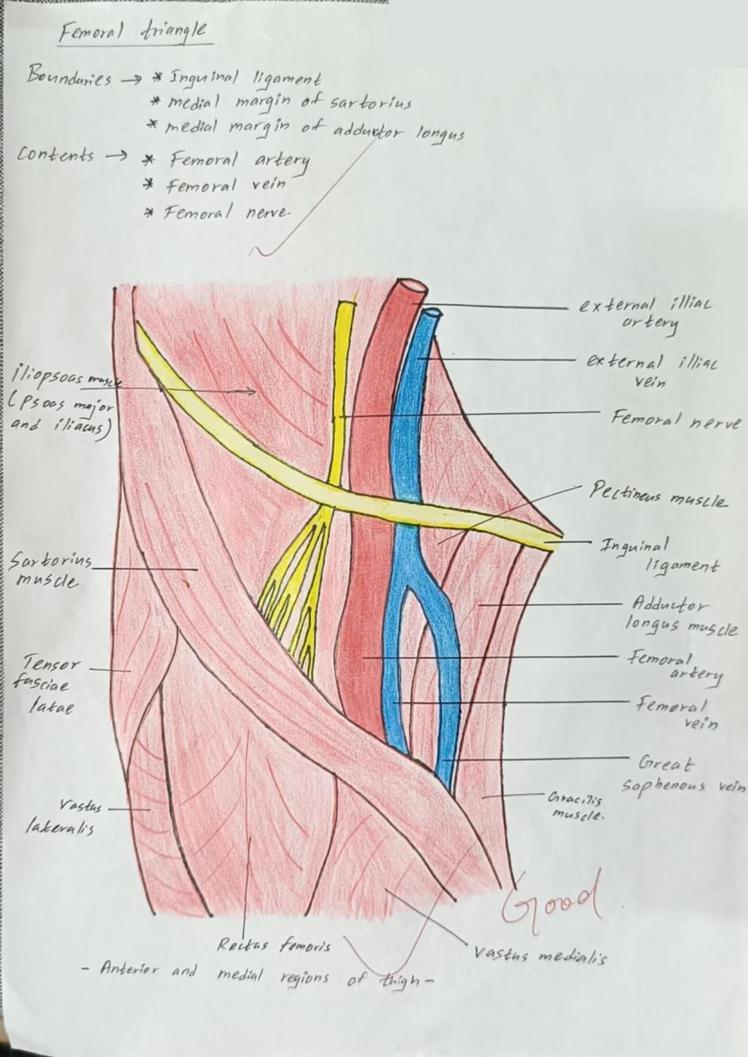
Draw the anterior and medial regions of the thigh. Label the boundaries and contents of the femoral triangle.
- Boundaries: inguinal ligament · medial margin of sartorius · medial margin of adductor longus
- Contents, lateral → medial: femoral nerve · femoral artery · femoral vein · lymphatics
- Iliopsoas (psoas major + iliacus) forming the lateral floor
- Pectineus and adductor longus forming the medial floor
- External iliac artery and vein becoming femoral above the ligament
- Great saphenous vein joining the femoral vein
- Sartorius · tensor fasciae latae · rectus femoris · vastus lateralis · vastus medialis · gracilis
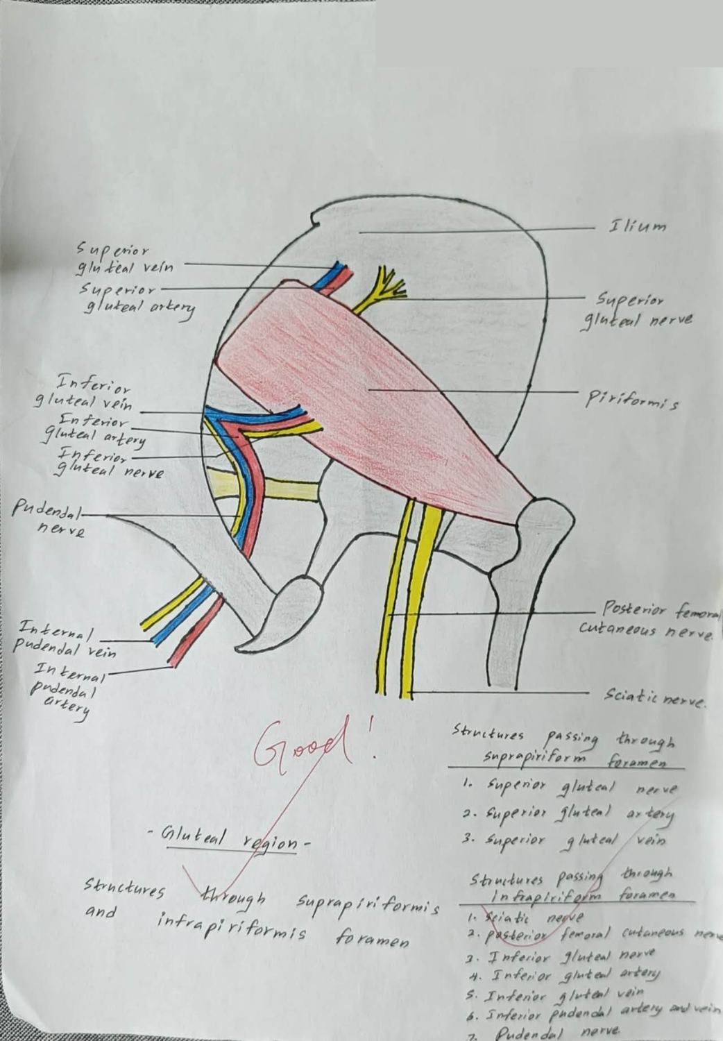
Draw the gluteal region. Show piriformis dividing the greater sciatic foramen, and label everything passing above and below it.
- Piriformis — sacrum to greater trochanter — dividing the greater sciatic foramen
- Suprapiriform (3), lateral → medial: superior gluteal nerve · artery · vein
- Infrapiriform (8), lateral → medial: sciatic nerve · posterior femoral cutaneous nerve · inferior gluteal nerve · inferior gluteal artery · inferior gluteal vein · internal pudendal artery · internal pudendal vein · pudendal nerve
- Sacrospinous ligament (makes the greater sciatic foramen)
- Sacrotuberous ligament (makes the lesser sciatic foramen)
- Gluteus maximus reflected; gluteus medius and minimus
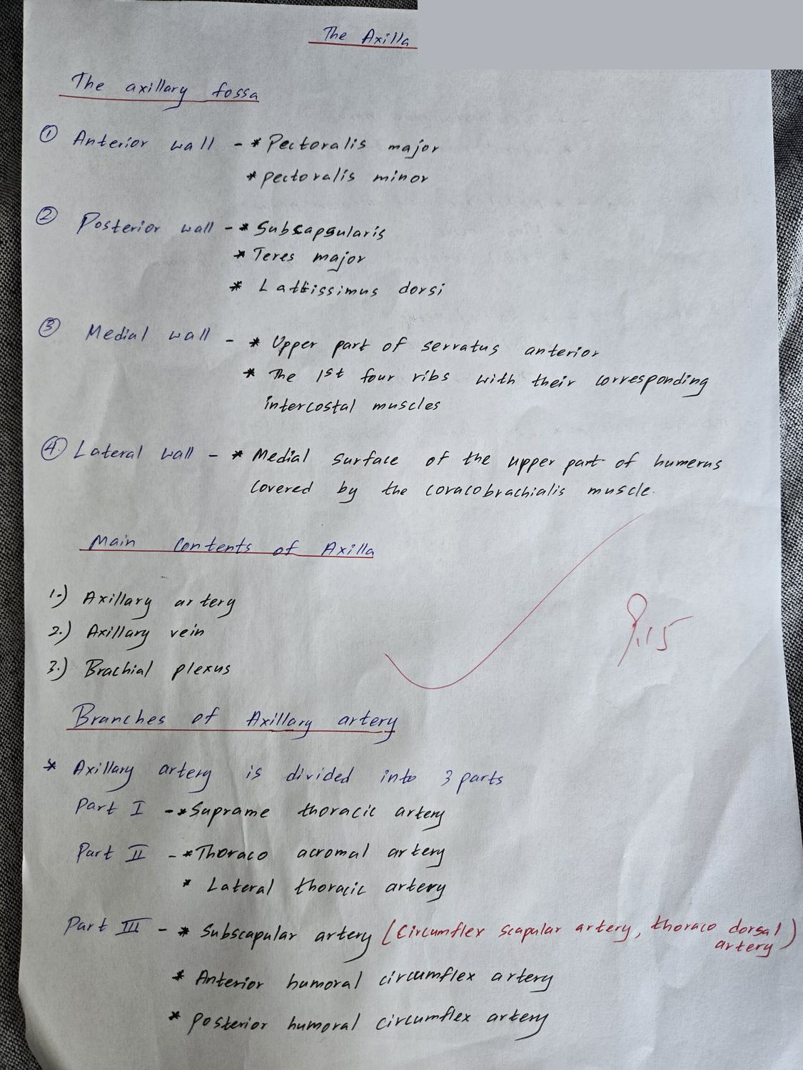
Draw the axilla. Label its four walls, apex and base, and the contents.
- Anterior wall: pectoralis major and minor, subclavius
- Posterior wall: subscapularis, latissimus dorsi, teres major
- Medial wall: upper 4–5 ribs with intercostal muscles, serratus anterior
- Lateral wall: intertubercular (bicipital) groove of the humerus
- Apex: between clavicle, first rib and upper border of scapula
- Contents: axillary artery and vein · cords and branches of the brachial plexus · axillary lymph nodes · axillary sheath · fat
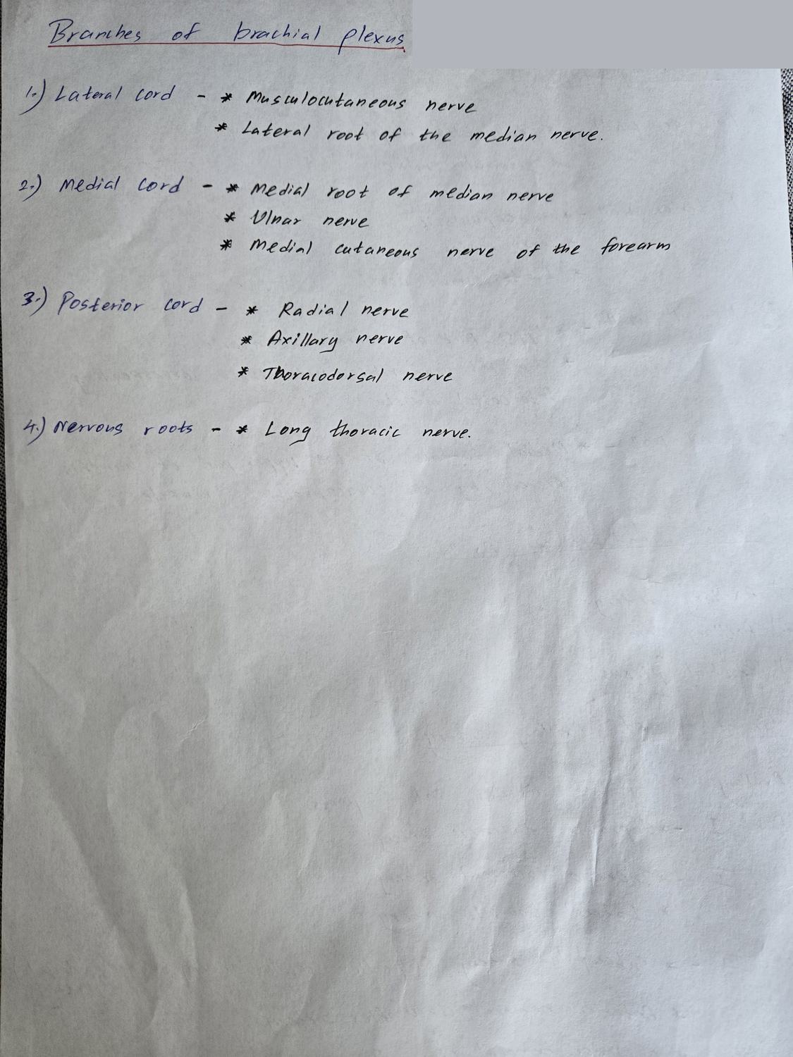
Draw the brachial plexus. Give the branches of each cord.
- Lateral cord: musculocutaneous nerve · lateral pectoral nerve · lateral root of the median nerve
- Medial cord: ulnar nerve · medial root of the median nerve · medial pectoral nerve · medial cutaneous nerves of arm and forearm
- Posterior cord: axillary nerve · radial nerve · upper and lower subscapular nerves · thoracodorsal nerve
- Roots C5–T1 · trunks (upper, middle, lower) · divisions · cords
- Long thoracic nerve (C5–7) from the roots — serratus anterior, winged scapula
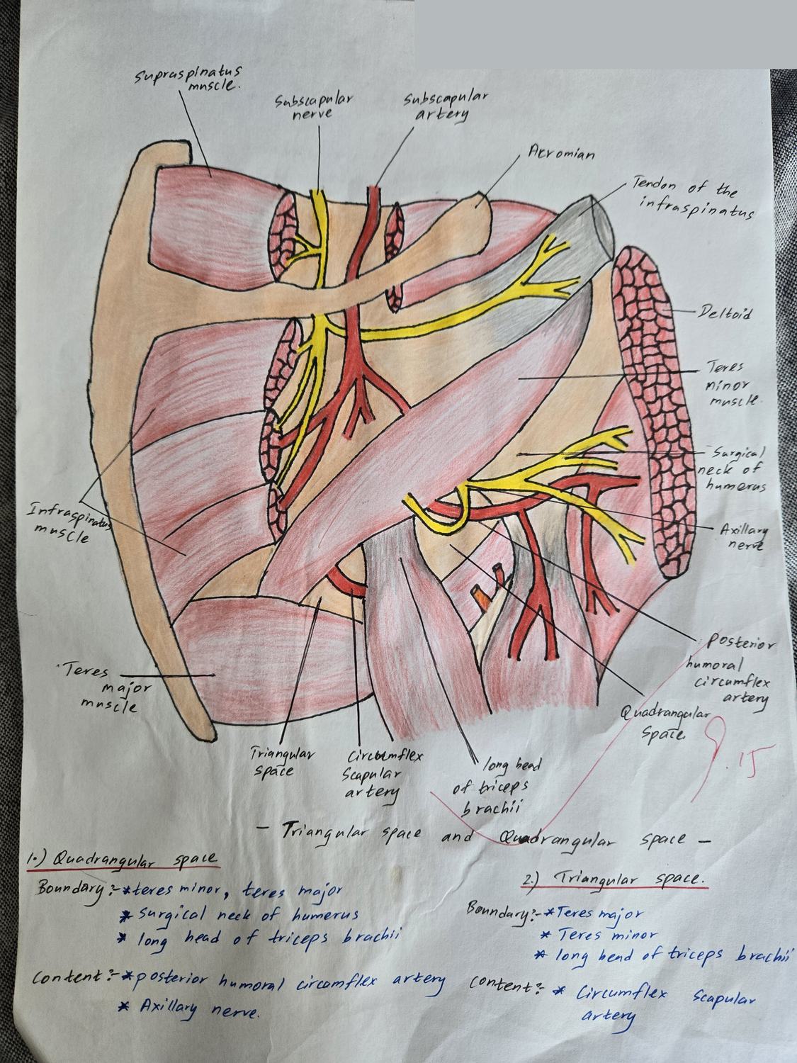
Draw the scapular region. Label the boundaries and contents of the quadrangular and triangular spaces.
- Quadrangular space — above: subscapularis, teres minor and shoulder joint capsule
- — below: teres major · medially: long head of triceps · laterally: surgical neck of humerus
- Quadrangular contents: axillary nerve · posterior circumflex humeral artery and vein
- Triangular space — above: teres minor · below: teres major · laterally: long head of triceps
- Triangular contents: circumflex scapular artery
- Supraspinatus · infraspinatus · subscapularis · deltoid
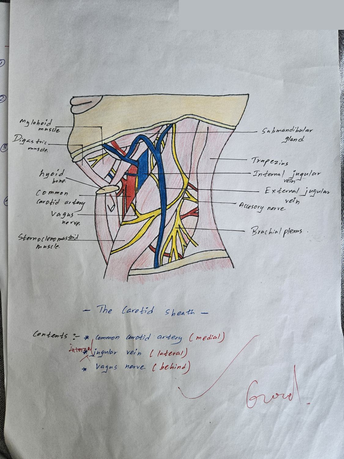
Draw a cross-section of the neck showing the carotid sheath. Label its contents and their relations.
- Common and internal carotid arteries — medially
- Internal jugular vein — laterally
- Vagus nerve [X] — posteriorly, in the angle between artery and vein
- Deep cervical lymph nodes along the sheath
- Formed from all three layers of deep cervical fascia — investing, pretracheal, prevertebral
- Sternocleidomastoid overlying it; ansa cervicalis on its anterior surface
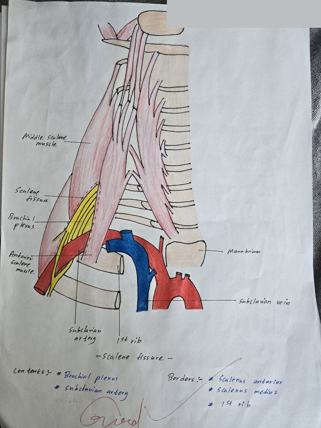
Draw the root of the neck. Label the scalene fissure and everything passing through and around it.
- Boundaries: scalenus anterior in front · scalenus medius behind · first rib below
- Contents: subclavian artery (third part) · roots of the brachial plexus
- ⚠️ Subclavian vein passes IN FRONT of scalenus anterior — not in the fissure
- Phrenic nerve descending on the front of scalenus anterior
- Cervical pleura and the apex of the lung below
- Thoracic duct on the left, arching to the venous angle
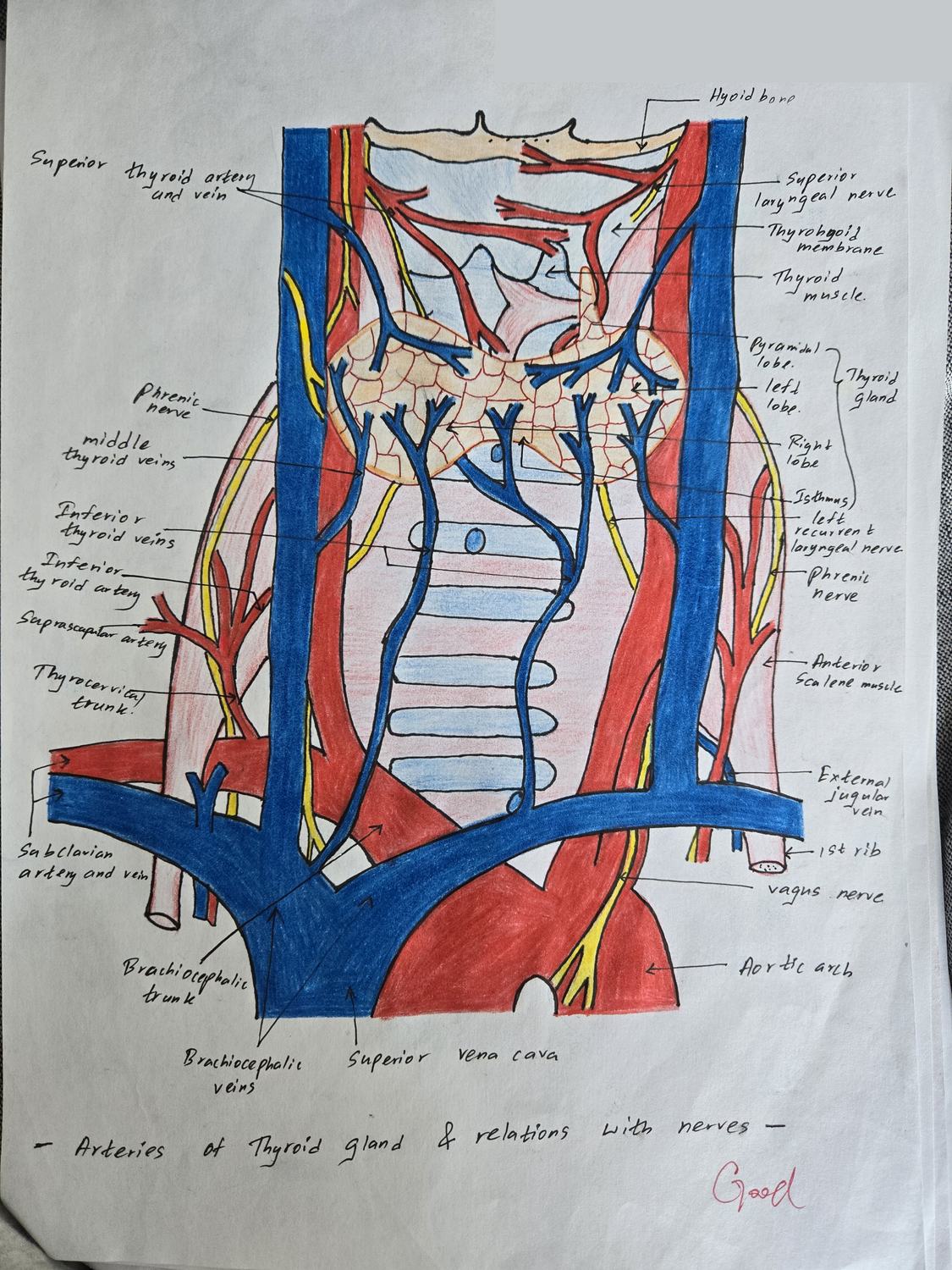
Draw the thyroid gland. Label its arteries, veins and the nerves related to them.
- Superior thyroid artery — from the external carotid artery — to the upper pole
- Inferior thyroid artery — from the thyrocervical trunk of the subclavian artery — to the lower pole
- Thyroidea ima artery (inconstant) — from the brachiocephalic trunk or arch of aorta
- Superior and middle thyroid veins → internal jugular vein
- Inferior thyroid veins → brachiocephalic veins
- External laryngeal nerve — runs with the superior thyroid artery (at risk in upper-pole ligation)
- Recurrent laryngeal nerve — runs with the inferior thyroid artery (at risk in lower-pole ligation)
- Isthmus over tracheal rings 2–4; hyoid bone and laryngeal cartilages above
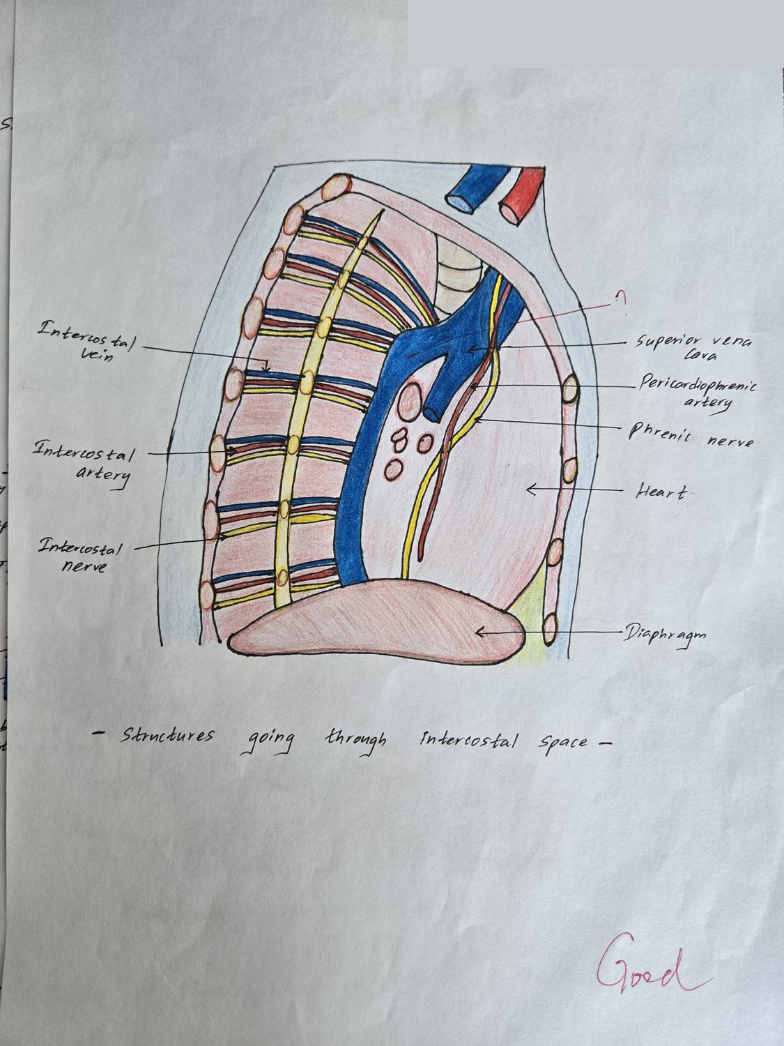
Draw an intercostal space in cross-section. Label the muscle layers and the neurovascular bundle in order.
- Neurovascular order in the costal groove, from ABOVE downwards: vein · artery · nerve (VAN)
- Bundle lies in the costal groove of the upper rib, between the internal and innermost intercostal muscles
- Three muscle layers: external intercostal · internal intercostal · innermost intercostal
- Posterior intercostal arteries — from the thoracic aorta (spaces 3–11)
- Anterior intercostal arteries — from the internal thoracic artery
- Parietal pleura innermost; endothoracic fascia
- Collateral branch running along the upper border of the rib below
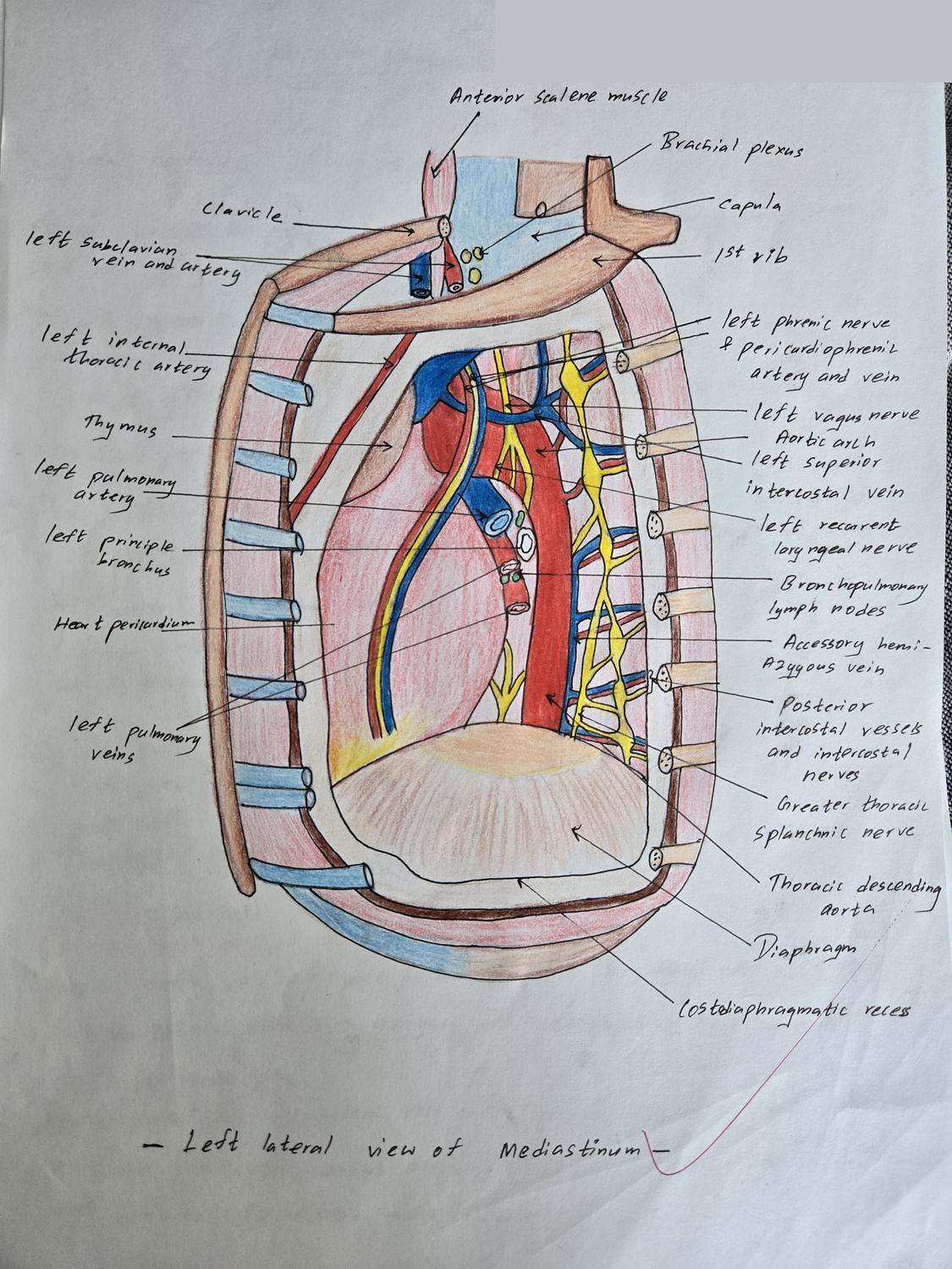
Draw the left side of the mediastinum as seen after removing the left lung. Label the structures.
- Arch of the aorta with its three branches; descending thoracic aorta
- Left phrenic nerve — passes anterior to the lung root
- Left vagus nerve — passes posterior to the lung root
- Left recurrent laryngeal nerve hooking under the arch of the aorta
- Left superior intercostal vein · hemiazygos and accessory hemiazygos veins
- Left subclavian and left common carotid arteries
- Thoracic duct · oesophagus · left sympathetic trunk
- Left lung root: bronchus, pulmonary artery and veins
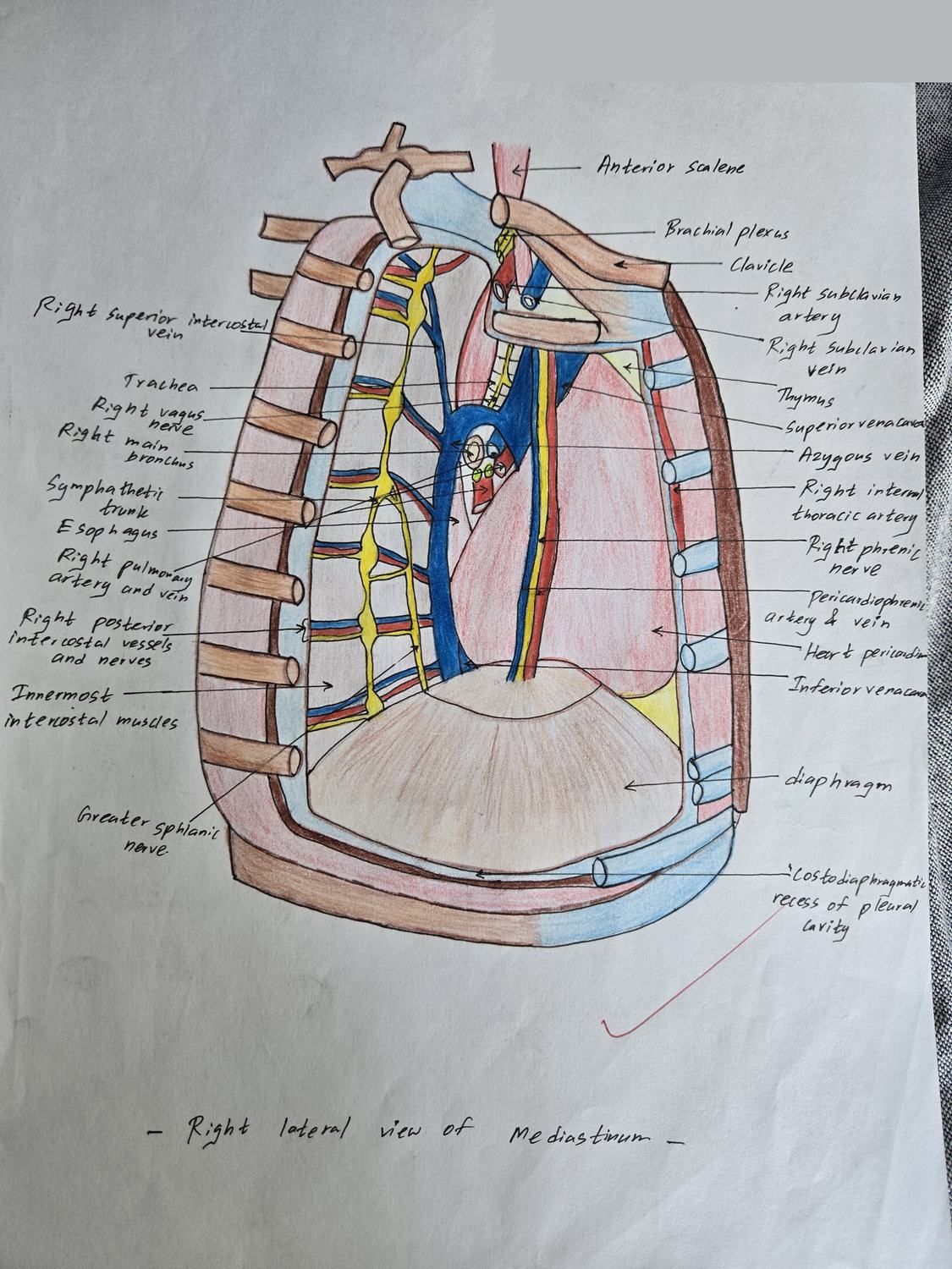
Draw the right side of the mediastinum as seen after removing the right lung. Label the structures.
- Arch of the azygos vein arching over the right lung root to the superior vena cava
- Superior vena cava and the right brachiocephalic vein
- Right phrenic nerve — anterior to the lung root
- Right vagus nerve — posterior to the lung root
- Oesophagus · trachea and its bifurcation
- Right sympathetic trunk and greater splanchnic nerve
- Right lung root: bronchus, pulmonary artery and veins
Draw the chambers of the heart. Label the internal features of each.
- Right atrium: crista terminalis · musculi pectinati · fossa ovalis · openings of SVC, IVC and coronary sinus
- Right ventricle: trabeculae carneae · papillary muscles · septomarginal trabecula (moderator band) · infundibulum
- Left atrium: four pulmonary vein openings · smooth wall · left auricle
- Left ventricle: thickest wall · two papillary muscles · aortic vestibule
- Tricuspid valve (3 cusps) and mitral/bicuspid valve (2 cusps)
- Pulmonary and aortic semilunar valves
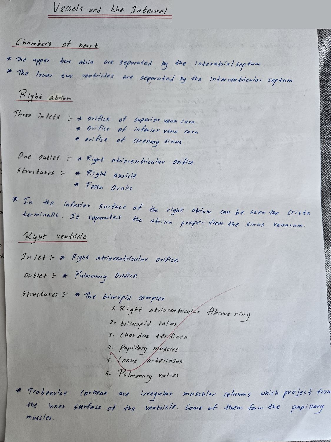
Give the inlets and outlets of each chamber of the heart, and the valves guarding them.
- Right atrium — inlets: superior vena cava · inferior vena cava · coronary sinus. Outlet: right atrioventricular orifice
- Right ventricle — inlet: right AV orifice (tricuspid valve). Outlet: pulmonary orifice (pulmonary valve)
- Left atrium — inlets: four openings of the pulmonary veins. Outlet: left AV orifice
- Left ventricle — inlet: left AV orifice (mitral valve). Outlet: aortic orifice (aortic valve)
- Tricuspid complex and bicuspid complex — ring, cusps, chordae tendineae, papillary muscles
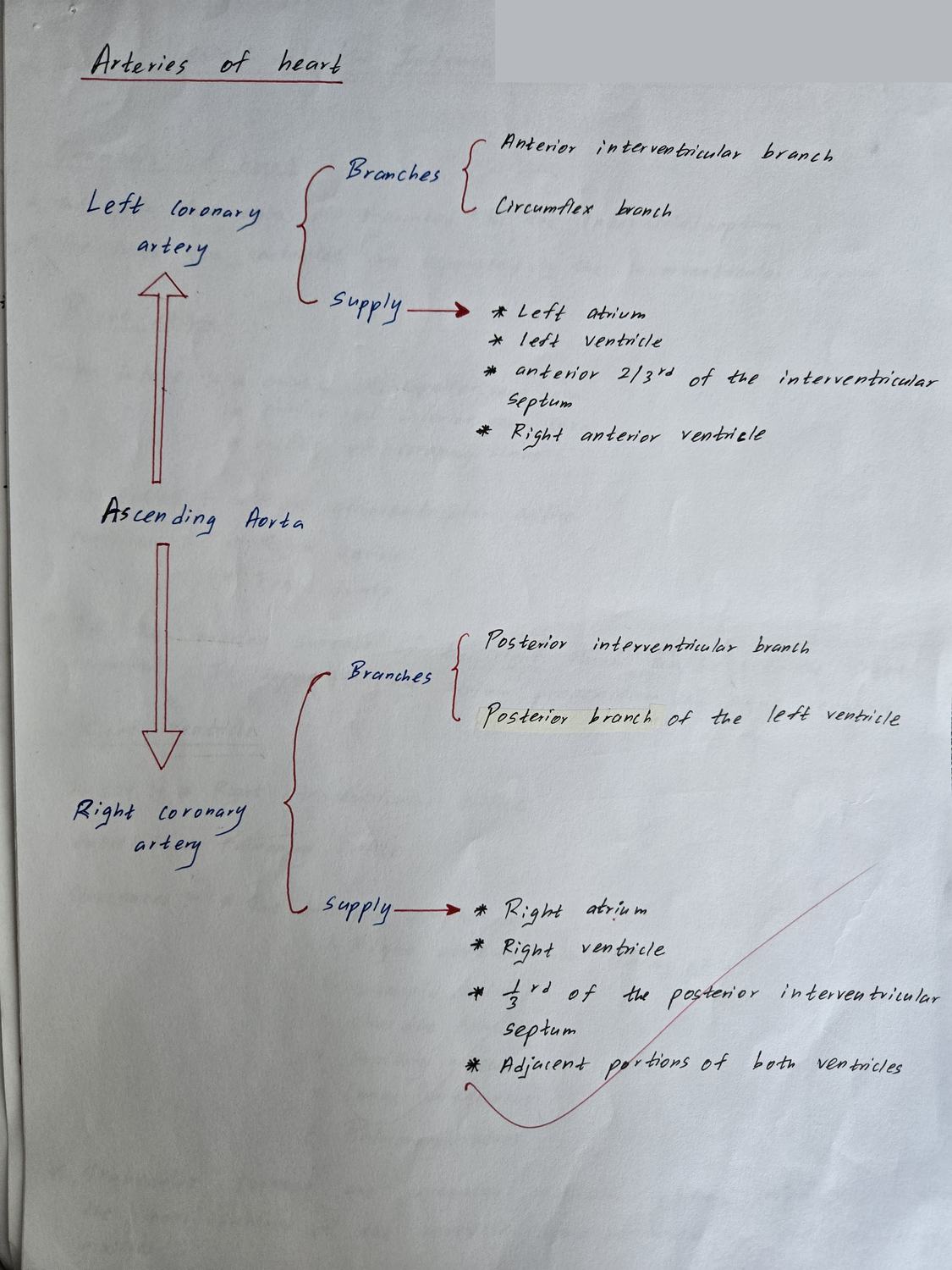
Draw the arteries of the heart. Where do they arise, and what are their branches?
- Right coronary artery — from the right aortic sinus
- RCA branches: right conus · anterior and posterior ventricular branches · right marginal artery · posterior interventricular (posterior descending) · SA and AV nodal branches
- Left coronary artery — from the left aortic sinus
- LCA branches: anterior interventricular (LAD) · circumflex · left marginal
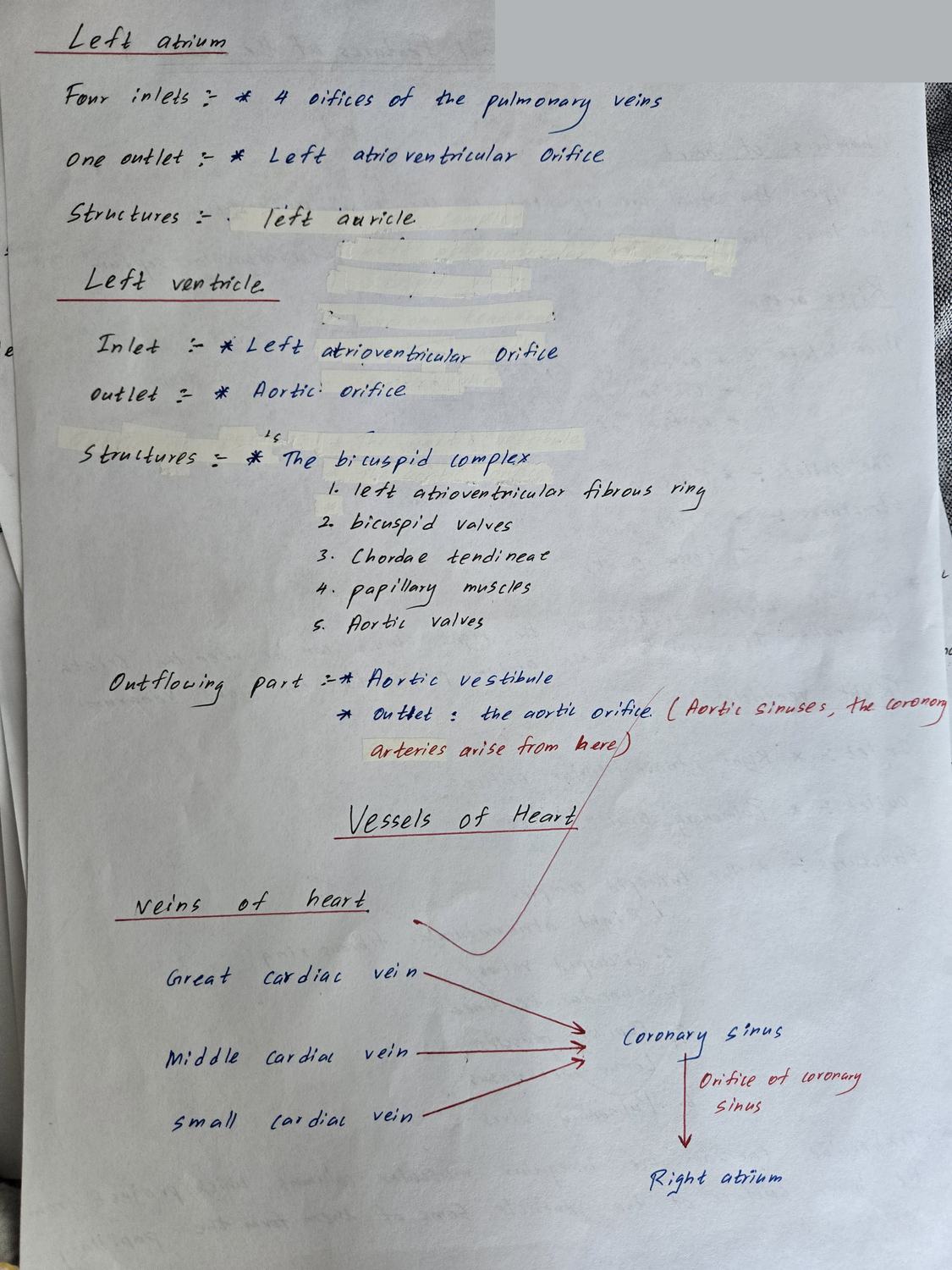
- Coronary sinus and the great, middle and small cardiac veins
- Atrioventricular crux — meeting of coronary, interatrial and interventricular grooves
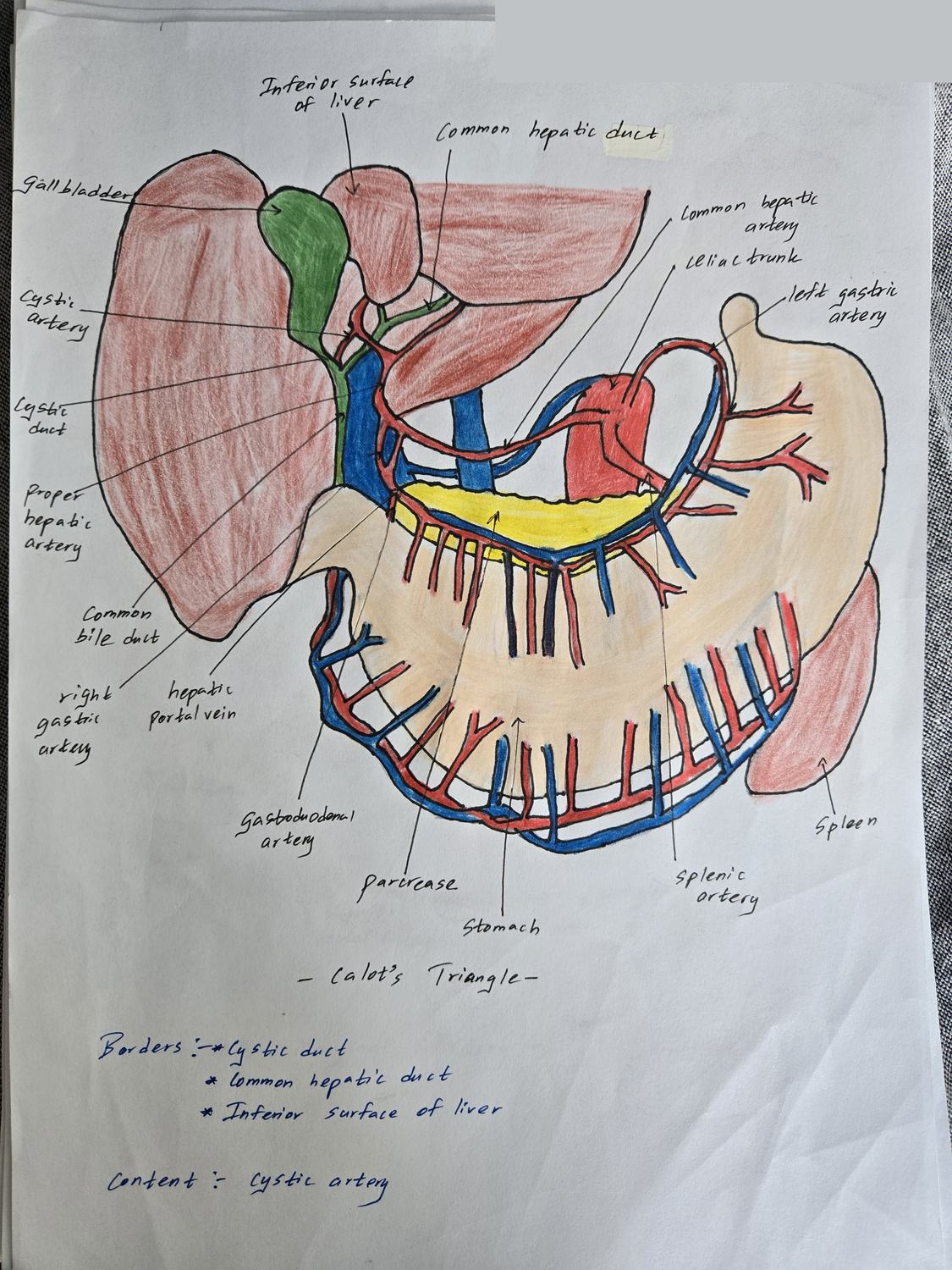
Draw the supracolic compartment showing the gall bladder triangle. Label its borders and contents, and the surrounding vessels.
- Borders: cystic duct (right) · common hepatic duct (left) · inferior surface of the liver (above)
- Content: cystic artery (usually from the right hepatic artery) · cystic lymph node
- Common bile duct · proper hepatic artery · hepatic portal vein (the portal triad)
- Celiac trunk → left gastric · splenic · common hepatic arteries
- Gastroduodenal artery · right and left gastric arteries · gastroepiploic arteries
- Gall bladder · pancreas · spleen · stomach
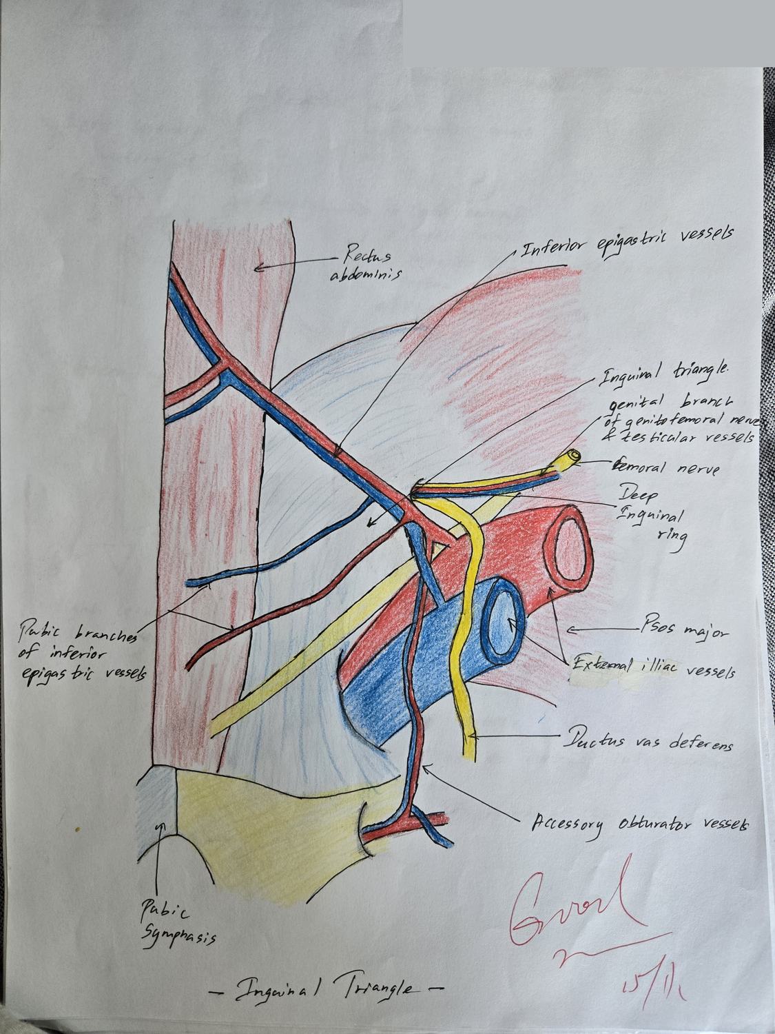
Draw the inguinal region from inside. Label the inguinal triangle and the inguinal canal.
- Inguinal triangle boundaries: inferior epigastric artery (laterally) · lateral border of rectus abdominis (medially) · inguinal ligament (inferiorly)
- Direct hernia bulges through the triangle, medial to the inferior epigastric vessels
- Indirect hernia enters the deep ring, lateral to those vessels
- Deep inguinal ring · superficial inguinal ring
- Inguinal canal walls — anterior: external oblique aponeurosis (+ internal oblique laterally); posterior: transversalis fascia + conjoint tendon; roof: arching internal oblique and transversus; floor: inguinal ligament
- Contents: spermatic cord (male) / round ligament of uterus (female) · ilioinguinal nerve
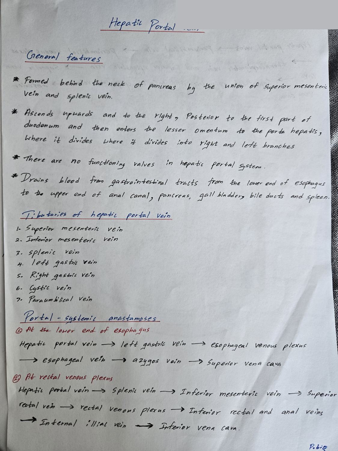
Draw the hepatic portal vein. Show how it is formed and name all its tributaries.
- Formed behind the neck of the pancreas by the union of the superior mesenteric vein and the splenic vein
- Tributaries: splenic vein · superior mesenteric vein · inferior mesenteric vein (into the splenic) · left gastric vein · right gastric vein · cystic vein · paraumbilical veins
- Runs in the free edge of the lesser omentum (hepatoduodenal ligament) with the bile duct and hepatic artery
- Divides into right and left branches at the porta hepatis
- No valves — which is why portal hypertension reverses flow
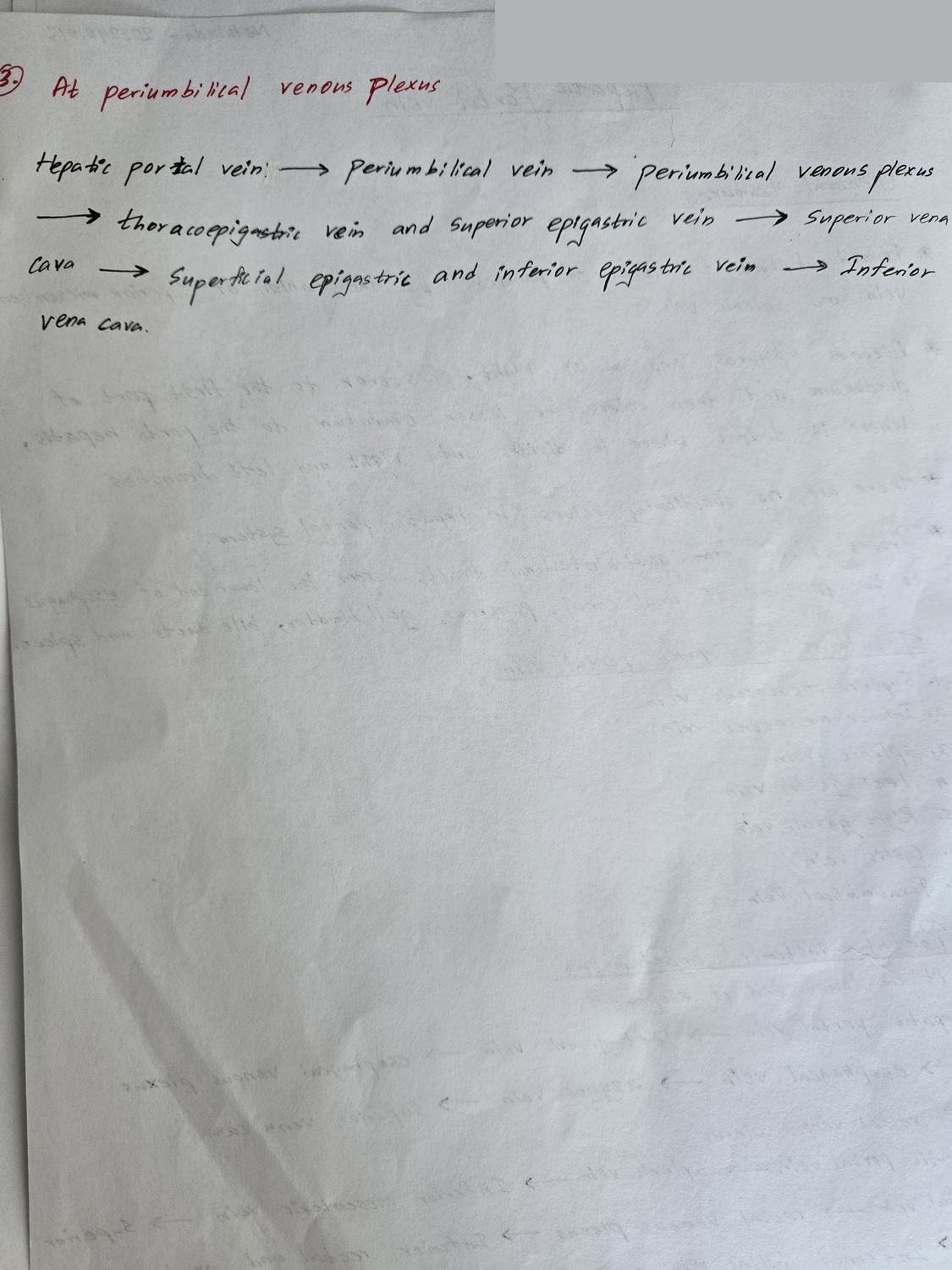
Name the sites of portosystemic anastomosis and what happens at each in portal hypertension.
- Lower oesophagus — left gastric vein ↔ oesophageal veins (azygos) → oesophageal varices
- Anal canal — superior rectal vein ↔ middle and inferior rectal veins → haemorrhoids
- Periumbilical — paraumbilical veins ↔ superficial epigastric veins → caput medusae
- Retroperitoneal / bare area of liver — colic and splenic veins ↔ retroperitoneal veins
- Portal vein has no valves, so obstruction reverses flow into these channels
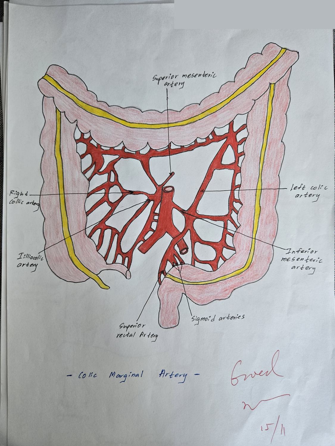
Draw the blood supply of the large intestine. Show the marginal artery and the branches of the superior and inferior mesenteric arteries.
- Superior mesenteric artery branches: inferior pancreaticoduodenal · jejunal and ileal · ileocolic · right colic · middle colic
- Inferior mesenteric artery branches: left colic · sigmoid · superior rectal
- Marginal artery (of Drummond) — the continuous anastomotic channel along the mesenteric border of the colon, linking SMA and IMA territories
- Watershed at the splenic flexure — the vulnerable point in ischaemic colitis
- Midgut/hindgut junction at the distal third of the transverse colon