The Gluteal Region
Orientation: a doorway, not a destination
The buttock looks like a solid slab of muscle, and for most of medicine that is all it is — a place to give an injection. Anatomically it is something much more interesting: it is a corridor. The pelvis is a closed bony bowl, and everything that has to get out of it to reach the perineum or the lower limb must squeeze through one of two gaps in its back wall. The gluteal region is where those structures surface, fan out, and go their separate ways.
That is why this unit is examined so heavily. Ask a student "what is in the buttock?" and you get a shrug. Ask "what passes above piriformis and what passes below it, in order?" and you are asking about the entire outflow of the sacral plexus. TMU asks the second question, and it asks it as a definition and as a drawing.
- Along the iliac crest, from the anterior superior iliac spine to the posterior superior iliac spine
- From the midpoint between the two posterior superior iliac spines
- To the tip of the coccyx
- A circular incision around the upper leg at the level of the tibial tuberosity
- A vertical cut down the middle of the back of the limb, then reflect the skin medially and laterally
Do not damage the sacrotuberous ligament — it is your landmark for the lesser sciatic foramen, and once cut you cannot put it back.
Under the skin lie the cluneal nerves — superior, medial and inferior — purely sensory branches supplying the skin of the buttock. Clunis is simply Latin for buttock, so the name is doing no more than telling you where they go.
- Why is the gluteal region called a corridor? → Everything leaving the pelvis for the limb or perineum passes through it
- Which ligament must not be cut during dissection? → The sacrotuberous ligament
- Name the three cutaneous nerves of the buttock? → Superior, medial and inferior cluneal nerves
Gluteus maximus — the lid
Gluteus maximus is the coarsest, thickest muscle in the body, and it functions as the lid over everything else in this region. Reflect it and the whole neurovascular corridor is exposed at once. Notice where it inserts: only part of it reaches bone at the gluteal tuberosity of the femur — the larger share goes into the iliotibial tract, the same band you met in Unit 1. That shared insertion is why the muscle can hold the knee straight from as far away as the hip.
Its action is worth thinking about rather than memorising. It extends and laterally rotates the thigh, but you barely use it in level walking. You recruit it when you climb stairs, rise from a chair, or run — any moment that demands powerful hip extension against gravity. And when the foot is planted, the muscle works in reverse, pulling the trunk upright over the fixed limb.
Origin: gluteal surface of the ilium and dorsal aspect of the sacrum.
Insertion: gluteal tuberosity of the femur and the iliotibial tract.
Action: extends and laterally rotates the thigh at the hip joint; raises the trunk
when the lower limb is fixed.
Nerve supply: inferior gluteal nerve.
- Which nerve supplies gluteus maximus? → Inferior gluteal nerve
- Its two insertions? → Gluteal tuberosity of femur + iliotibial tract
- When do you actually recruit it? → Stairs, standing from sitting, running — powerful hip extension
Beneath the lid: two layers of muscle
Lift gluteus maximus and you find the region is built in two storeys. The superficial group — gluteus medius and gluteus minimus, with tensor fasciae latae in front — are the abductors. The deep group — piriformis, obturator internus, the two gemelli and quadratus femoris — are a row of small lateral rotators running almost horizontally from the pelvis to the greater trochanter.
The important structural point, and the one that makes the rest of this unit make sense: the nerves and vessels run in the plane between those two layers. They are not buried in muscle; they lie in a fascial sheet sandwiched between the superficial and deep groups, which is precisely why reflecting gluteus maximus exposes them all at once.
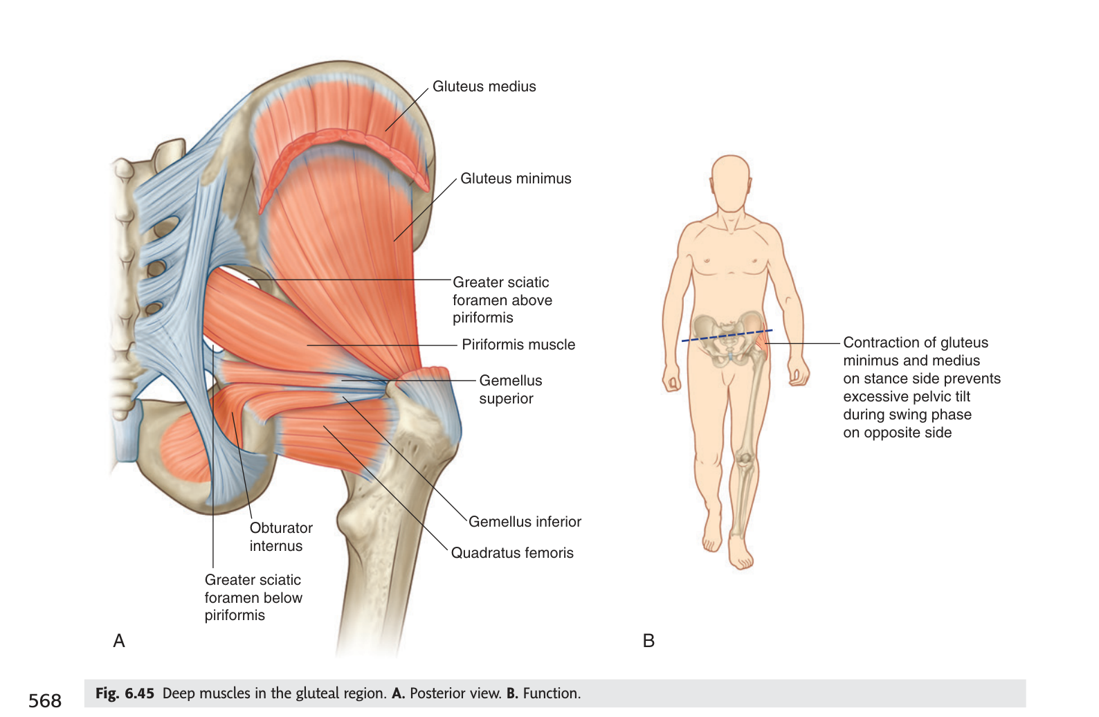
Gluteus medius and minimus do not lift your leg sideways in ordinary walking — they do the opposite. When you stand on one leg, they contract on the supporting side to stop the pelvis dropping on the swinging side. Damage the superior gluteal nerve and that stabiliser fails: when the patient stands on the affected leg, the pelvis sags on the opposite side. That is Trendelenburg's sign, and the classic iatrogenic cause is a badly placed intramuscular injection — which is exactly why the next-but-one section matters.
- Which two muscle layers are there, and what lies between them? → Superficial abductors and deep lateral rotators, with the nerves and vessels in between
- Name the deep lateral rotators? → Piriformis · obturator internus · superior and inferior gemelli · quadratus femoris
- Nerve to gluteus medius and minimus? → Superior gluteal nerve
- Which side does the pelvis drop in Trendelenburg's sign? → The opposite (unsupported) side
Piriformis — the key to the whole region ★★★
If you learn one structure in this unit, learn this one. Piriformis arises from the front of the sacrum, passes out of the pelvis through the greater sciatic foramen, and inserts on the greater trochanter. In doing so it does something no other muscle does: it fills the middle of the greater sciatic foramen and splits it into two smaller openings — a suprapiriform foramen above it and an infrapiriform foramen below it.
Think of it as a bar placed horizontally across an open doorway. The doorway was one space; now there is a gap above the bar and a gap below it, and every structure that wants through has to commit to one or the other. Learn which structures chose which gap and you have learned the gluteal region.
Origin: anterior surface of the sacrum. Insertion: greater
trochanter of the femur. Action: laterally rotates the thigh at the hip joint.
Nerve: branches from the sacral plexus.
Key relationship: divides the greater sciatic foramen into the suprapiriform and
infrapiriform foramina.
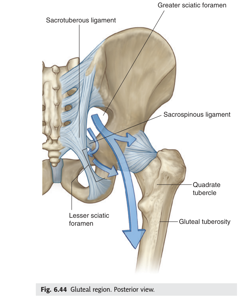
The hip bone has two notches in its back edge, not two holes. Two ligaments turn them into holes: the sacrospinous ligament closes off the greater sciatic notch to make the greater sciatic foramen, and the sacrotuberous ligament closes the lesser notch to make the lesser sciatic foramen. Piriformis then subdivides the greater one. So three ligament-and-muscle facts generate every opening in this region.
- What does piriformis divide, and into what? → The greater sciatic foramen, into suprapiriform and infrapiriform foramina
- Which ligaments create the two sciatic foramina? → Sacrospinous (greater) and sacrotuberous (lesser)
- Origin and insertion of piriformis? → Anterior surface of sacrum → greater trochanter
The suprapiriform foramen ★★
The gap above piriformis is the simple one. Only one neurovascular bundle uses it, and the three parts of that bundle keep the same order you would expect of any nerve travelling with its vessels.
The opening between the upper border of piriformis and the upper margin of the greater sciatic
foramen.
Contents, from lateral to medial: the superior gluteal nerve, the
superior gluteal artery and the superior gluteal vein.
Above piriformis, everything is superior: superior gluteal Nerve, Artery, Vein. One word, three structures, lateral to medial. If a structure in this region is called "superior gluteal", it went above.
- Three contents of the suprapiriform foramen? → Superior gluteal nerve, artery and vein
- In what order? → Lateral to medial: nerve, artery, vein
- What does the superior gluteal nerve supply? → Gluteus medius, gluteus minimus and tensor fasciae latae
The infrapiriform foramen ★★★
The gap below piriformis is the crowded one — eight structures in a single row — and it is the drawing TMU sets as homework. Do not try to memorise eight items as a flat list. Read the row as three groups, going from lateral to medial.
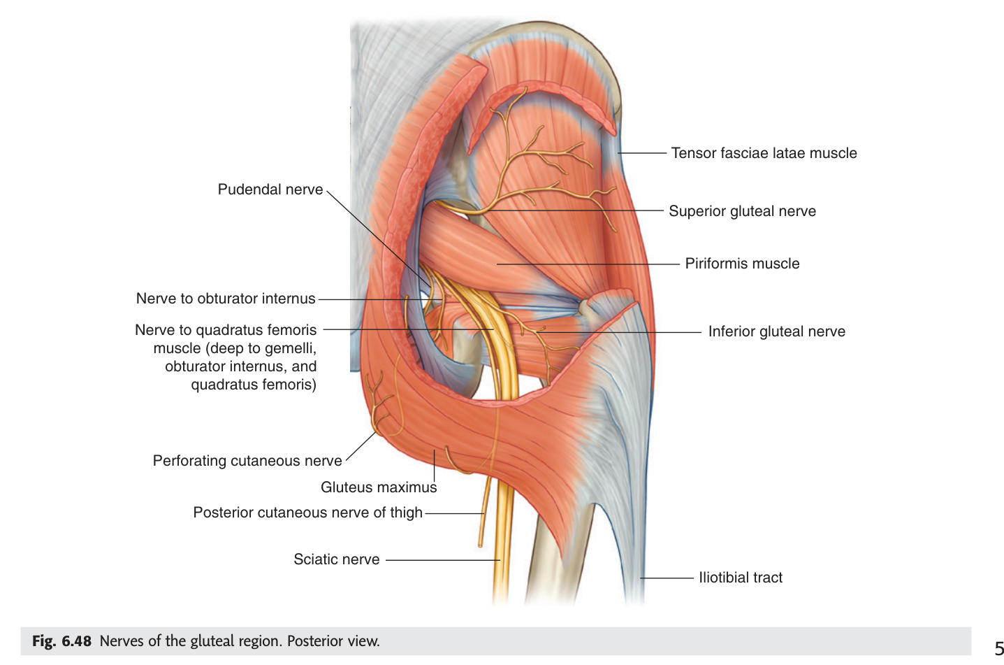
Furthest lateral is the traffic heading for the back of the thigh: the sciatic nerve, and immediately beside it the posterior femoral cutaneous nerve. Next comes the bundle that stays in the buttock to supply gluteus maximus: the inferior gluteal nerve, artery and vein. Most medial is the traffic bound for the perineum: the internal pudendal artery and vein and the pudendal nerve. Lateral to medial is therefore thigh → buttock → perineum, which is also the order of how far each has to travel.
The opening between the lower border of piriformis and the sacrospinous ligament / upper margin of
the lesser sciatic foramen.
Contents, from lateral to medial: sciatic nerve · posterior femoral cutaneous
nerve · inferior gluteal nerve · inferior gluteal artery · inferior gluteal vein · internal pudendal
artery · internal pudendal vein · pudendal nerve.
To the thigh — sciatic n., posterior femoral cutaneous n.
To the buttock — inferior gluteal n., a., v.
To the perineum — internal pudendal a., v., pudendal n.
Three groups is memorable; eight items is not.
The pudendal nerve and internal pudendal vessels do something odd: they leave the pelvis through the infrapiriform foramen, hook around the sacrospinous ligament, and immediately re-enter through the lesser sciatic foramen to reach the perineum. They are barely in the buttock at all — they merely pass behind it. This detour is what makes a pudendal nerve block possible: the needle is guided to the ischial spine, where the nerve is at its most predictable, to provide anaesthesia for the second stage of labour.
- How many structures pass the infrapiriform foramen? → Eight
- Which is most lateral? → The sciatic nerve
- Which is most medial? → The pudendal nerve
- Which structures leave and immediately re-enter through the lesser sciatic foramen? → Pudendal nerve and internal pudendal vessels
- Where is a pudendal nerve block placed? → At the ischial spine
The sciatic nerve
The sciatic nerve is the thickest nerve in the body — about as wide as your little finger where it crosses the buttock. It carries fibres from L4 to S3 and it is really two nerves travelling in one sheath: a tibial division and a common fibular (peroneal) division, which usually separate only at the top of the popliteal fossa.
You need its surface projection, because that line is what tells a clinician where not to put a needle. Mark the midpoint between the posterior superior iliac spine and the ischial tuberosity, then mark a point midway between the ischial tuberosity and the greater trochanter. The nerve runs along a curved line joining those points, then descends down the midline of the back of the thigh.
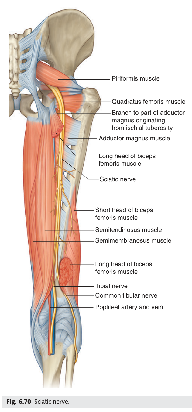
In most people the whole sciatic nerve passes below piriformis. In a minority the nerve divides inside the pelvis and the common fibular part pierces the muscle, or passes above it. Those people are vulnerable to piriformis syndrome — buttock pain radiating down the leg, worse on sitting and on internal rotation of the hip, produced by the muscle compressing the nerve rather than by any disc problem. Anatomical variation is the entire explanation for why the syndrome exists.
- Root value of the sciatic nerve? → L4–S3
- Its two terminal divisions, and where? → Tibial and common fibular nerves, at the upper angle of the popliteal fossa
- Which muscles does it supply in the thigh? → Biceps femoris, semitendinosus, semimembranosus (+ hamstring part of adductor magnus)
- What causes piriformis syndrome? → A variant course through or above piriformis, with muscular compression
The gluteal arteries
Both gluteal arteries are branches of the internal iliac artery inside the pelvis, and each simply follows its matching nerve out of the appropriate foramen — the superior gluteal artery above piriformis, the inferior gluteal artery below it. Once you know the foramina, you already know the arteries.
What makes them clinically interesting is that they anastomose freely with the medial and lateral circumflex femoral arteries and with the first perforating branch of the deep femoral artery. This cruciate anastomosis around the hip provides a collateral route if the external iliac or femoral artery is blocked — one of the reasons the lower limb tolerates proximal arterial disease better than you might expect.
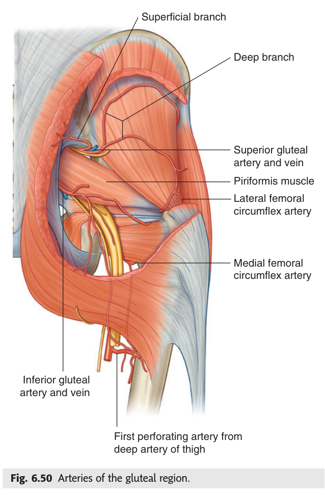
- Parent vessel of both gluteal arteries? → The internal iliac artery
- Which foramen does each use? → Superior gluteal above piriformis, inferior gluteal below
- Why does the anastomosis matter? → It provides collateral flow if the femoral or external iliac artery is occluded
Where to put the needle ★★
This is the one piece of gluteal anatomy every doctor and nurse uses, and it follows directly from everything above. The sciatic nerve leaves below piriformis and runs down the middle of the buttock; the superior gluteal nerve runs forwards above piriformis. Both are therefore in the middle and lower parts of the region. So the safe territory is the part furthest from both.
Divide the buttock into quadrants with a vertical line and a horizontal line. The upper lateral quadrant — specifically its anterior corner — is the safe injection site. A needle placed there enters gluteus medius, anterosuperior to the margin of gluteus maximus, well away from the sciatic nerve and from the other gluteal nerves and vessels.
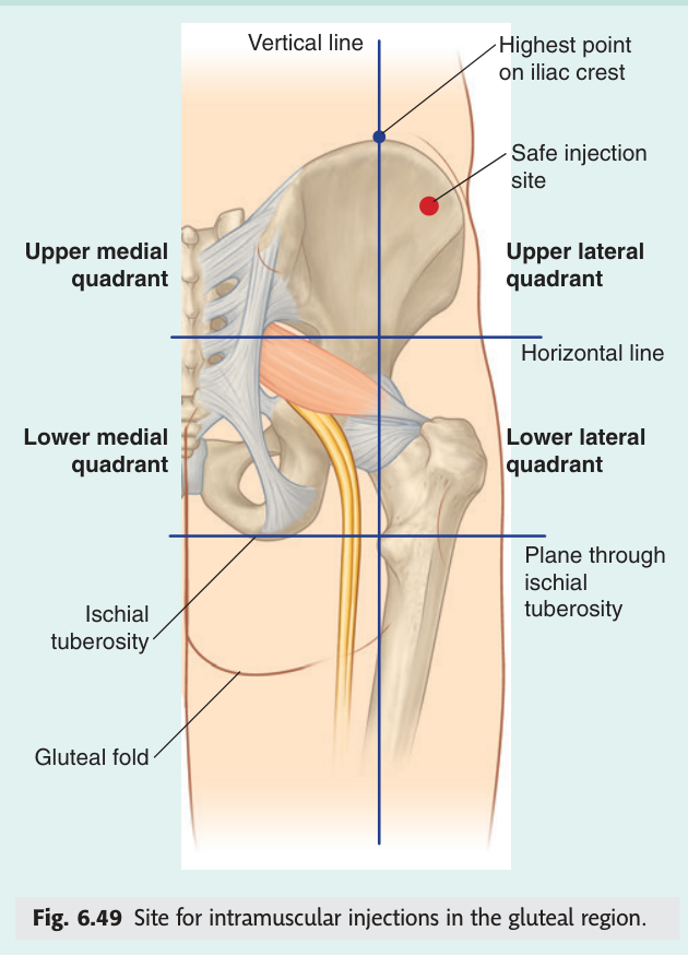
An injection placed in the lower medial quadrant can strike the sciatic nerve directly. The result is immediate burning pain down the limb followed by weakness of the hamstrings and of everything below the knee, with foot drop. It is permanent in a proportion of cases and it is entirely preventable — which is why this quadrant rule is drilled into every clinical student, and why it appears on this paper.
- Which quadrant is safe for intramuscular injection? → The upper lateral quadrant, at its anterior corner
- Which muscle does the needle enter? → Gluteus medius
- What is injured if you inject too low and medially? → The sciatic nerve — foot drop and hamstring weakness
Revision layer
The two foramina — the table to draw from memory
| Suprapiriform foramen | Infrapiriform foramen |
|---|---|
| Superior gluteal nerve | Sciatic nerve (most lateral) |
| Superior gluteal artery | Posterior femoral cutaneous nerve |
| Superior gluteal vein | Inferior gluteal nerve |
| Inferior gluteal artery | |
| Inferior gluteal vein | |
| Internal pudendal artery | |
| Internal pudendal vein | |
| 3 structures | Pudendal nerve (most medial) — 8 structures |
Muscles of the gluteal region and their nerves
| Group | Muscle | Nerve | Main action |
|---|---|---|---|
| Superficial | Gluteus maximus | Inferior gluteal nerve | Extends, laterally rotates thigh |
| Gluteus medius | Superior gluteal nerve | Abducts thigh; steadies pelvis in gait | |
| Gluteus minimus | Superior gluteal nerve | Abducts and medially rotates thigh | |
| Deep (lateral rotators) | Piriformis | Branches of sacral plexus | Laterally rotates thigh |
| Obturator internus | Nerve to obturator internus | Laterally rotates thigh | |
| Superior gemellus | Nerve to obturator internus | Laterally rotates thigh | |
| Inferior gemellus | Nerve to quadratus femoris | Laterally rotates thigh | |
| Quadratus femoris | Nerve to quadratus femoris | Laterally rotates thigh |
Obturator externus is grouped with the medial compartment by innervation (obturator nerve) though it acts as a lateral rotator — TMU's slide lists it with the posterior hip group.
- Draw the greater sciatic foramen with piriformis across it, and label both openings
- List the 8 contents of the infrapiriform foramen, lateral to medial
- List the 3 contents of the suprapiriform foramen
- Give origin, insertion, action and nerve of gluteus maximus and of piriformis
- State the safe injection quadrant and what is damaged if you miss it