Anterior & Medial Regions of the Thigh
Why this year feels different
Last year you learned the body as a set of systems. You followed the arterial tree from the aorta outward, then went back to the beginning and followed the nerves, then the muscles, then the veins. Each pass was clean and logical, and each pass ignored the others.
A surgeon cannot work that way. When a scalpel goes through the front of the thigh, it does not meet "the arterial system" — it meets skin, then fat, then a tough white sheet of fascia, then a nerve lying beside an artery lying beside a vein, all wrapped together and all in a fixed order. Regional anatomy teaches you that order. The question stops being "where does the femoral artery go?" and becomes "if I cut here, what will I hit, and what is immediately medial to it?"
That shift explains why the TMU paper is shaped the way it is. Thirty percent of it asks you to define a named space — a triangle, a canal, a fissure — and a full-mark definition is always the same two things: boundaries and contents. Another ten percent puts a dissection photograph in front of you and asks you to name the numbered structures. Neither of those rewards a beautiful understanding of physiology. They reward knowing precisely what lies beside what.
Read the prose first so the geography makes sense, then close the page and try to redraw the femoral triangle from memory — three borders, floor, and four contents in order. If you can draw it, you can define it. Every named space in this subject yields to the same drill.
The layers, from skin inward
Every region in this course is opened in the same sequence, and knowing the sequence by heart saves you in the dissection room and in the picture question. From the outside in: skin, then superficial fascia, then deep fascia, then skeletal muscle, then the vessels and nerves running between and beneath the muscles, and finally bone.
The two fascial layers behave completely differently, and the difference matters clinically. Superficial fascia is loose and fatty — it is where you find the subcutaneous veins and the cutaneous nerves, and it is why a superficial infection can spread diffusely through a region. Deep fascia is dense, white and tough. It wraps muscles into compartments and it does not stretch. That inelasticity is exactly why bleeding or swelling inside a deep fascial compartment raises the pressure until circulation fails — the anatomy of compartment syndrome is written into this one structural fact.
- Anterior superior iliac spine → pubic tubercle (along the inguinal ligament)
- Down the front of the thigh to 5 cm below the tibial tuberosity
- Transverse cut between the medial and lateral malleoli
The midpoint of the inguinal ligament is your key surface landmark — the femoral artery is palpable there, and almost everything in this unit is described relative to it.
- Name the six layers from skin to bone? → Skin · superficial fascia · deep fascia · muscle · vessels & nerves · bone
- Which layer carries the great saphenous vein? → Superficial fascia
- Why does deep fascia cause compartment syndrome? → It is dense and inelastic, so pressure inside cannot escape
The great saphenous vein
The great saphenous vein is the longest vein in the body, and it is the first thing you meet once the skin is off. It matters far beyond this dissection: it is the vein a surgeon harvests for coronary artery bypass grafting, the vein stripped or ablated in varicose vein disease, and the vein cut down onto in an emergency when no other access can be found.
Its course is worth memorising as a journey rather than a list. It begins on the medial end of the dorsal venous arch of the foot. It then does the single thing that makes it findable in a shocked, collapsed patient: it passes in front of the medial malleolus — the bony bump on the inside of the ankle. That position is constant. From there it runs up the medial side of the leg and thigh, and finally dives through the saphenous opening to join the femoral vein about 3–4 cm below the inguinal ligament.
In a patient who has lost so much blood that peripheral veins have collapsed, you can still reach the great saphenous vein reliably: make a small transverse incision one finger-breadth anterior and superior to the medial malleolus. The vein is always there. This is one of the few places in the body where anatomy is so constant that you can cut blind and expect to find a vessel.
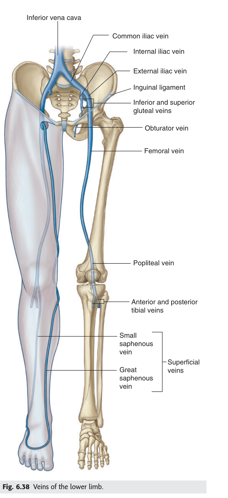
Near its termination the great saphenous vein collects five named tributaries. They are worth learning because they all drain territory outside the limb — the lower abdominal wall, the perineum, the gluteal region — which is why the groin becomes a crossroads for collateral venous flow when deeper veins are blocked.
- Superficial epigastric vein — from the lower anterior abdominal wall
- Superficial iliac circumflex vein — from the flank, running below the inguinal ligament
- External pudendal vein — from the external genitalia
- Superficial medial femoral vein — from the medial thigh
- Superficial lateral femoral vein — from the lateral thigh
Think of the groin as a five-road junction: two come from the thigh itself (medial and lateral femoral), one from above (superficial epigastric), one from the side (superficial iliac circumflex), and one from between the legs (external pudendal).
- Where does the GSV begin? → Medial end of the dorsal venous arch of the foot
- What is its constant surface landmark at the ankle? → It passes in front of the medial malleolus
- Where and into what does it end? → Femoral vein, 3–4 cm below the inguinal ligament
- Name its five tributaries? → Superficial epigastric · superficial iliac circumflex · external pudendal · superficial medial femoral · superficial lateral femoral
Fascia lata, iliotibial tract and the saphenous opening
Strip away the superficial fascia and you expose a thick white membrane wrapping the whole thigh like a tight stocking. This is the fascia lata. On the lateral side it thickens into a strong longitudinal band, the iliotibial tract, into which the tensor fasciae latae in front and most of the gluteus maximus behind both insert. Working through that shared tendon, the two muscles hold the knee extended once other muscles have straightened it, and they steady the hip by stopping the top of the femur drifting laterally out of the acetabulum.
There is one prominent hole in this stocking. Just below the medial end of the inguinal ligament, the fascia lata has an oval defect — the saphenous opening (also called the saphenous ring or fossa ovalis of the thigh). Its purpose is simple: it is the doorway through which the great saphenous vein leaves the superficial fascia and dives down to reach the femoral vein.
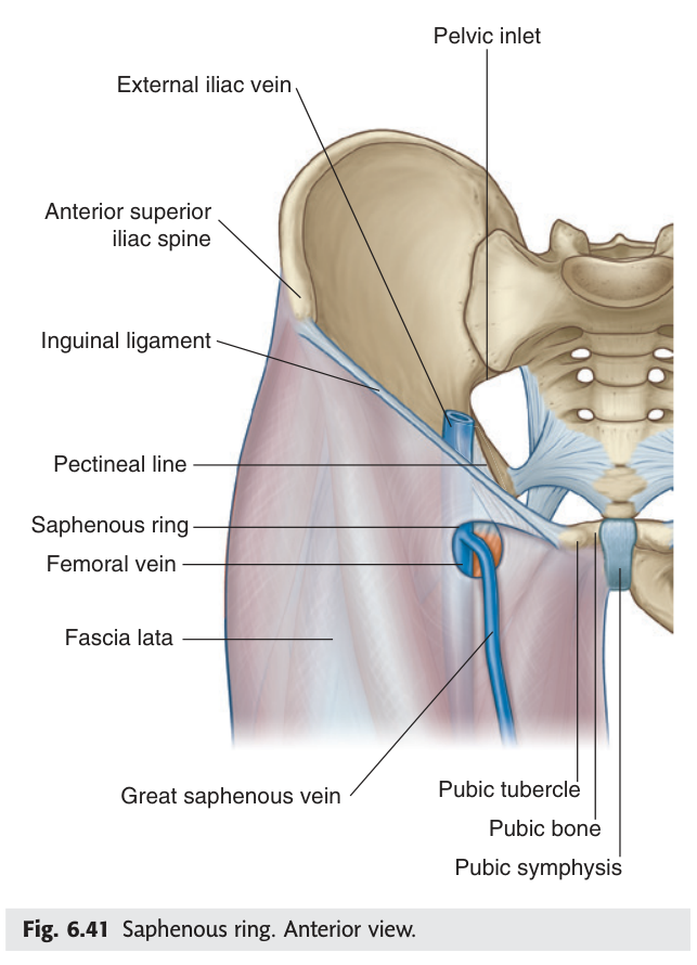
Also lying in this superficial plane is the lateral femoral cutaneous nerve, which becomes subcutaneous roughly 2–4 cm below the anterior superior iliac spine. Remember that measurement — it is where the nerve can be trapped.
Compression of the lateral femoral cutaneous nerve where it passes near the anterior superior iliac spine produces burning, numbness and tingling over the outer thigh, with no weakness at all — because this nerve is purely sensory. Tight belts, heavy tool belts, pregnancy and obesity are the usual culprits. The purely sensory presentation is the diagnostic clue.
- What is the deep fascia of the thigh called? → Fascia lata
- Which two muscles insert into the iliotibial tract? → Tensor fasciae latae and gluteus maximus
- What passes through the saphenous opening? → The great saphenous vein, to join the femoral vein
- Where does the lateral femoral cutaneous nerve become superficial? → 2–4 cm below the ASIS
The anterior group of thigh muscles
The front of the thigh is dominated by one enormous extensor mass and one long ribbon-like muscle crossing it. The mass is the quadriceps femoris, and its job is to straighten the knee — which is why it is the muscle that fails first and visibly wastes when a knee is injured or immobilised. It has four heads: rectus femoris in the middle (the only one that also crosses the hip, so it flexes the hip as well), and vastus medialis, vastus lateralis and vastus intermedius wrapping the shaft of the femur.
Crossing all of them diagonally is the sartorius, the longest muscle in the body. It runs from the anterior superior iliac spine down and medially to the upper tibia, and its name comes from sartor, Latin for tailor — the cross-legged posture a tailor sits in is exactly what this muscle produces. For your purposes its most important property is that its medial margin forms the lateral border of the femoral triangle.
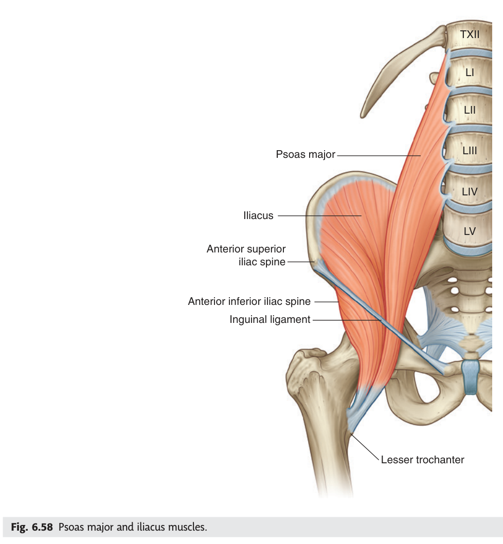
Behind these, descending from the abdomen, is the iliopsoas — really two muscles, psoas major from the lumbar vertebrae and iliacus from the iliac fossa, which fuse and insert together on the lesser trochanter. It is the most powerful hip flexor, and it forms the lateral part of the floor of the femoral triangle.
- Four heads of quadriceps femoris? → Rectus femoris · vastus medialis · vastus lateralis · vastus intermedius
- Which head also flexes the hip, and why? → Rectus femoris — it is the only one crossing the hip joint
- Which border of the femoral triangle does sartorius form? → The lateral border (its medial margin)
- What two muscles make up iliopsoas? → Psoas major + iliacus
The femoral triangle ★★★
This is the single most examined structure in the lower limb, and it has appeared as a definition question in essentially every past paper we have. Understand it once, properly, and you will never lose those marks.
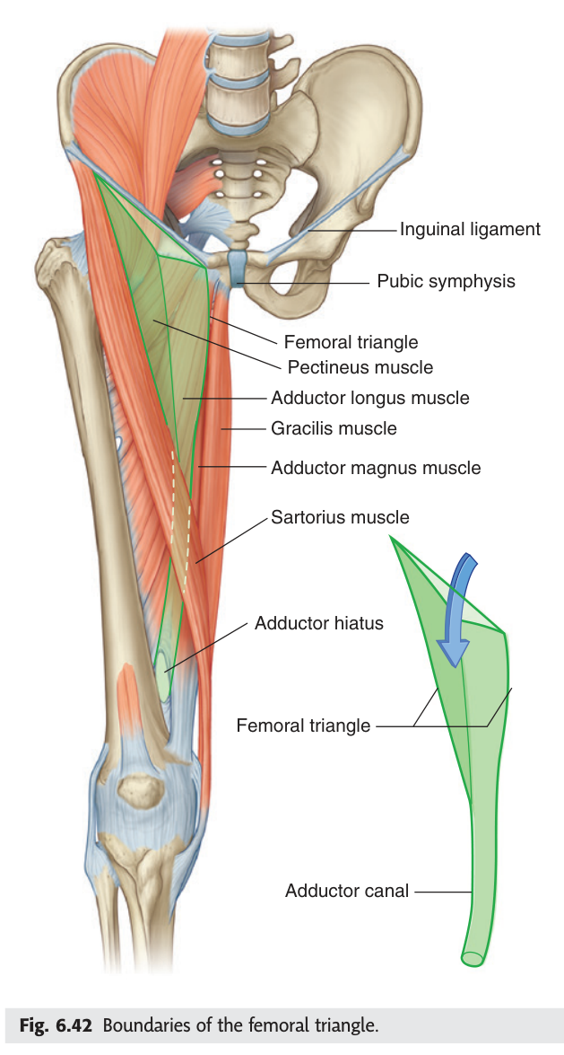
Picture the junction where the abdomen becomes the thigh. The inguinal ligament runs across it like a taut rope from the anterior superior iliac spine to the pubic tubercle. Below that rope, two muscles run diagonally away from each other — sartorius heading down-and-medially, adductor longus heading down-and-laterally. Where those three lines meet they enclose a wedge-shaped hollow pointing downward. That hollow is the femoral triangle, and it is the gateway through which every major vessel and nerve enters the lower limb.
Boundaries. Superiorly (base): the inguinal ligament. Laterally: the
medial margin of sartorius. Medially: the medial margin of adductor longus. The
apex points inferiorly and is continuous with the adductor canal.
Floor (from lateral to medial): iliopsoas and pectineus, with
adductor longus medially.
Roof: fascia lata, pierced by the saphenous opening.
Contents (lateral → medial): femoral nerve, femoral artery, femoral vein,
lymphatics (in the femoral canal).
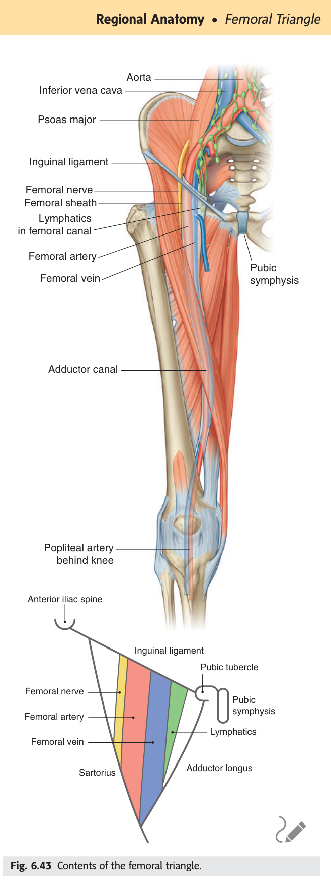
From lateral to medial: N-A-V-E-L — Nerve, Artery, Vein, Empty space, Lymphatics. The navel sits in the midline, so read the mnemonic towards the midline — that fixes the direction and stops you reversing it under pressure.
Your TMU slide states the same order from the opposite end — "from medial to lateral: femoral vein, femoral artery, femoral nerve". Both are correct. In the exam, state which direction you are reading, or you will be marked wrong for a right answer.
To take blood or place a central line in the groin you feel the femoral artery pulse at the mid-inguinal point, then aim just medial to it — because the vein is always medial to the artery. Aim lateral instead and you will hit the artery, and beyond it the nerve. The mnemonic is a procedural instruction, not trivia.
- Three boundaries of the femoral triangle? → Inguinal ligament (base) · medial margin of sartorius (lateral) · medial margin of adductor longus (medial)
- What forms its floor? → Iliopsoas and pectineus, with adductor longus medially
- Contents from lateral to medial? → Femoral nerve · artery · vein · lymphatics
- What does the apex become? → The adductor canal
- Where do you aim a femoral venous puncture? → Just medial to the palpated arterial pulse
Femoral sheath and femoral canal ★★
Inside the femoral triangle the vessels are not lying loose. They are carried in a funnel-shaped sleeve of fascia called the femoral sheath, which is simply the transversalis fascia and iliac fascia of the abdomen prolapsing downward into the thigh as the vessels pass under the inguinal ligament. Think of it as the abdominal lining being dragged along like a sock pulled onto a leg.
The sheath is divided internally into three compartments. Laterally sits the femoral artery; in the middle, the femoral vein; and medially, a small conical space containing only lymphatics and a little fat. That medial compartment is the femoral canal, and its upper opening — the femoral ring — is a genuine weak point in the lower abdominal wall.
Abdominal contents can push down through the femoral ring into the femoral canal, appearing as a lump below and lateral to the pubic tubercle. (An inguinal hernia appears above and medial to it — that single relationship distinguishes the two at the bedside.) Femoral hernias are commoner in women because of the wider female pelvis, and they strangulate far more readily than inguinal hernias because the femoral ring is a small, rigid, unyielding opening. A femoral hernia is therefore a surgical emergency more often than not.
- What is the femoral sheath continuous with above? → Transversalis fascia and iliac fascia of the abdomen
- Its three compartments contain? → Femoral artery (lateral) · femoral vein (middle) · lymphatics (medial = femoral canal)
- Is the femoral nerve inside it? → No — it lies lateral to and outside the sheath
- How do you distinguish femoral from inguinal hernia? → Femoral = below & lateral to pubic tubercle; inguinal = above & medial
Passing under the inguinal ligament ★★
The space beneath the inguinal ligament is not one tunnel but two, divided by a fascial partition called the iliopectineal arch which runs from the ligament down to the iliopubic eminence. Everything travelling between abdomen and thigh must choose a side. This is the Chinese and European descriptive scheme your TMU lecturer uses, and it appears in the paper as a definition — Gray's does not name these two spaces, so learn them from the slide.
The lateral compartment beneath the inguinal ligament, bounded laterally by the ilium and medially by the iliopectineal arch. Transmits the iliopsoas muscle, the femoral nerve, and the lateral femoral cutaneous nerve.
The medial compartment beneath the inguinal ligament, bounded laterally by the iliopectineal arch and medially by the lacunar ligament. Transmits the femoral artery, the femoral vein, and the femoral canal with its lymphatics.
The names give the answer away if you trust them. Musculorum = muscles, so the muscular lacuna carries the muscle (iliopsoas) — and nerves travel with muscle. Vasorum = vessels, so the vascular lacuna carries the vessels. Lateral is muscular, medial is vascular; and that fits the NAVEL order you already know, since the nerve sits most laterally of all.
- What divides the two lacunae? → The iliopectineal arch
- Contents of the lacuna musculorum? → Iliopsoas · femoral nerve · lateral femoral cutaneous nerve
- Contents of the lacuna vasorum? → Femoral artery · femoral vein · femoral canal (lymphatics)
- Which is lateral? → The muscular lacuna
The femoral artery
The femoral artery is simply the external iliac artery renamed: the moment it passes under the inguinal ligament it takes a new name, and the moment it passes through the adductor hiatus at the other end of the thigh it becomes the popliteal artery. Names change at doorways — that principle repeats throughout this course.
Its most important branch arises 2–4 cm below the inguinal ligament: the deep femoral artery (profunda femoris), which dives backwards to supply the bulk of the thigh musculature and, through its perforating branches, the posterior compartment. The superficial continuation of the femoral artery then travels on through the adductor canal largely to supply the leg and foot. So the deep branch feeds the thigh; the parent vessel is really in transit.
Because the femoral artery is large, superficial and lies over the femoral head — a firm surface to compress against — it is the standard entry point for coronary angiography, angioplasty and many endovascular procedures. Compression against the femoral head is what allows haemostasis afterwards.
- What does the femoral artery become, and where? → Popliteal artery, at the adductor hiatus
- Where does the deep femoral artery arise? → 2–4 cm below the inguinal ligament
- Where is the femoral pulse felt? → Midway between ASIS and pubic symphysis, just below the inguinal ligament
The adductor canal ★★
Follow the femoral vessels down from the apex of the femoral triangle and they disappear into a narrow fascial tunnel on the medial side of the middle third of the thigh. This is the adductor canal, also called Hunter's canal after the eighteenth-century surgeon John Hunter, who tied the femoral artery within it to treat popliteal aneurysm. It is triangular in cross-section and roofed by a strong fascial sheet stretched between vastus medialis and the adductors, with sartorius lying on top.
Three structures run in it, and the order — front to back: saphenous nerve, femoral artery, femoral vein — is the examinable detail. Note what happens to the saphenous nerve: it is the only structure that does not continue through to the popliteal fossa. It leaves the canal to become superficial and supply skin on the medial side of the leg and foot, while the artery and vein carry on through the adductor hiatus to become the popliteal vessels.
A fascial tunnel in the medial third of the thigh, extending from the apex of the femoral triangle
to the adductor hiatus in adductor magnus.
Boundaries: anterolaterally vastus medialis; posteriorly adductor
longus above and adductor magnus below; roofed by a fibrous membrane covered by
sartorius.
Contents (anterior → posterior): saphenous nerve, femoral artery, femoral
vein.
Anaesthetists inject local anaesthetic into this canal for knee surgery. It numbs the saphenous nerve — giving good analgesia over the medial knee — while largely sparing the motor branches to quadriceps, so the patient can still straighten the leg and mobilise early. This is precisely why the block has replaced the older femoral nerve block for many knee operations: the anatomy of what runs in the canal determines what gets blocked.
- Where does the adductor canal begin and end? → Apex of femoral triangle → adductor hiatus
- Its three contents, front to back? → Saphenous nerve · femoral artery · femoral vein
- Which content leaves the canal without reaching the popliteal fossa? → The saphenous nerve
- Which muscle roofs it? → Sartorius, over a fibrous membrane
The medial group of thigh muscles
The medial compartment holds the adductors — the muscles that pull the thigh towards the midline, and the muscles a footballer tears as a "groin strain". There are five: pectineus, adductor longus, adductor brevis, adductor magnus and gracilis. Gracilis is the odd one out in shape, a thin strap running all the way to the tibia, which is why it crosses the knee as well as the hip and is often harvested as a graft.
The compartment is supplied by the obturator nerve, which enters alongside the obturator artery and vein through the obturator canal. But there are two exceptions you must know, because examiners live on exceptions.
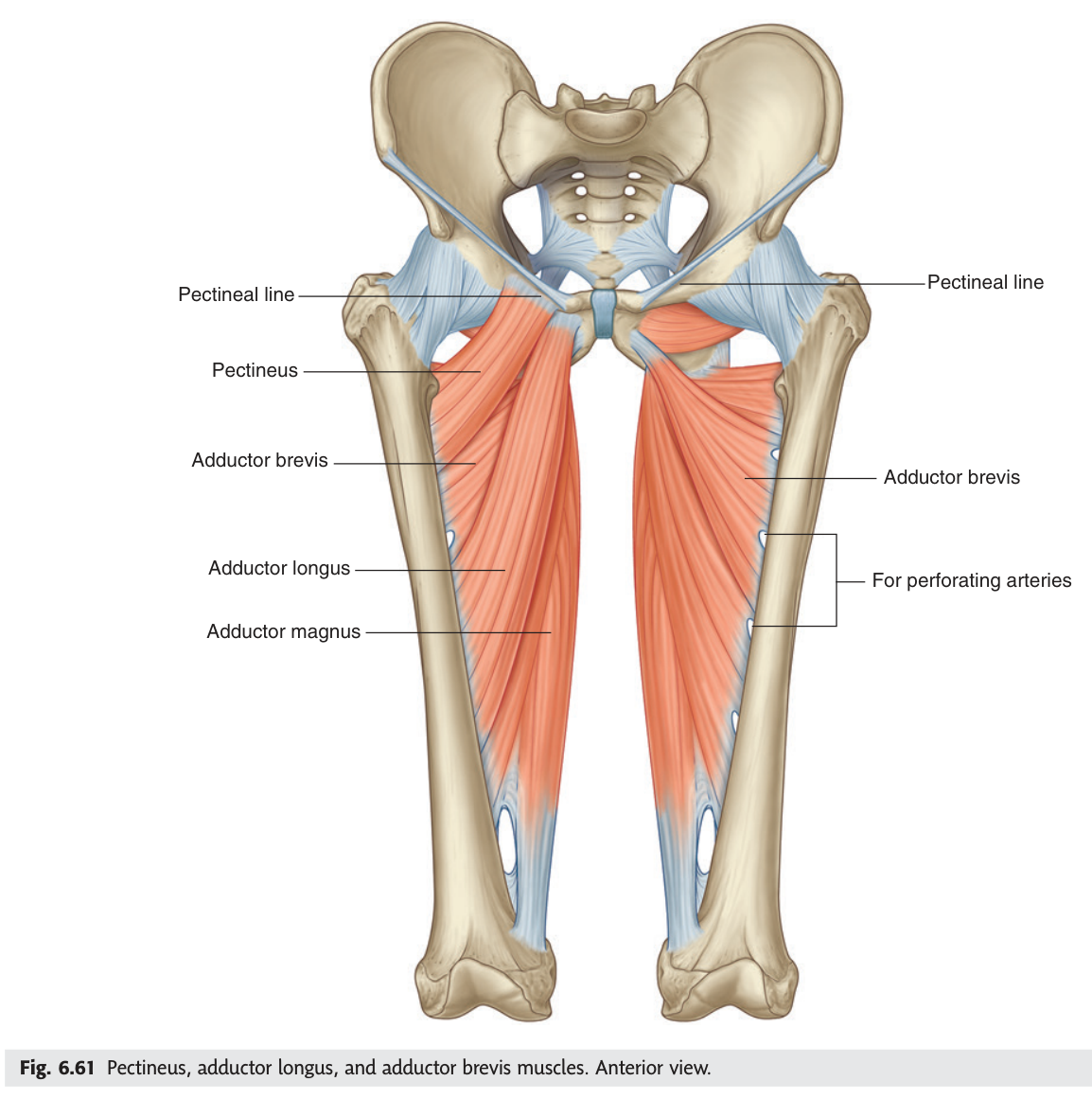
Adductor magnus has a gap in its lower attachment, the adductor hiatus, through which the femoral vessels pass to the back of the knee. This is a common site for arterial injury in femoral shaft fractures, because the vessel is relatively tethered as it passes through the opening and cannot move out of the way of displaced bone.
- Name the five medial-compartment muscles? → Pectineus · adductor longus · adductor brevis · adductor magnus · gracilis
- What is the compartment's main nerve? → Obturator nerve
- Which muscle is the femoral-nerve exception? → Pectineus
- Which has dual supply, and from what? → Adductor magnus — obturator + tibial division of sciatic
Revision layer — the tables to memorise
Everything above was to make it make sense. What follows is the cram layer: the exact lists that earn marks. Cover the right-hand column and recite.
Muscles of the thigh and their innervation — the standard 10-mark question
| Group | Muscles | Nerve | Main action |
|---|---|---|---|
| Anterior | Sartorius; Quadriceps femoris (rectus femoris, vastus medialis, vastus lateralis, vastus intermedius) | Femoral nerve | Extend knee (rectus femoris also flexes hip) |
| Medial | Pectineus; adductor longus; adductor brevis; adductor magnus; gracilis | Obturator nerve except pectineus = femoral; adductor magnus hamstring part = tibial |
Adduct thigh at hip |
| Posterior (Unit 3) |
Biceps femoris; semitendinosus; semimembranosus | Sciatic nerve | Extend hip, flex knee |
| Hip — anterior | Iliopsoas (psoas major + iliacus); tensor fasciae latae | Femoral n. / branches of lumbar plexus | Flex hip |
The four named spaces of this unit
| Space | Boundaries | Contents |
|---|---|---|
| Femoral triangle | Base: inguinal ligament · Lateral: medial margin of sartorius · Medial: medial margin of adductor longus · Floor: iliopsoas + pectineus (+ adductor longus) | Femoral nerve → artery → vein → lymphatics (lateral to medial) |
| Adductor canal | Anterolateral: vastus medialis · Posterior: adductor longus then magnus · Roof: fibrous membrane + sartorius | Saphenous nerve → femoral artery → femoral vein (front to back) |
| Lacuna musculorum | Lateral compartment under inguinal ligament; medial limit = iliopectineal arch | Iliopsoas · femoral nerve · lateral femoral cutaneous nerve |
| Lacuna vasorum | Medial compartment under inguinal ligament; lateral limit = iliopectineal arch, medial = lacunar ligament | Femoral artery · femoral vein · femoral canal (lymphatics) |
Numbers examiners ask for
| Measurement | Value |
|---|---|
| Great saphenous vein joins femoral vein | 3–4 cm below the inguinal ligament |
| Deep femoral artery arises | 2–4 cm below the inguinal ligament |
| Lateral femoral cutaneous nerve becomes superficial | 2–4 cm below the ASIS |
| Tributaries of the great saphenous vein | 5 |
| Muscles of the medial compartment | 5 |
- Draw the femoral triangle: three borders, floor, four contents in order
- Draw the adductor canal: two walls, roof, three contents front-to-back
- List the thigh muscles by group with their nerve, including both exceptions
- Trace the great saphenous vein from the foot to the femoral vein, naming its five tributaries
- State what passes through each lacuna under the inguinal ligament