Infracolic Compartment
Two arteries and one vein
TMU's slide for this lecture reduces the entire infracolic compartment to a single phrase: two arteries, one vein. The two arteries are the superior and inferior mesenteric; the one vein is the hepatic portal vein that collects everything they deliver.
The logic behind the division is embryological and it makes the branch lists predictable. The midgut — from the descending duodenum to the distal third of the transverse colon — is supplied by the superior mesenteric artery. The hindgut — from there to the upper rectum — is supplied by the inferior mesenteric artery. Once you know where the boundary is, you can predict which vessel supplies any given piece of bowel.
- Which artery supplies the midgut? → The superior mesenteric artery
- Where does midgut territory end? → At the distal third of the transverse colon
- Which vein drains the whole gut? → The hepatic portal vein
The superior mesenteric artery ★★★
The superior mesenteric artery arises from the front of the aorta at L1, just below the celiac trunk, and runs down between the layers of the mesentery. Its branches divide neatly by which side they leave from — and that is how to remember them.
From the LEFT side (to the small intestine): the jejunal and ileal arteries — twelve to fifteen of them, forming arcades and vasa recta.
From the RIGHT side (to the large intestine): the middle colic, right colic and ileocolic arteries — the ileocolic giving the appendicular artery.
First branch: the inferior pancreaticoduodenal artery.
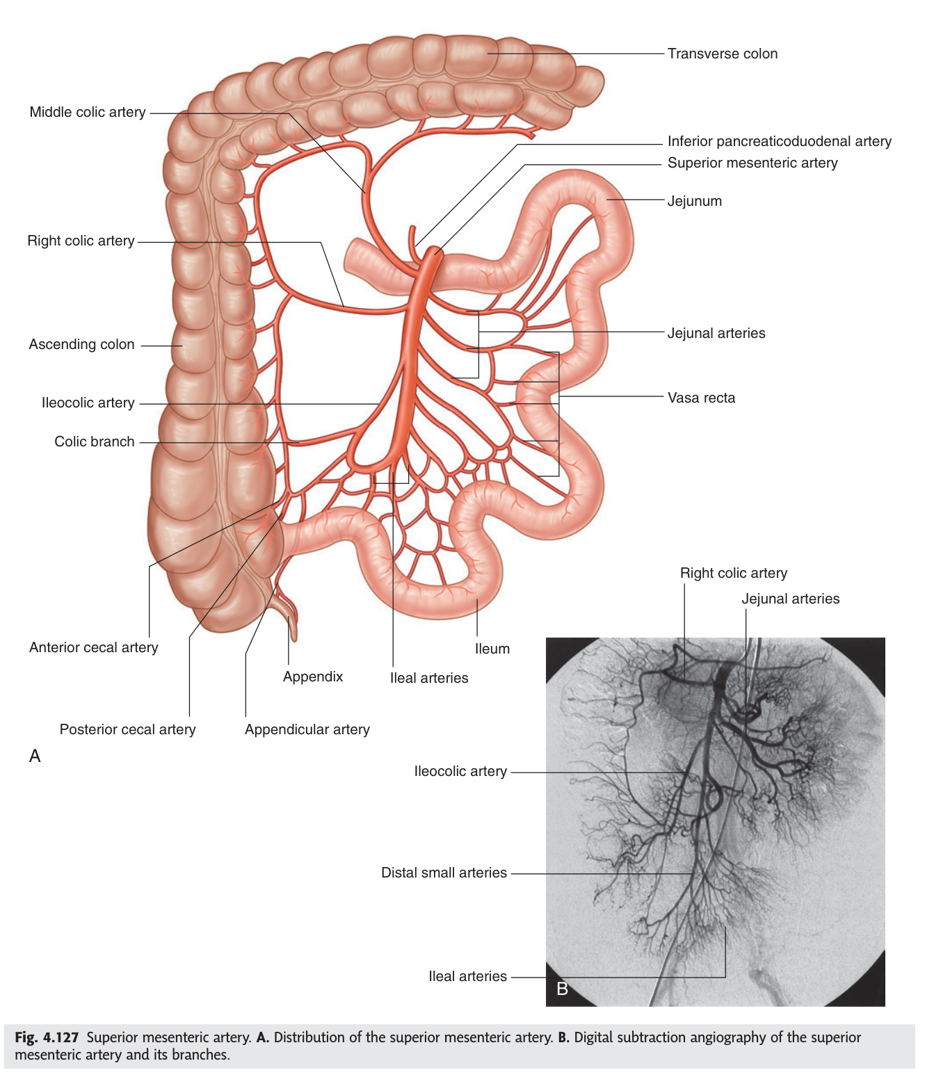
In the jejunum there are one or two large arcades with long straight vasa recta; in the ileum there are four or five tiers of smaller arcades with short vasa recta. A surgeon can tell jejunum from ileum by holding the mesentery up to the light. The mesentery is also thicker and fattier at the ileal end. Anatomy that can be read at a glance in theatre.
An embolus lodging in the SMA infarcts almost the entire small intestine and the right colon at once. The presentation is famously deceptive: severe central abdominal pain with a strikingly soft abdomen — pain out of all proportion to the physical signs — because the peritoneum is not yet inflamed. By the time guarding appears the bowel is dead. Mortality remains very high, and the reason is that the SMA territory has few collaterals.
- At what level does the SMA arise? → L1, just below the celiac trunk
- Its branches to the right? → Middle colic · right colic · ileocolic (→ appendicular)
- Its branches to the left? → Jejunal and ileal arteries
- How do jejunal and ileal arcades differ? → Jejunum: few arcades, long vasa recta · Ileum: many arcades, short vasa recta
The inferior mesenteric artery ★★★
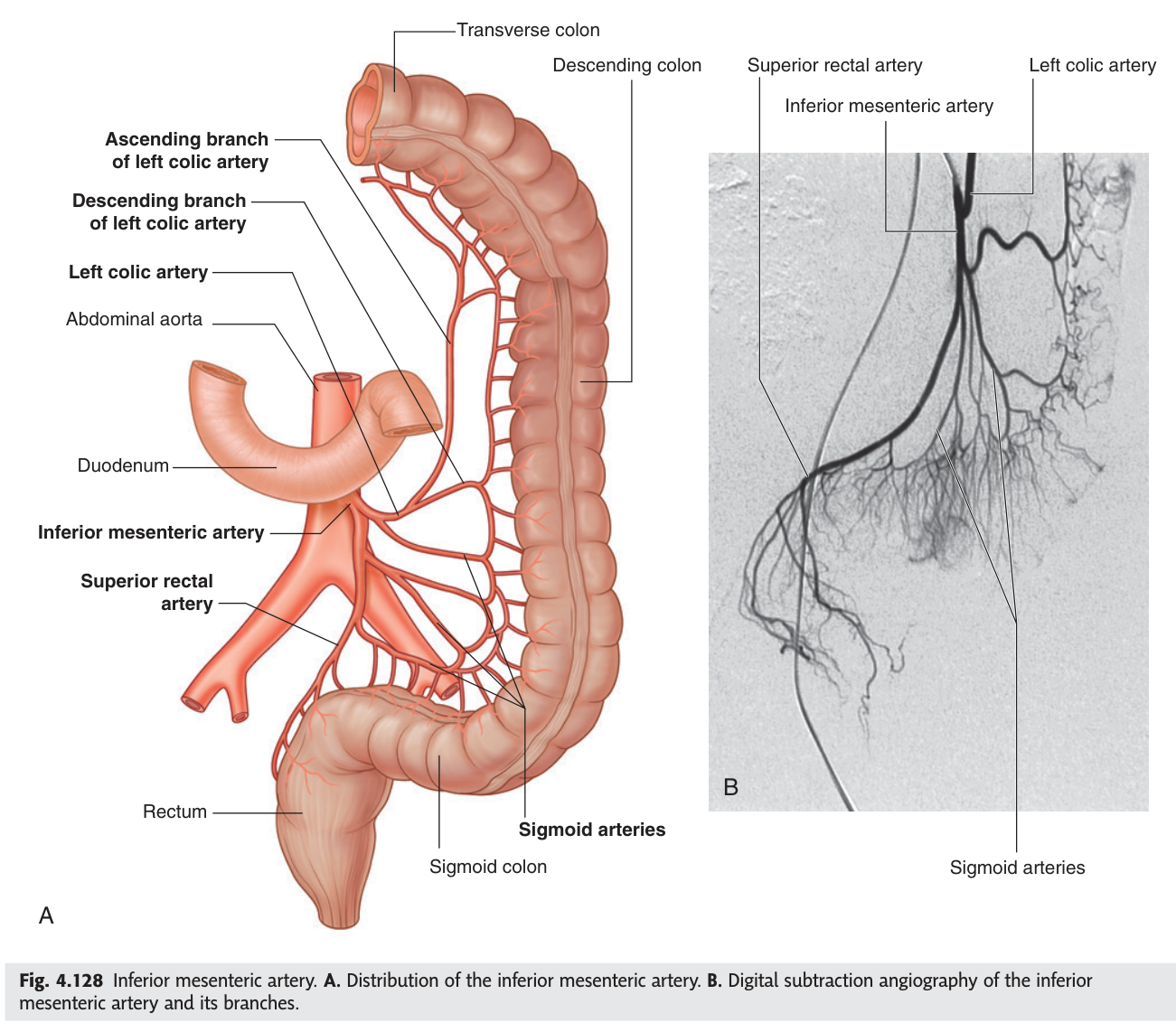
The inferior mesenteric artery arises from the front of the aorta at L3 and supplies the hindgut. It has only three branches, and TMU has asked for them directly.
1. Left colic artery — to the descending colon.
2. Sigmoid arteries — two to three, to the sigmoid colon.
3. Superior rectal artery — the terminal continuation, to the upper rectum.
- At what level does the IMA arise? → L3
- Its three branches? → Left colic · sigmoid · superior rectal
- What is its terminal branch? → The superior rectal artery
- What does it supply? → The hindgut — distal third of transverse colon to upper rectum
The marginal artery and the watershed
Along the whole inner border of the colon, the terminal branches of every colic artery join end to end to form one continuous vessel — the marginal artery (of Drummond). It runs from the ileocaecal junction to the rectosigmoid, linking SMA and IMA territory.
The continuous anastomotic channel along the mesenteric border of the colon, formed by the anastomosing terminal branches of the ileocolic, right colic, middle colic, left colic and sigmoid arteries.
It connects the superior and inferior mesenteric arterial territories and supplies the colon through short vasa recta.
The marginal artery is weakest where the two territories meet — at the splenic flexure, the junction of midgut and hindgut supply. In a period of low blood pressure — cardiac arrest, major haemorrhage, aortic surgery — this watershed area is the first to become ischaemic.
The result is ischaemic colitis, classically presenting with left-sided abdominal pain and bloody diarrhoea in an elderly patient after a hypotensive episode. The site of the disease is predicted entirely by the anatomy of the anastomosis.
- What is the marginal artery? → The continuous anastomotic channel along the colon linking SMA and IMA territory
- Where is the watershed? → The splenic flexure, at the midgut–hindgut junction
- What happens there in hypotension? → Ischaemic colitis
The hepatic portal vein ★★★
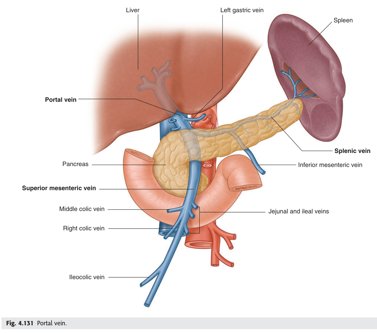
Everything the gut absorbs must be inspected by the liver before it reaches the general circulation. The hepatic portal vein is the vessel that enforces that rule — a vein that begins in capillaries and ends in capillaries, carrying about 75% of the liver's blood supply. TMU asked for its composition and tributaries in the 2021–22 paper, worth nine marks.
Formed behind the neck of the pancreas, at about L2, by the union of the superior mesenteric vein and the splenic vein.
Tributaries:
• Superior mesenteric vein
• Splenic vein
• Inferior mesenteric vein — usually joining the splenic
• Left gastric vein
• Right gastric vein
• Cystic vein
• Paraumbilical veins
It ascends in the free edge of the lesser omentum (hepatoduodenal ligament), behind the bile duct and hepatic artery, and divides at the porta hepatis into right and left branches. It has no valves.
- How is the portal vein formed? → Union of the superior mesenteric and splenic veins, behind the neck of the pancreas
- Name its tributaries? → SMV · splenic · IMV · left and right gastric · cystic · paraumbilical veins
- Where does it run? → In the free edge of the lesser omentum, behind the bile duct and hepatic artery
- Does it have valves? → No — which is why portal hypertension reverses flow
Portosystemic anastomoses ★★
Because the portal vein has no valves, obstruction to flow through the liver — most often cirrhosis — forces blood backwards into the places where the portal and systemic venous systems meet. Those junctions are normally trivial. Under pressure they become the whole clinical picture of portal hypertension.
| Site | Portal vein | Systemic vein | Clinical result |
|---|---|---|---|
| Lower oesophagus | Left gastric vein | Oesophageal veins → azygos | Oesophageal varices — may bleed catastrophically |
| Anal canal | Superior rectal vein | Middle and inferior rectal veins | Haemorrhoids |
| Periumbilical | Paraumbilical veins | Superficial epigastric veins | Caput medusae |
| Retroperitoneal | Colic veins | Retroperitoneal veins | Usually silent |
All four anastomoses dilate in portal hypertension, but only one regularly kills. The veins at the lower oesophagus lie immediately beneath a thin mucosa, in a tube that food passes through under pressure. When they rupture the patient vomits large volumes of blood, and mortality from a first variceal bleed remains high.
Haemorrhoids from the same cause are unpleasant but rarely dangerous; caput medusae is a sign, not a threat. Position determines lethality.
- Why does portal hypertension reverse flow? → The portal vein has no valves
- The four sites of portosystemic anastomosis? → Lower oesophagus · anal canal · periumbilical · retroperitoneal
- Which portal vein is involved at the oesophagus? → The left gastric vein
- What is caput medusae? → Dilated periumbilical veins radiating from the umbilicus
Revision layer
The three gut arteries
| Artery | Level | Territory | Branches |
|---|---|---|---|
| Celiac trunk | T12 | Foregut — to descending duodenum | Left gastric · common hepatic · splenic |
| Superior mesenteric | L1 | Midgut — to distal ⅓ of transverse colon | Inferior pancreaticoduodenal · jejunal and ileal · middle colic · right colic · ileocolic (→ appendicular) |
| Inferior mesenteric | L3 | Hindgut — to upper rectum | Left colic · sigmoid · superior rectal |
Hepatic portal vein
| Detail | |
|---|---|
| Formed by | Superior mesenteric vein + splenic vein, behind the neck of the pancreas (L2) |
| Tributaries | SMV · splenic · IMV · left gastric · right gastric · cystic · paraumbilical |
| Course | Free edge of the lesser omentum, behind bile duct and hepatic artery |
| Ends | Right and left branches at the porta hepatis |
- Give all branches of the SMA and IMA with their levels of origin
- Describe the marginal artery and explain the splenic flexure watershed
- State how the portal vein is formed and name all its tributaries
- Name the four portosystemic anastomoses with their clinical results
- Explain why SMA occlusion presents with pain out of proportion to signs