Retroperitoneal Space
What the retroperitoneal space contains
The retroperitoneal space lies between the parietal peritoneum in front and the fascia covering the muscles of the posterior abdominal wall behind. It is not a cavity but a plane full of fat, and it holds the organs that never migrated into the peritoneal sac.
Kidneys · suprarenal glands · ureters · the descending and inferior parts of the duodenum · the pancreas · the abdominal aorta and inferior vena cava with their branches and tributaries · lymph nodes · the lumbar plexus and sympathetic trunks.
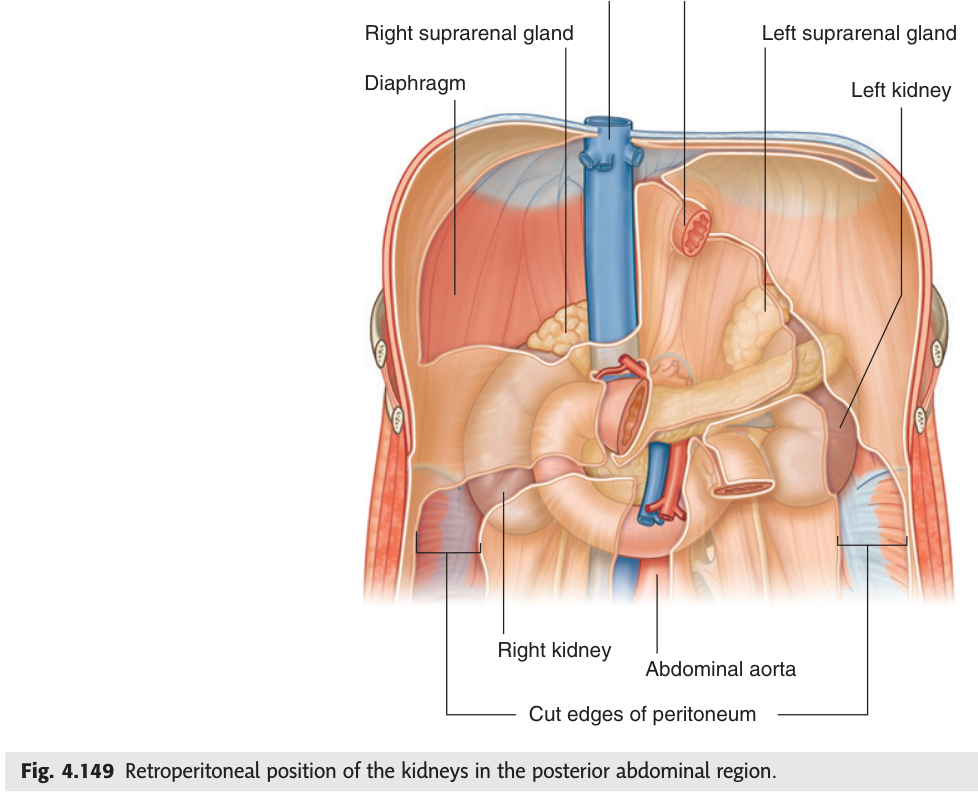
- What are the boundaries of the retroperitoneal space? → Parietal peritoneum in front, posterior abdominal wall fascia behind
- Name six retroperitoneal structures? → Kidneys · suprarenals · ureters · pancreas · duodenum (2nd–4th parts) · aorta and IVC
The coverings of the kidney ★★★
The kidney is wrapped in three layers, and TMU has asked for them from inner to outer in a past paper. Getting the direction right is half the mark.
1. Fibrous capsule — the tough membrane directly on the kidney surface, strippable from the healthy organ.
2. Adipose capsule (perirenal fat) — the fatty layer that cushions it.
3. Renal fascia — the outermost fibrous sheath, with anterior and posterior layers enclosing both the kidney and the suprarenal gland; outside it lies pararenal fat.
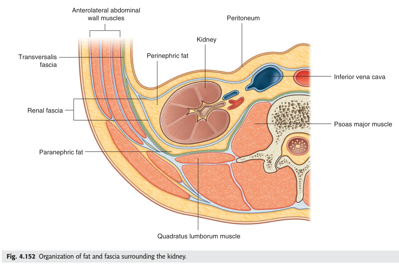
The kidney is held in position mainly by the perirenal fat and the renal fascia, not by its vessels. Rapid, substantial weight loss removes that packing and the kidney can descend — nephroptosis — sometimes kinking the ureter and causing intermittent loin pain relieved by lying down.
The same fascial planes explain why a perinephric abscess stays confined to the renal fascia and tracks downward rather than spreading freely.
- The three coverings, inner to outer? → Fibrous capsule · adipose capsule · renal fascia
- Which is closest to the kidney? → The fibrous capsule
- What holds the kidney in place? → Perirenal fat and the renal fascia
- What is nephroptosis? → Descent of the kidney after loss of perirenal fat
The renal pedicle ★★★
The renal pedicle is the bundle of structures entering and leaving the hilum: the renal artery, the renal vein and the renal pelvis. TMU's slide marks this ★★★ because the order differs depending on which axis you read it along — and the exam asks for both.
From FRONT backwards: renal vein → renal artery → renal pelvis.
From ABOVE downwards: renal artery → renal vein → renal pelvis.
Whichever direction you read, the renal pelvis is most posterior and most inferior — it is always the last of the three. Only the artery and vein swap: the vein is in front, the artery is above. Remember it as "vein in front, artery on top, pelvis behind and below."
That position has two consequences: the left renal vein can be compressed between the two arteries (the nutcracker phenomenon), and it is the reason the left testicular / ovarian vein drains into the left renal vein rather than into the IVC directly.
The left testicular vein drains into the left renal vein at a right angle, giving a longer column of blood and higher pressure; the right drains obliquely straight into the IVC. That is why varicoceles are far commoner on the left.
It also means a new left varicocele in an older man can be the first sign of a renal carcinoma obstructing the left renal vein — which is why it is imaged rather than dismissed.
- The pedicle from front to back? → Renal vein · renal artery · renal pelvis
- From above downwards? → Renal artery · renal vein · renal pelvis
- Why is the left renal vein longer? → It must cross the aorta to reach the right-sided IVC
- Where does the left testicular vein drain? → Into the left renal vein
The suprarenal glands ★★★
The suprarenal (adrenal) glands cap the upper poles of the kidneys, inside the renal fascia. Their blood supply is one of the strangest arrangements in the body, and TMU asked for it as a ten-mark question in 2021–22: three arteries, one vein.
THREE arteries, from three different sources:
• Superior suprarenal artery — from the inferior phrenic artery
• Middle suprarenal artery — from the abdominal aorta directly
• Inferior suprarenal artery — from the renal artery
ONE vein on each side, draining differently:
• Right suprarenal vein → the inferior vena cava (short, direct)
• Left suprarenal vein → the left renal vein
The asymmetry has the same explanation as the renal veins: the IVC lies on the right, so the right gland can drain straight into it, while the left has to route through the left renal vein.
The triple arterial supply reflects the gland's importance — it is an endocrine organ that must never lose its blood supply, so it is fed from above, from the middle and from below. Three sources in, asymmetric drainage out.
The right suprarenal vein is short and wide and enters the IVC directly, often at an awkward angle behind the liver. During a right adrenalectomy it can tear off flush with the cava, producing sudden major haemorrhage from a vessel that is hard to reach and hard to clamp. The left vein, being longer and draining into the renal vein, is far more forgiving.
- The three suprarenal arteries and their sources? → Superior — inferior phrenic · Middle — abdominal aorta · Inferior — renal artery
- Where does the right suprarenal vein drain? → Directly into the inferior vena cava
- Where does the left drain? → Into the left renal vein
- Why is the drainage asymmetrical? → The IVC lies to the right, so the left side routes via the renal vein
Branches of the abdominal aorta
The abdominal aorta runs from the aortic hiatus at T12 to its bifurcation at L4, and its branches sort into three tidy groups. TMU's slide divides the visceral branches into paired and unpaired, and the unpaired ones have been asked directly.
| Group | Branches |
|---|---|
| Unpaired visceral | Celiac trunk (T12) · superior mesenteric (L1) · inferior mesenteric (L3) |
| Paired visceral | Middle suprarenal · renal · testicular (or ovarian) |
| Paired parietal | Inferior phrenic · four lumbar arteries |
| Terminal | Two common iliac arteries, at L4 |
The testicular or ovarian arteries deserve a note. They arise from the anterior wall of the aorta and run down across the ureter — a long course explained by the gonad's origin high on the posterior abdominal wall, dragging its artery down as it descends. The vessel is a fossil of the journey.
- The three unpaired visceral branches and their levels? → Celiac trunk T12 · SMA L1 · IMA L3
- The paired visceral branches? → Middle suprarenal · renal · testicular/ovarian
- Where does the aorta bifurcate? → L4, into the two common iliac arteries
- Why do the gonadal arteries have such a long course? → The gonads develop high on the posterior abdominal wall and descend
The lumbar plexus
The lumbar plexus is formed from the anterior rami of L1–L4 within the substance of psoas major. Its branches emerge from the muscle's borders, and which border tells you which nerve it is — which is exactly how TMU has examined it.
| Emerges from | Nerves |
|---|---|
| Lateral border of psoas major | Iliohypogastric · ilioinguinal · lateral cutaneous nerve of the thigh · femoral nerve |
| Medial border of psoas major | Obturator nerve · lumbosacral trunk |
| Anterior surface | Genitofemoral nerve |
A nerve trunk formed by part of the anterior ramus of the fourth lumbar nerve — emerging from the medial border of psoas major — joining the anterior ramus of the fifth lumbar nerve.
It descends into the pelvis in front of the sacroiliac joint and joins the sacral plexus, providing the anatomical bridge by which L4 and L5 fibres reach the sciatic nerve.
- Root value of the lumbar plexus, and where is it formed? → L1–L4, within psoas major
- Which nerves emerge laterally? → Iliohypogastric · ilioinguinal · lateral femoral cutaneous · femoral
- Which emerge medially? → Obturator nerve and lumbosacral trunk
- What forms the lumbosacral trunk? → Part of L4 joining L5
Revision layer
This is the last unit of the subject. The tables below are the ones the retroperitoneal questions come from — and several of them have appeared verbatim in the 2021–22 paper.
Kidney — coverings and pedicle
| Answer | |
|---|---|
| Coverings, inner → outer | Fibrous capsule · adipose capsule · renal fascia |
| Pedicle, front → back | Renal vein · renal artery · renal pelvis |
| Pedicle, above → below | Renal artery · renal vein · renal pelvis |
Suprarenal gland — three in, one out
| Vessel | Source / destination |
|---|---|
| Superior suprarenal artery | Inferior phrenic artery |
| Middle suprarenal artery | Abdominal aorta |
| Inferior suprarenal artery | Renal artery |
| Right suprarenal vein | Inferior vena cava |
| Left suprarenal vein | Left renal vein |
Abdominal aorta — branches
| Group | Branches |
|---|---|
| Unpaired visceral | Celiac trunk (T12) · superior mesenteric (L1) · inferior mesenteric (L3) |
| Paired visceral | Middle suprarenal · renal · testicular/ovarian |
| Paired parietal | Inferior phrenic · lumbar arteries |
| Terminal | Common iliac arteries (L4) |
- Give the kidney coverings and the renal pedicle in both directions
- Give the suprarenal blood supply and explain the asymmetric venous drainage
- List the branches of the abdominal aorta by group with levels
- Define the lumbosacral trunk and say which border of psoas it emerges from
- Explain why varicoceles are commoner on the left