Supracolic Compartment
What lies above the colon
The supracolic compartment is everything above the transverse colon and its mesocolon: the liver, the stomach, the spleen and the superior part of the duodenum. Almost all of it is supplied by one short, fat artery — the celiac trunk — and almost every exam question about this region is really a question about that vessel's branches.
The reason for that unity is embryological. Everything from the lower oesophagus to the middle of the duodenum is foregut, and the foregut has one artery. Learn the boundary — the celiac trunk stops where the superior mesenteric takes over, at the descending part of the duodenum — and the whole abdomen divides into three tidy arterial territories.
- What defines the supracolic compartment? → Everything above the transverse colon and mesocolon
- Which organs are in it? → Liver, stomach, spleen, superior duodenum
- Which artery supplies the foregut? → The celiac trunk
- Where does foregut supply end? → At the descending part of the duodenum
The celiac trunk ★★★
The celiac trunk is a short, wide artery arising from the front of the abdominal aorta at about T12, immediately below the aortic hiatus. After a centimetre or two it explodes into three branches — and that trifurcation is one of the most reliably examined structures in the abdomen.
1. Left gastric artery — to the lesser curvature; gives oesophageal branches.
2. Common hepatic artery — which divides into:
• Proper hepatic artery → right and left hepatic branches (the cystic artery usually arises from the right hepatic) and the right gastric artery
• Gastroduodenal artery → right gastro-omental (gastroepiploic) and superior pancreaticoduodenal arteries
3. Splenic artery — runs along the upper border of the pancreas, giving pancreatic branches, then short gastric and left gastro-omental (gastroepiploic) arteries.
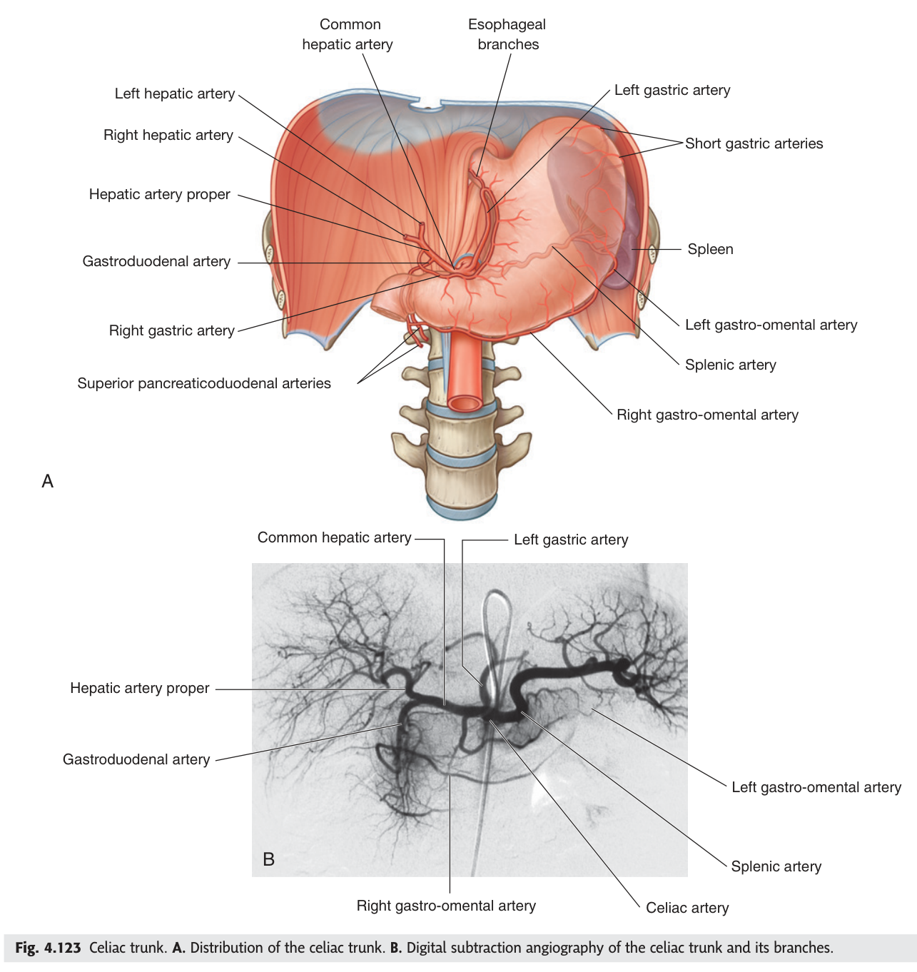
The gastroduodenal artery runs directly behind the first part of the duodenum. An ulcer on the posterior wall erodes into it and causes torrential haemorrhage — one of the classic causes of massive upper gastrointestinal bleeding.
An ulcer on the anterior wall of the same segment does something quite different: it perforates into the peritoneal cavity. Same organ, same disease, opposite complication, decided entirely by which wall is involved.
- At what vertebral level does the celiac trunk arise? → About T12
- Its three branches? → Left gastric · common hepatic · splenic
- The two branches of the common hepatic artery? → Proper hepatic and gastroduodenal
- Why does a posterior duodenal ulcer bleed massively? → It erodes the gastroduodenal artery behind it
Blood supply of the stomach ★★
The stomach is supplied by five arteries, and they are laid out with beautiful regularity: a pair running along each curvature, plus one group to the fundus. All five trace back to the celiac trunk. TMU has asked for them, with their origins, in a past paper.
| Curvature | Arteries | From |
|---|---|---|
| Lesser curvature | Left gastric artery | Celiac trunk directly |
| Right gastric artery | Proper hepatic artery | |
| Greater curvature | Left gastro-omental (gastroepiploic) | Splenic artery |
| Right gastro-omental (gastroepiploic) | Gastroduodenal artery | |
| Fundus | Short gastric arteries | Splenic artery |
Along the lesser curvature the two gastric arteries meet; along the greater curvature the two gastro-omental arteries meet; and the short gastric arteries reach the fundus from the spleen. Left-sided vessels come from the celiac trunk or splenic artery; right-sided vessels come from the hepatic side. Left from the left, right from the right.
Because the two arteries on each curvature anastomose end to end, and the short gastrics add a third route, the stomach has one of the richest collateral supplies in the body. You can ligate several of its arteries during a partial gastrectomy and the remaining stomach survives. Gastric ischaemia is correspondingly rare, in sharp contrast to the intestine.
- The five arteries of the stomach? → Left and right gastric · left and right gastro-omental · short gastric
- Which two run along the lesser curvature? → Left and right gastric arteries
- Where does the left gastro-omental arise? → The splenic artery
- Where does the right gastro-omental arise? → The gastroduodenal artery
Calot's triangle ★★★
Under the liver, where the gallbladder drains into the biliary tree, three structures enclose a small triangle. It is only a few centimetres across, and it is the most dangerous few centimetres in general surgery.
A small triangular region on the inferior surface of the liver.
Right (laterally): the cystic duct.
Left (medially): the common hepatic duct.
Above: the inferior surface of the liver.
Contents: the cystic artery — usually a branch of the right hepatic artery — together with the right hepatic artery itself and a cystic lymph node.
Note that this is a TMU / continental term: it is not named in Gray's or Snell's, though the surgical literature uses it universally. Write it as the TMU slide does, and recognise "cystohepatic triangle" when you read around.
During a cholecystectomy the surgeon must find and clip the cystic artery and the cystic duct inside this triangle — and nothing else. The disaster is mistaking the common bile duct for the cystic duct and dividing it, which is the leading cause of bile duct injury in laparoscopic cholecystectomy and can require major reconstructive surgery.
The safeguard is the critical view of safety: the triangle is cleared of all fat and fibrous tissue until only two structures are seen entering the gallbladder, and only then is anything clipped. The whole protocol exists because of this one small triangle.
- The three borders of Calot's triangle? → Cystic duct · common hepatic duct · inferior surface of the liver
- Its principal content? → The cystic artery, usually from the right hepatic artery
- Why does it matter surgically? → Mistaking the common bile duct for the cystic duct causes bile duct injury
- Is it named in Gray's? → No — it is a TMU/surgical term; recognise 'cystohepatic triangle'
Revision layer
The celiac trunk — the whole tree in one table
| Branch | Sub-branches |
|---|---|
| Left gastric | Oesophageal branches; runs along the lesser curvature |
| Common hepatic | Proper hepatic → right hepatic (→ cystic), left hepatic, right gastric Gastroduodenal → right gastro-omental, superior pancreaticoduodenal |
| Splenic | Pancreatic branches · splenic branches · short gastric · left gastro-omental |
Arteries of the stomach — with origins
| Artery | Origin | Territory |
|---|---|---|
| Left gastric | Celiac trunk | Lesser curvature, upper part; oesophagus |
| Right gastric | Proper hepatic artery | Lesser curvature, lower part |
| Left gastro-omental | Splenic artery | Greater curvature, left part |
| Right gastro-omental | Gastroduodenal artery | Greater curvature, right part |
| Short gastric | Splenic artery | Fundus |
- Draw the celiac trunk with every branch and sub-branch
- Name the five arteries of the stomach and where each arises
- Define Calot's triangle with borders and contents
- Explain why a posterior duodenal ulcer bleeds and an anterior one perforates
- Explain why the stomach rarely infarcts