Peritoneum & Exploration of the Viscera
What the peritoneum actually is
The peritoneum is the same trick as the pleura and pericardium, on a larger scale: a closed serous sac with organs pushed into it from outside. The layer lining the wall is the parietal peritoneum; the layer reflected over the organs is the visceral peritoneum; between them is the peritoneal cavity, holding only a film of fluid.
Where the membrane passes from the wall to an organ it forms a double fold — a mesentery, omentum or ligament — and that fold is how the organ gets its blood supply. Every mesentery is a road for vessels, nerves and lymphatics. That is the single idea that makes peritoneal anatomy make sense: the folds are not decorative, they are the plumbing.
The parietal peritoneum is supplied by somatic nerves — the lower intercostal and subcostal nerves — so it localises pain precisely and produces guarding and rebound tenderness. The visceral peritoneum has only autonomic supply and gives poorly localised, midline pain.
This is exactly why appendicitis migrates: it begins as vague periumbilical pain (visceral, referred to the T10 dermatome) and shifts to sharp, localised right iliac fossa pain once the inflamed appendix irritates the parietal peritoneum. The classic history is a lesson in peritoneal innervation.
- The two layers of peritoneum? → Parietal (lining the wall) and visceral (covering the organs)
- What is a mesentery for, besides support? → It carries the blood vessels, nerves and lymphatics to the organ
- Why does appendicitis pain migrate? → Visceral peritoneal pain is central and vague; parietal irritation localises it to the right iliac fossa
Intraperitoneal versus retroperitoneal ★★
Organs are classified by their relationship to the membrane. An intraperitoneal organ is almost completely wrapped in visceral peritoneum and suspended by a mesentery — it can move. A retroperitoneal organ lies behind the peritoneum with only its front covered — it is fixed.
| Intraperitoneal (mobile, have a mesentery) | Retroperitoneal (fixed) |
|---|---|
| Stomach · spleen · liver · gallbladder | Kidneys and ureters · suprarenal glands |
| Jejunum and ileum · transverse colon · sigmoid colon | Most of the duodenum · pancreas (except tail) |
| First part of duodenum · appendix · caecum (usually) | Ascending and descending colon |
| Ovary and uterine tube | Abdominal aorta and inferior vena cava · rectum (lower part) |
- Define an intraperitoneal organ? → Almost completely covered by visceral peritoneum and suspended by a mesentery
- Which parts of the colon are intraperitoneal? → Transverse and sigmoid
- Which part of the duodenum is intraperitoneal? → Only the first part
- Name four retroperitoneal structures? → Kidneys, suprarenals, pancreas (except tail), aorta and IVC
The two compartments
The transverse colon and its mesocolon stretch across the abdomen like a shelf, dividing the peritoneal cavity into two floors: the supracolic compartment above, containing the liver, stomach, spleen and first part of the duodenum, and the infracolic compartment below, containing the small intestine and the rest of the colon.
This division is not academic tidiness — it decides where pus and blood collect. The two compartments communicate down the paracolic gutters, the channels lateral to the ascending and descending colon, and it is along those gutters that infection tracks.
The hepatorenal recess, between the liver and the right kidney, is the lowest part of the peritoneal cavity in a supine patient. Fluid — blood after trauma, or pus from a perforation — collects there first.
That is why it is the first place a FAST scan looks in a trauma patient, and why a subphrenic abscess after appendicitis so often sits there: pus travels up the right paracolic gutter into Morison's pouch.
- What divides the two compartments? → The transverse colon and transverse mesocolon
- What is in the supracolic compartment? → Liver, stomach, spleen, first part of duodenum
- What connects the two compartments? → The paracolic gutters
- Where is the lowest point of the peritoneal cavity when supine? → The hepatorenal recess (Morison's pouch)
The greater omentum
The greater omentum is a large apron of four layers of peritoneum hanging down from the greater curvature of the stomach, draping over the transverse colon and the coils of small intestine, then turning back up to attach to the transverse colon and mesocolon. It is usually laden with fat.
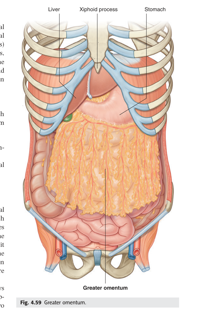
The greater omentum is mobile and richly supplied with immune cells. When a viscus perforates or an organ becomes inflamed, the omentum migrates toward it and wraps around it, walling off the infection and preventing generalised peritonitis. Surgeons find it plastered over an inflamed appendix or a perforated ulcer often enough that the nickname has stuck.
In children the omentum is short and thin, which is one reason appendicitis perforates and spreads more readily in the young.
- What does the greater omentum hang from? → The greater curvature of the stomach
- How many layers of peritoneum? → Four
- What is its protective function? → It migrates to and walls off inflamed or perforated viscera
The lesser omentum ★★★
The lesser omentum is a double fold running from the lesser curvature of the stomach and the first part of the duodenum up to the inferior surface of the liver — specifically the porta hepatis and the fissure for the ligamentum venosum. TMU asked for its position, composition and contents in the 2021–22 paper, worth seven marks.
A two-layered fold of peritoneum extending from the lesser curvature of the stomach and the first part of the duodenum to the inferior surface of the liver.
It consists of two ligaments:
1. Hepatogastric ligament — the larger, medial part, from the lesser curvature to the liver.
2. Hepatoduodenal ligament — the thickened free right edge, from the first part of the duodenum to the liver.
The hepatoduodenal ligament contains the PORTAL TRIAD:
• the proper hepatic artery — on the left
• the common bile duct — on the right
• the hepatic portal vein — behind the other two
together with lymphatics, lymph nodes and the hepatic nerve plexus.
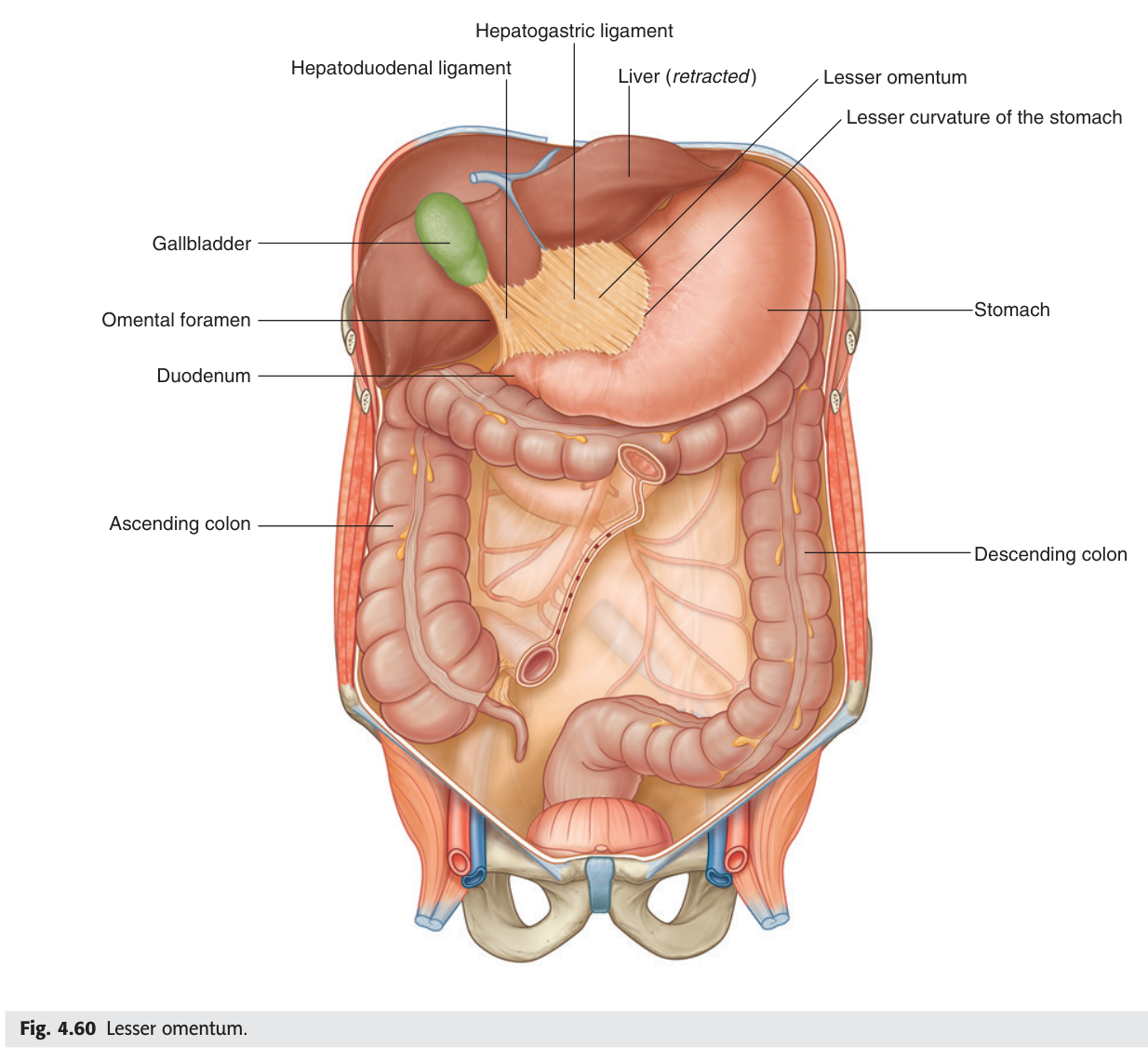
In the hepatoduodenal ligament: Duct on the right, Artery on the left, Vein behind. Duct–Artery–Vein, right–left–behind.
The left gastric artery also runs in the lesser omentum along the lesser curvature, with the right gastric artery.
- Where does the lesser omentum run between? → Lesser curvature of the stomach and first part of duodenum → inferior surface of the liver
- Its two ligaments? → Hepatogastric and hepatoduodenal
- The three contents of the free edge? → Proper hepatic artery (left) · common bile duct (right) · hepatic portal vein (behind)
- What else is in it? → Lymphatics, nodes and the hepatic nerve plexus
The omental bursa and epiploic foramen ★★
Behind the stomach and the lesser omentum lies a flattened space, the omental bursa (lesser sac). It exists because the stomach rotates during development and traps a pocket of peritoneal cavity behind itself. It communicates with the main peritoneal cavity through one small opening — the epiploic (omental) foramen.
The opening connecting the greater and lesser sacs of the peritoneal cavity.
Anteriorly: the free edge of the lesser omentum — the hepatoduodenal ligament with the portal triad.
Posteriorly: the inferior vena cava.
Superiorly: the caudate lobe of the liver.
Inferiorly: the first part of the duodenum.
In a liver injury with torrential bleeding, the surgeon puts a finger through the epiploic foramen and pinches the free edge of the lesser omentum between finger and thumb. That compresses the hepatic artery and portal vein at once and stops almost all inflow to the liver — buying time to find the bleeding point.
This is the clearest possible demonstration of why boundaries matter: the manoeuvre works only because the artery and vein are in front of the foramen and the IVC is behind it.
- What does the epiploic foramen connect? → The greater and lesser sacs
- Its anterior boundary? → The free edge of the lesser omentum, containing the portal triad
- Its posterior boundary? → The inferior vena cava
- What is the Pringle manoeuvre? → Compressing the free edge through the foramen to occlude hepatic inflow
Revision layer
Intraperitoneal vs retroperitoneal — the MCQ table
| Organ | Status |
|---|---|
| Stomach, spleen, liver, gallbladder | Intraperitoneal |
| Jejunum, ileum, transverse colon, sigmoid colon, appendix | Intraperitoneal |
| Duodenum | 1st part intraperitoneal; 2nd, 3rd, 4th retroperitoneal |
| Ascending and descending colon | Retroperitoneal |
| Kidneys, ureters, suprarenal glands | Retroperitoneal |
| Pancreas | Retroperitoneal except the tail |
| Abdominal aorta, inferior vena cava | Retroperitoneal |
The lesser omentum in one box
| Detail | |
|---|---|
| Attachments | Lesser curvature of stomach + 1st part of duodenum → inferior surface of liver |
| Parts | Hepatogastric ligament · hepatoduodenal ligament (free right edge) |
| Contents of free edge | Proper hepatic artery (left) · common bile duct (right) · hepatic portal vein (behind) |
| Behind the free edge | The epiploic (omental) foramen |
- Classify the abdominal organs as intra- or retroperitoneal
- Describe the lesser omentum: attachments, two ligaments, contents of the free edge
- Give all four boundaries of the epiploic foramen
- Explain the Pringle manoeuvre from the anatomy
- Explain why appendicitis pain migrates