Abdominal Wall & Inguinal Region
The nine layers of the lateral abdominal wall ★★★
The abdominal wall has to contain the viscera, move with breathing, and generate the pressure needed for coughing and childbirth. It does that with three flat muscle sheets whose fibres run in three different directions — the same principle as plywood, and for the same reason: layers crossed at angles are far stronger than the sum of their parts.
TMU has asked for the layers from superficial to deep in a past paper, and the answer is nine. Two of them catch students out, because the superficial fascia here is not one layer but two.
1. Skin
2. Superficial fascia — fatty layer (Camper's fascia)
3. Superficial fascia — membranous layer (Scarpa's fascia)
4. External oblique abdominis
5. Internal oblique abdominis
6. Transversus abdominis
7. Transversalis fascia
8. Extraperitoneal tissue (fat)
9. Parietal peritoneum
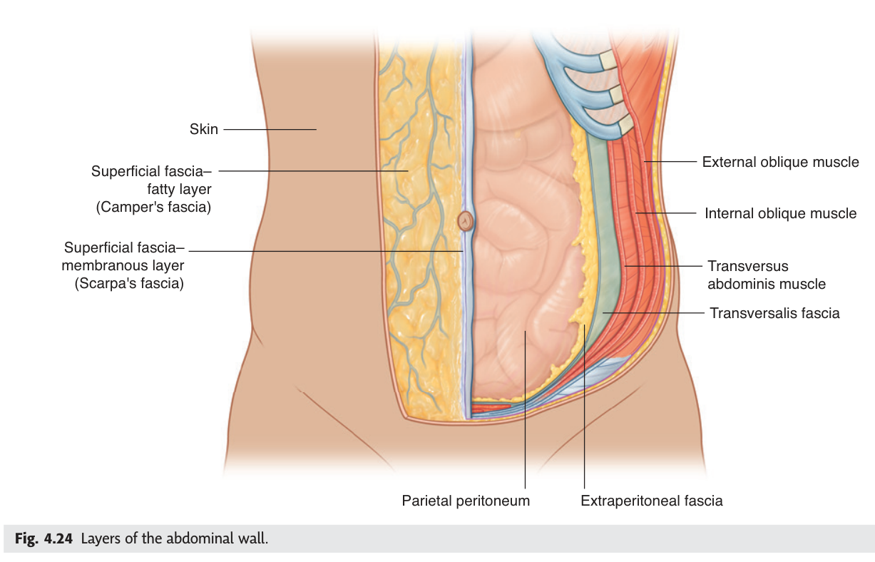
The three muscle sheets run in a fixed sequence you can check on yourself: external oblique fibres run downward and forward (hands-in-pockets direction), internal oblique at right angles to it, and transversus abdominis horizontally. The neurovascular plane lies between the internal oblique and transversus — the same arrangement as the intercostal space, because the abdominal wall is developmentally continuous with it.
Scarpa's membranous layer does not stop at the abdomen. It continues into the perineum as Colles' fascia and into the scrotum and penis, but is firmly attached to the fascia lata just below the inguinal ligament.
So when the urethra ruptures, extravasated urine spreads up the abdominal wall and into the scrotum and penis — but never down into the thigh. The pattern of swelling tells the surgeon exactly which fascial plane has been breached.
- The nine layers superficial to deep? → Skin · Camper's · Scarpa's · external oblique · internal oblique · transversus abdominis · transversalis fascia · extraperitoneal tissue · peritoneum
- The two parts of the superficial fascia? → Fatty (Camper's) and membranous (Scarpa's)
- Where does the neurovascular plane lie? → Between internal oblique and transversus abdominis
- Why does extravasated urine not enter the thigh? → Scarpa's fascia is attached to the fascia lata below the inguinal ligament
The rectus sheath and linea alba
In the midline the three aponeuroses interlace and wrap around rectus abdominis, forming the rectus sheath. Where the sheaths of the two sides meet in the midline they form the linea alba — literally the white line — running from the xiphoid process to the pubic symphysis.
A fibrous median raphe in the midline of the anterior abdominal wall, extending from the xiphoid process to the pubic symphysis.
It is formed by the interlacing aponeuroses of the external oblique, internal oblique and transversus abdominis of both sides, and separates the two rectus abdominis muscles.
Clinical: it is relatively avascular and crossed by no nerves, which is why it is chosen for a midline laparotomy incision.
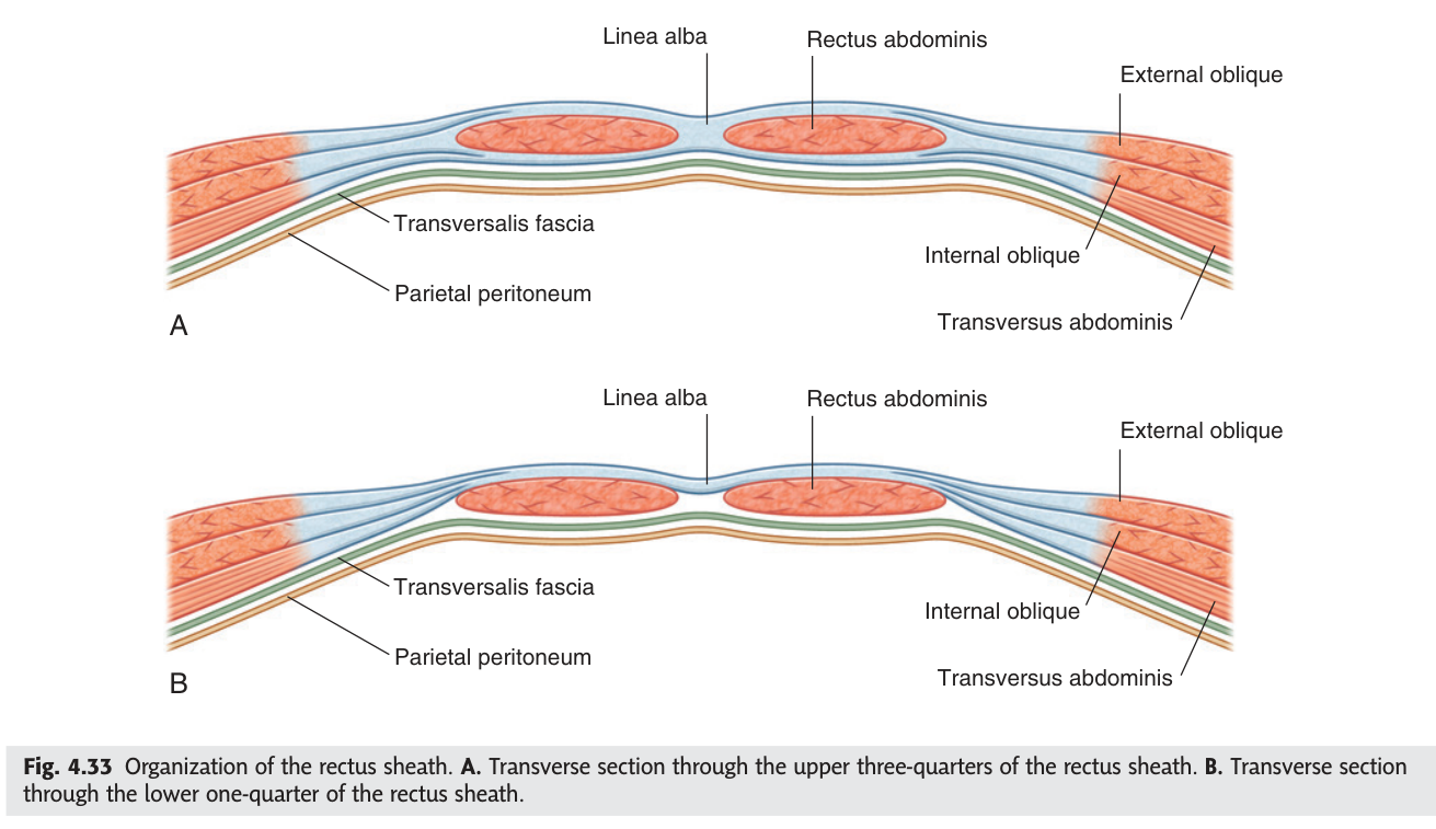
The sheath is not the same all the way down, and the change happens at the arcuate line, roughly a third of the way from the umbilicus to the pubis. Above it, the internal oblique aponeurosis splits to pass both in front of and behind the muscle, so there is a posterior wall. Below it, all three aponeuroses pass in front, and the posterior wall is only transversalis fascia.
The inferior epigastric artery enters the sheath below the arcuate line and ascends to anastomose with the superior epigastric artery from the internal thoracic. A violent cough or a sudden contraction can tear it. Because the sheath is a closed compartment, blood collects as a painful tender mass that does not move when the muscle is tensed — Fothergill's sign, which distinguishes it from an intra-abdominal mass.
- What is the linea alba formed by? → Interlacing aponeuroses of all three flat muscles of both sides
- Why is it used for laparotomy? → Relatively avascular, and no nerves crossed
- What changes at the arcuate line? → Below it all three aponeuroses pass in front; there is no posterior sheath
- Which two arteries anastomose in the sheath? → Superior epigastric (from internal thoracic) and inferior epigastric (from external iliac)
The inguinal canal ★★★
The inguinal canal is a paradox: a deliberate weak point in the strongest part of the abdominal wall. It exists because the testis develops on the posterior abdominal wall and must migrate to the scrotum, dragging its vessels and duct through the wall. The canal is the tunnel it leaves behind — which is why inguinal hernias are so much commoner in men.
It runs about 4 cm, obliquely, above and parallel to the medial half of the inguinal ligament, from the deep inguinal ring to the superficial inguinal ring. TMU asks for its four walls as a fill-in-the-blank, and the past paper answer is below.
Anterior wall: aponeurosis of the external oblique, reinforced laterally by muscle fibres of the internal oblique.
Posterior wall: transversalis fascia, reinforced medially by the conjoint tendon.
Superior wall (roof): arching fibres of the internal oblique and transversus abdominis.
Inferior wall (floor): the inguinal ligament.
Contents: the spermatic cord in the male, the round ligament of the uterus in the female, and the ilioinguinal nerve in both.
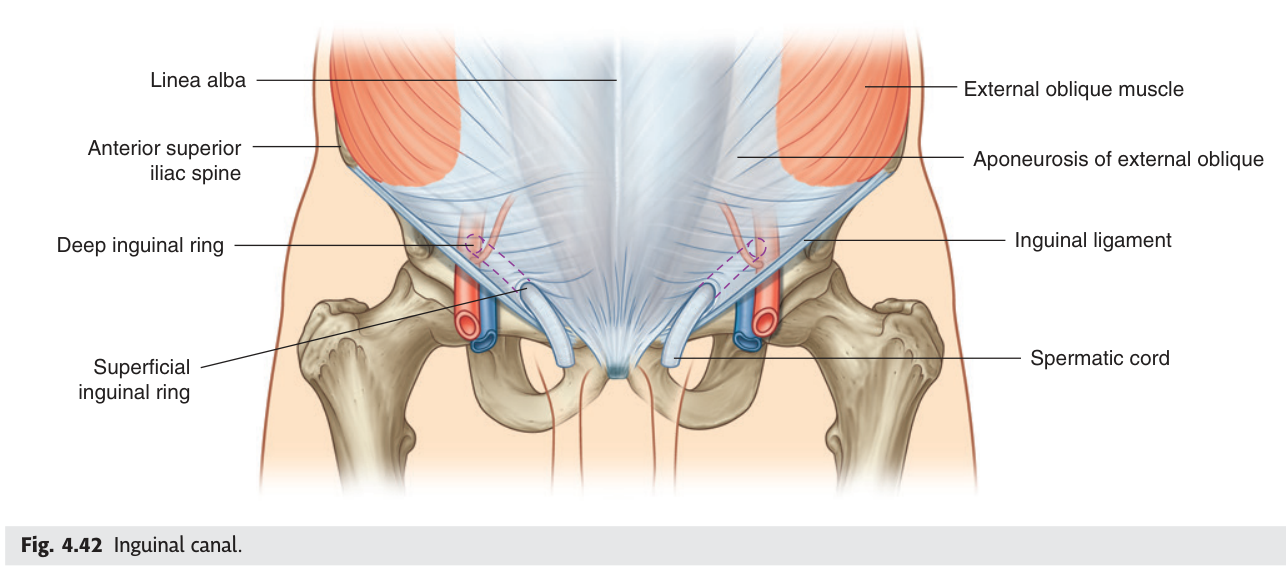
Notice the symmetry: the muscle that is weak at one end is reinforced at the other. The anterior wall is strong laterally, where the deep ring lies; the posterior wall is strong medially, where the superficial ring lies. So wherever a hernia tries to push through, there is muscle behind it — and the whole canal collapses when you cough, because the arching roof descends toward the floor. That is the shutter mechanism, and it is why most people never herniate.
- The four walls of the inguinal canal? → Anterior: external oblique aponeurosis (+ internal oblique laterally) · Posterior: transversalis fascia (+ conjoint tendon medially) · Roof: arching internal oblique and transversus · Floor: inguinal ligament
- Its contents in the male and female? → Spermatic cord / round ligament of uterus, plus the ilioinguinal nerve in both
- Why does the canal exist? → It is the path of testicular descent
- What is the shutter mechanism? → The arching roof descends on coughing, closing the canal
The inguinal triangle ★★★
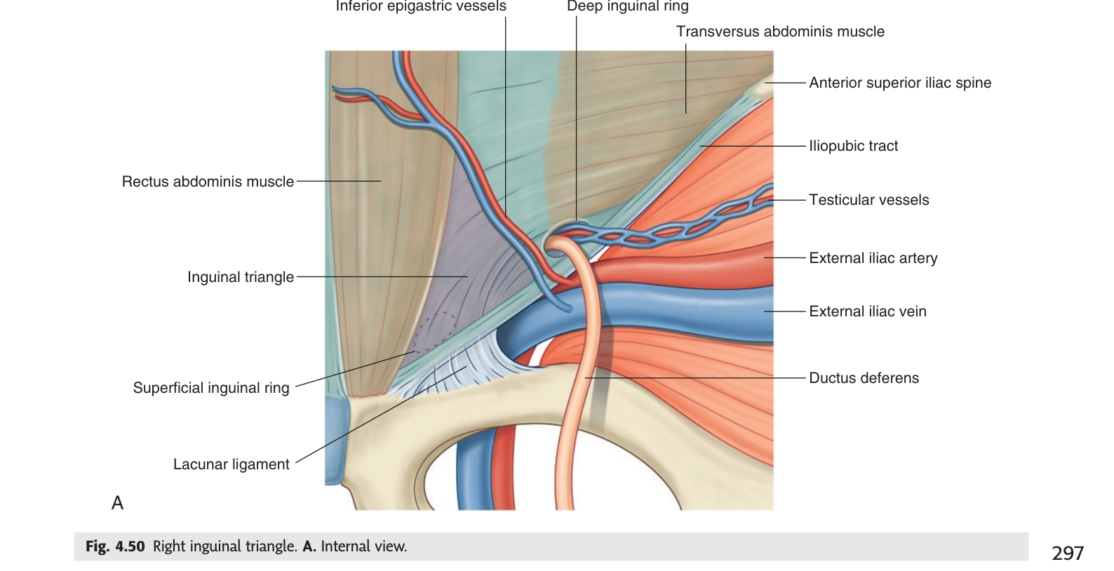
Within the posterior wall of the canal there is a triangular area with no muscle behind it at all — only transversalis fascia and peritoneum. This is the inguinal triangle, and it is the site of a direct hernia.
A triangular area of potential weakness in the lower anterior abdominal wall.
Laterally: the inferior epigastric artery.
Medially: the lateral border of rectus abdominis.
Inferiorly: the inguinal ligament.
No structure passes through it — it is defined by its weakness. A direct inguinal hernia bulges forward through it, medial to the inferior epigastric vessels.
- The three boundaries of the inguinal triangle? → Inferior epigastric artery laterally · lateral border of rectus abdominis medially · inguinal ligament inferiorly
- What passes through it? → Nothing — it is an area of weakness
- Which hernia occurs through it? → A direct inguinal hernia
- What is its other name? → Hesselbach's triangle
Direct versus indirect hernia
One vessel separates the two kinds of inguinal hernia, and getting it right is worth marks in every anatomy and surgery exam you will ever sit. The inferior epigastric artery is the landmark.
| Indirect | Direct | |
|---|---|---|
| Relation to inferior epigastric artery | LATERAL to it | MEDIAL to it |
| Route | Through the deep ring, along the whole canal | Forward through the inguinal triangle; does not traverse the whole canal |
| Cause | Congenital — persistent processus vaginalis | Acquired — weakened abdominal musculature |
| Typical patient | Younger; commonest overall | Older men |
| Coverings | All three coverings of the spermatic cord | Usually only external spermatic fascia |
| Into the scrotum? | Commonly | Rarely |
Medial = Direct. Both words are about the middle — a direct hernia comes straight forward through the triangle, medial to the vessels. The indirect one takes the long way round, from lateral.
Do not forget Unit 1. A femoral hernia descends through the femoral canal and appears below and lateral to the pubic tubercle, whereas both inguinal hernias appear above and medial to it. That single bony landmark separates the two families, and femoral hernias matter more because the femoral ring is small and rigid, so they strangulate far more readily.
- Which hernia is lateral to the inferior epigastric artery? → Indirect
- Which is medial? → Direct
- Which is congenital? → Indirect, from a persistent processus vaginalis
- How is a femoral hernia distinguished? → It appears below and lateral to the pubic tubercle
Revision layer
The nine layers — the past-paper list
| # | Layer |
|---|---|
| 1 | Skin |
| 2 | Superficial fascia — fatty layer (Camper's) |
| 3 | Superficial fascia — membranous layer (Scarpa's) |
| 4 | External oblique abdominis |
| 5 | Internal oblique abdominis |
| 6 | Transversus abdominis |
| 7 | Transversalis fascia |
| 8 | Extraperitoneal tissue |
| 9 | Parietal peritoneum |
Inguinal canal — walls and contents
| Wall | Formed by |
|---|---|
| Anterior | External oblique aponeurosis; internal oblique muscle laterally |
| Posterior | Transversalis fascia; conjoint tendon medially |
| Roof | Arching fibres of internal oblique and transversus abdominis |
| Floor | Inguinal ligament |
| Contents | Spermatic cord (male) / round ligament of uterus (female) · ilioinguinal nerve |
- List the nine layers of the lateral abdominal wall in order
- Give the four walls and contents of the inguinal canal
- Define the inguinal triangle with all three boundaries
- Distinguish direct, indirect and femoral hernia by their landmarks
- Explain what changes at the arcuate line and why it matters