Lungs, Great Vessels & Lateral Mediastinum
The two lungs compared
The lungs are not mirror images, and the differences all come from one fact: the heart sits mostly on the left. The right lung is shorter and wider — shorter because the liver pushes the right dome of the diaphragm up, wider because the heart is not in its way. The left lung is longer and narrower, and carries a cardiac notch and the tongue-like lingula where the heart indents it.
| Right lung | Left lung | |
|---|---|---|
| Shape | Shorter and wider | Longer and narrower |
| Lobes | Three — superior, middle, inferior | Two — superior, inferior |
| Fissures | Oblique and horizontal | Oblique only |
| Special features | — | Cardiac notch and lingula |
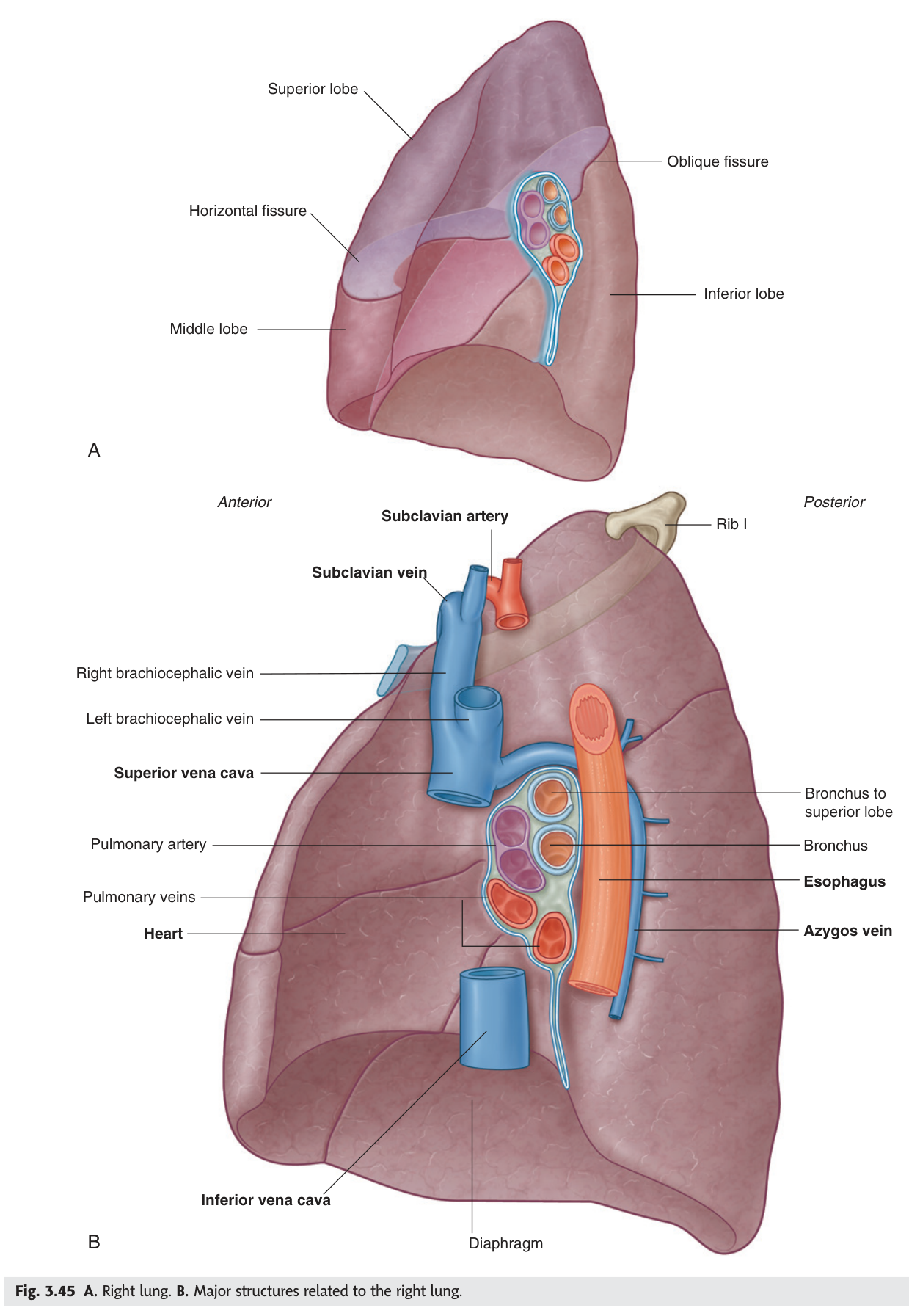
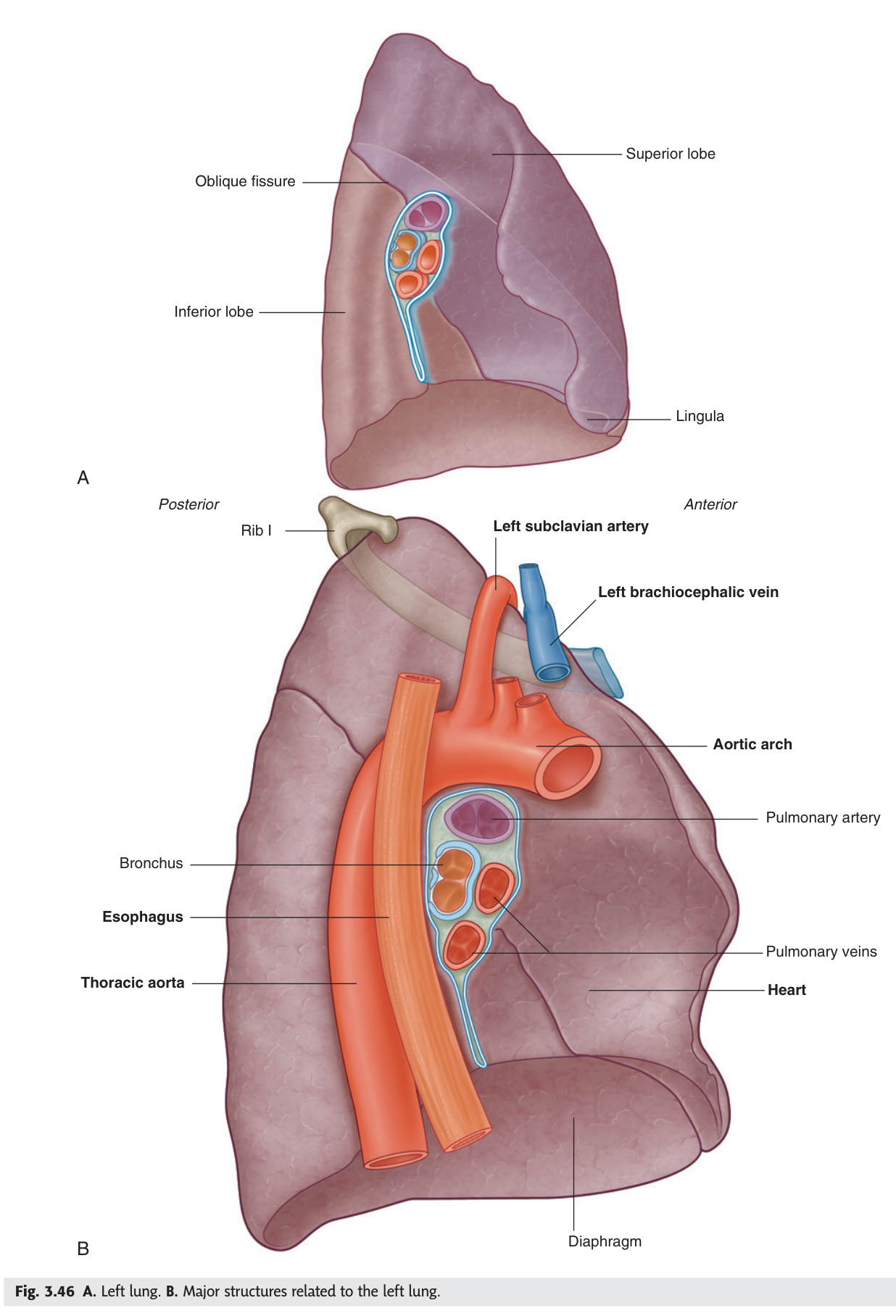
The right main bronchus is wider, shorter and more vertical than the left, because the heart pushes the left bronchus into a more horizontal course. An inhaled peanut, tooth or piece of food therefore lodges in the right bronchial tree far more often — typically in the posterior segment of the right upper lobe if the patient is supine, or the basal segments if upright. Aspiration pneumonia follows the same anatomy.
- How many lobes and fissures has each lung? → Right: 3 lobes, oblique + horizontal fissures · Left: 2 lobes, oblique only
- Which lung is shorter and wider, and why? → The right — the liver raises the diaphragm on that side
- What are the cardiac notch and lingula? → Features of the left lung where the heart indents it
- Why do inhaled objects usually go right? → The right main bronchus is wider, shorter and more vertical
The root of the lung ★★
The root (hilum) is the stalk connecting each lung to the mediastinum — the principal bronchus, the pulmonary artery, the two pulmonary veins, bronchial vessels, lymphatics and nerves, all wrapped in a sleeve of pleura that hangs down below as the pulmonary ligament.
Within the root the arrangement is fixed enough to be examined, and it differs slightly between the two sides. From anterior to posterior on both sides the order is pulmonary vein, pulmonary artery, bronchus. From above downwards it differs: on the right the bronchus is highest (the eparterial bronchus to the upper lobe passes above the artery); on the left the artery is highest, because the aortic arch pushes it up over the bronchus.
Right = Bronchus on top; Left = Artery on top — because the aortic arch on the left hooks over the bronchus and drags the pulmonary artery up with it. Anterior to posterior is the same on both sides: vein, artery, bronchus.
- What passes through the lung root? → Principal bronchus · pulmonary artery · two pulmonary veins · bronchial vessels · lymphatics · nerves
- Order from anterior to posterior? → Vein · artery · bronchus (both sides)
- What is highest on the right? → The bronchus
- What is highest on the left, and why? → The pulmonary artery — the aortic arch pushes it up
The right lateral mediastinum ★★★
Remove the right lung and you are looking at the right side of the mediastinum — one of the two views TMU sets as a picture question. Learn it as a scene rather than a list: the arch of the azygos vein curves forward over the lung root to reach the superior vena cava, and everything else is arranged around that landmark.
Arch of the azygos vein — ascends on the right of the vertebral bodies to T4, then arches over the right lung root to enter the superior vena cava.
Superior vena cava and the right brachiocephalic vein.
Right phrenic nerve — descends on the right of the SVC and passes in front of the lung root, with the pericardiacophrenic vessels, to the diaphragm.
Right vagus nerve — lies behind the SVC on the side of the trachea and passes behind the lung root.
Oesophagus and trachea; the thoracic duct ascending with the aorta on its left and the azygos vein on its right.
Right sympathetic trunk (about twelve ganglia) lateral to the azygos vein, and the greater splanchnic nerve from roots T6–T9.
The one relationship the examiners test again and again: the phrenic nerve passes ANTERIOR to the lung root; the vagus passes POSTERIOR. It is true on both sides. Remember it as "phrenic to the front" — and note that this makes anatomical sense, because the phrenic is heading for the diaphragm alongside the pericardium, while the vagus is heading for the oesophagus behind.
- What arches over the right lung root? → The arch of the azygos vein, entering the SVC
- Where does the right phrenic nerve pass? → In front of the lung root
- Where does the right vagus pass? → Behind the lung root
- Root value of the greater splanchnic nerve? → T6–T9
The left lateral mediastinum ★★★
Remove the left lung and the dominating landmark is different: the arch of the aorta curls over the left lung root exactly as the azygos arch does on the right. That symmetry is the easiest way to remember which side you are looking at.
Arch of the aorta — joins the descending aorta at the left side of the body of T4. Its concavity is connected to the left pulmonary artery by the arterial ligament (ligamentum arteriosum).
Left phrenic nerve with pericardiacophrenic vessels — in front of the lung root.
Left vagus nerve — crosses the left side of the aortic arch, passes behind the lung root, and forms the oesophageal plexus.
Left recurrent laryngeal nerve — leaves the vagus at the arch, hooks under it lateral to the arterial ligament, and ascends in the groove between trachea and oesophagus.
Descending thoracic aorta, oesophagus between pericardium and aorta, the upper thoracic duct behind the origin of the left subclavian artery, the hemiazygos and accessory hemiazygos veins, and the left sympathetic trunk.
You met this nerve in Unit 11 from the thyroid's point of view. Here you can see why it takes such a long route: it is hooked around the aortic arch at the ligamentum arteriosum, deep inside the chest.
That is why a bronchial carcinoma at the left hilum, an aortic arch aneurysm, or a grossly enlarged left atrium in mitral stenosis can all present with hoarseness. A hoarse voice with no laryngeal cause sends the clinician to the chest — and this figure is the reason.
- What arches over the left lung root? → The arch of the aorta
- At what level does the arch become the descending aorta? → The left side of the body of T4
- What connects the arch to the left pulmonary artery? → The arterial ligament (ligamentum arteriosum)
- Where does the left recurrent laryngeal nerve hook? → Under the arch, lateral to the arterial ligament
- Which veins are found on the left? → Hemiazygos and accessory hemiazygos
The arch of the aorta ★★
The aorta is described in three parts — ascending aorta, arch of the aorta, descending (thoracic then abdominal) aorta — and the arch is the part that gives the branches to the head, neck and upper limbs. TMU has asked for those three branches, in order, in a past paper.
1. Brachiocephalic trunk — which divides into the right common carotid and right subclavian arteries.
2. Left common carotid artery.
3. Left subclavian artery.
There is no left brachiocephalic trunk, and students often expect one. The reason is positional: the arch is already on the left, so the left carotid and subclavian arteries can come straight off it. The right-sided vessels have to reach across the midline, and they share a common stem to do so. Asymmetry of the arch explains the asymmetry of its branches — and, as you saw in Unit 11, the asymmetry of the recurrent laryngeal nerves too.
A congenital narrowing of the aorta, classically just distal to the left subclavian artery near the ligamentum arteriosum. Blood reaches the lower body by collateral flow — chiefly through the internal thoracic and posterior intercostal arteries, which enlarge and erode the under-surfaces of the ribs, producing the rib notching seen on a chest X-ray.
Clinically: hypertension and strong pulses in the arms, weak delayed femoral pulses, and radio-femoral delay. The chest wall's double blood supply, from Unit 12, is what keeps the patient alive.
- The three branches of the aortic arch, right to left? → Brachiocephalic trunk · left common carotid · left subclavian
- What does the brachiocephalic trunk divide into? → Right common carotid and right subclavian arteries
- Why is there no left brachiocephalic trunk? → The arch is already on the left, so those vessels arise directly
- What causes rib notching in coarctation? → Enlarged collateral intercostal arteries eroding the ribs
The azygos system
The azygos system is the venous drainage of the thoracic wall, and it is the back-up route between the superior and inferior venae cavae. Azygos means "unpaired" — there is one main vessel, on the right.
| Vein | Course | Drains |
|---|---|---|
| Azygos | Ascends on the right of the vertebral bodies to T4, then arches over the right lung root into the SVC | Right posterior intercostal veins |
| Hemiazygos | Ascends on the left, crosses the midline behind the aorta at about T8 to join the azygos | Lower left posterior intercostal veins |
| Accessory hemiazygos | Descends on the left upper vertebrae, crosses to join the azygos | Upper left posterior intercostal veins |
Because the azygos system connects the inferior vena cava below with the superior vena cava above, it provides a collateral pathway if either is obstructed. In superior vena cava obstruction — most often by a bronchial carcinoma — blood from the head and arms reverses down dilated chest wall veins into the azygos system and the IVC. The patient has a swollen, plethoric face and neck with distended non-pulsatile neck veins and visible chest wall collaterals. The anatomy is doing the compensating.
- On which side does the azygos vein ascend? → The right
- Where does it end? → Arching over the right lung root into the superior vena cava
- At what level does the hemiazygos cross the midline? → About T8
- Why does the azygos system matter clinically? → It is the collateral route between IVC and SVC in caval obstruction
Revision layer
Right vs left lateral mediastinum — the comparison the picture question wants
| RIGHT side | LEFT side | |
|---|---|---|
| Arches over the lung root | Arch of the azygos vein | Arch of the aorta |
| Great vein | Superior vena cava | — |
| Phrenic nerve | In front of the lung root | In front of the lung root |
| Vagus nerve | Behind the lung root | Behind the lung root; forms oesophageal plexus |
| Recurrent laryngeal | Hooks under the right subclavian artery (in the neck) | Hooks under the aortic arch |
| Veins of the wall | Azygos vein | Hemiazygos and accessory hemiazygos |
| Also present | Trachea, oesophagus, thoracic duct, sympathetic trunk | Descending aorta, oesophagus, thoracic duct, sympathetic trunk |
Lung root arrangement
| Axis | Right | Left |
|---|---|---|
| Anterior → posterior | Vein · artery · bronchus | Vein · artery · bronchus |
| Superior → inferior | Bronchus · artery · vein | Artery · bronchus · vein |
- Draw both lateral mediastinal views and label every structure
- State the phrenic and vagus relations to the lung root
- Give the three branches of the aortic arch in order
- Compare the two lungs — lobes, fissures, shape, special features
- Trace the azygos vein and explain its role in caval obstruction