Mediastinum, Pericardium & Heart
Dividing the mediastinum ★★★
Remove both lungs and what is left in the middle of the chest is the mediastinum — everything between the two mediastinal pleurae. It is not an organ but a compartment, and like the neck it only becomes learnable once it is divided up.
Anteriorly: sternum and costal cartilages. Posteriorly: the thoracic vertebrae. Laterally: the mediastinal pleura on each side. Above: the thoracic inlet. Below: the diaphragm.
It is divided by an imaginary horizontal plane running from the sternal angle to the lower border of the fourth thoracic vertebra — the plane of Louis. Above that plane is the superior mediastinum; below it, the inferior mediastinum, which is further split into three by the pericardium.
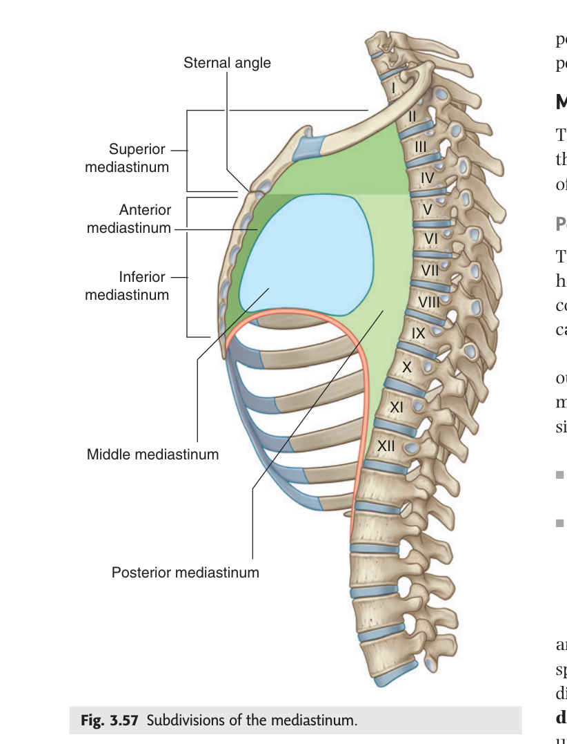
| Division | Position | Principal contents |
|---|---|---|
| Superior | Above the sternal angle plane | Arch of the aorta and its three branches · brachiocephalic veins and SVC · trachea · oesophagus · thoracic duct · vagus and phrenic nerves · thymus |
| Anterior | Between the sternum and the pericardium | Thymus (or its remnant) · fat · lymph nodes |
| Middle | The pericardium and its contents | Heart in the pericardial sac · origins of the great vessels · phrenic nerves |
| Posterior | Between the pericardium and the vertebral column | Descending thoracic aorta · oesophagus · thoracic duct · azygos system · sympathetic trunks and splanchnic nerves |
That single horizontal plane at the sternal angle (T4/T5) marks: the division of the mediastinum, the beginning and end of the arch of the aorta, the bifurcation of the trachea, and the level of the second costal cartilage — the landmark you count ribs from. One palpable ridge, four facts.
- Boundaries of the mediastinum? → Sternum in front · thoracic vertebrae behind · mediastinal pleurae laterally · thoracic inlet above · diaphragm below
- What plane divides superior from inferior? → Sternal angle to the lower border of T4
- The three parts of the inferior mediastinum? → Anterior, middle and posterior
- What is in the middle mediastinum? → The heart in the pericardial sac, with a phrenic nerve on each side
The pericardium
The heart sits inside a double bag. The outer layer is the tough fibrous pericardium, which fuses below with the central tendon of the diaphragm and above with the adventitia of the great vessels — anchoring the heart in place. Inside it lies the serous pericardium, in two layers: a parietal layer lining the fibrous sac, and a visceral layer (the epicardium) stuck to the heart itself.
Between the two serous layers is the pericardial cavity, containing about 50 mL of fluid — just enough to let the heart change shape sixty times a minute without friction.
The fibrous pericardium is tough and does not stretch acutely. Blood entering the cavity — from a stab wound, a ruptured ventricle after infarction, or an aortic dissection — has nowhere to go, so pressure rises fast and squeezes the heart. As little as 100–200 mL accumulating quickly can stop filling altogether.
The signs are Beck's triad: falling blood pressure, rising jugular venous pressure, and muffled heart sounds. Treatment is pericardiocentesis — a needle under the xiphoid aimed at the left shoulder. Slowly accumulating fluid, by contrast, can reach a litre or more, because the sac has time to stretch.
- The two components of the pericardium? → Fibrous pericardium and serous pericardium
- The two layers of serous pericardium? → Parietal (lining the fibrous sac) and visceral (epicardium)
- Normal volume of pericardial fluid? → About 50 mL
- Beck's triad? → Hypotension · raised JVP · muffled heart sounds
The pericardial sinuses ★★
As the heart develops, the serous pericardium reflects around the great vessels in two groups — the arteries together, the veins together. Between and behind those reflections it leaves two spaces, and both are useful to a surgeon.
Transverse pericardial sinus — a passage lying behind the aorta and pulmonary trunk and in front of the superior vena cava and left atrium. It is the gap between the arterial and venous reflections.
Oblique pericardial sinus — a blind cul-de-sac behind the left atrium, bounded by the reflections around the pulmonary veins and inferior vena cava.
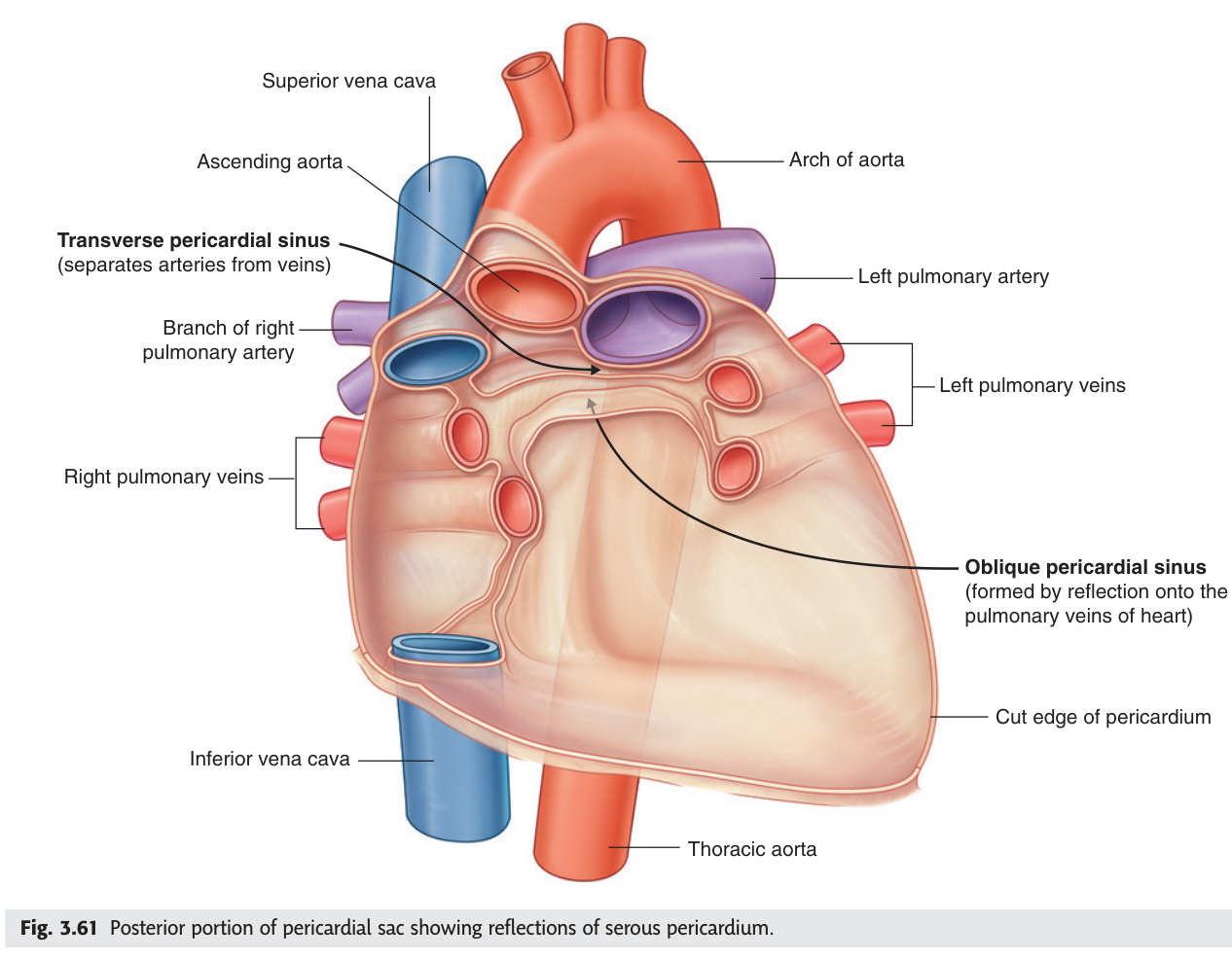
In cardiac surgery the surgeon passes a finger — and then a ligature — through the transverse sinus, behind the aorta and pulmonary trunk. Tightening it clamps both arterial outflows at once, which is exactly what is needed to establish cardiopulmonary bypass. A developmental quirk of pericardial reflection turns out to be a surgical handle.
- What lies in front of the transverse sinus? → The aorta and pulmonary trunk
- What lies behind it? → The superior vena cava and left atrium
- Where is the oblique sinus? → A blind recess behind the left atrium
- Surgical use of the transverse sinus? → To pass a ligature around both great arterial trunks in bypass surgery
External features of the heart
TMU's slide gives the heart's external anatomy as a formula worth memorising: the heart has an apex, a base, two surfaces, three borders and four grooves.
| Feature | Detail |
|---|---|
| Apex | Directed down, forward and to the left; formed by the left ventricle; lies in the 5th left intercostal space, 1–2 cm medial to the midclavicular line |
| Base | Faces backward, upward and to the right; formed mainly by the left atrium |
| Two surfaces | Anterior (sternocostal) · inferior (diaphragmatic) |
| Three borders | Right — right atrium · Left — mainly left ventricle · Inferior — right ventricle |
| Four grooves | Coronary (atrioventricular) · anterior interventricular · posterior interventricular · posterior interatrial |
The apex beat is where you place your fingers at the bedside — the 5th left intercostal space in the midclavicular line. If it is displaced downward and laterally, the left ventricle is enlarged. That is the whole clinical value of the apex, and it comes straight from knowing which chamber forms it.
- Which chamber forms the apex? → The left ventricle
- Where is the apex, in surface terms? → 5th left intercostal space, 1–2 cm medial to the midclavicular line
- Which chamber forms the base? → Mainly the left atrium
- Which forms the inferior border? → The right ventricle
- Name the four grooves? → Coronary · anterior interventricular · posterior interventricular · posterior interatrial
Right atrium and right ventricle ★★
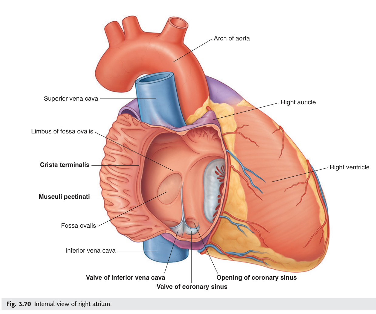
Every chamber is described the same way in the TMU exam: inlets, outlet, and internal structures. Learn that template and all four chambers become one question asked four times.
Three inlets: orifice of the superior vena cava · orifice of the inferior vena cava · orifice of the coronary sinus.
One outlet: the right atrioventricular orifice.
Internal features: the crista terminalis, a muscular ridge separating the smooth sinus venarum behind from the rough atrium proper in front, running from the SVC to the IVC orifice; musculi pectinati in the rough part and the right auricle; and the fossa ovalis on the interatrial septum — the remnant of the fetal foramen ovale.
Inlet: right atrioventricular orifice, guarded by the tricuspid valve. Outlet: the pulmonary orifice, guarded by the pulmonary valve.
Internal features: trabeculae carneae; papillary muscles; the septomarginal trabecula (moderator band) carrying part of the conducting system; and the smooth-walled outflow tract, the conus arteriosus (infundibulum).
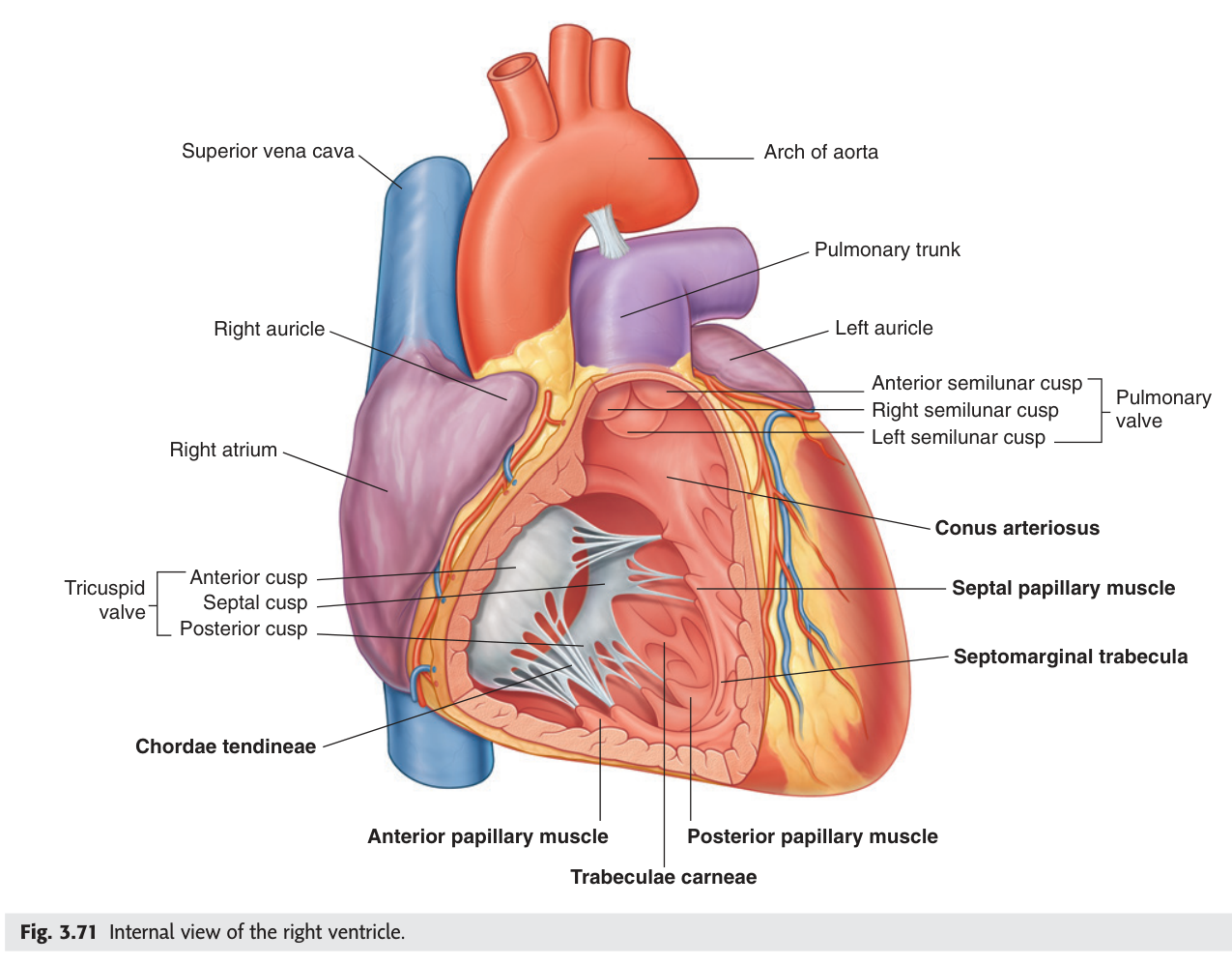
The fibrous ring, the tricuspid valve cusps, the chordae tendineae and the papillary muscles, considered as one functional unit.
Function: to prevent backward flow of blood from the right ventricle into the right atrium during ventricular contraction. The bicuspid (mitral) complex is its counterpart on the left.
When the ventricle contracts, blood pushes the cusps upward until they meet and seal. Left to themselves they would simply invert, like an umbrella in the wind. The papillary muscles contract at the same moment and pull on the chordae tendineae, holding the cusps at exactly the closed position. This is why papillary muscle rupture after a heart attack causes sudden, catastrophic valve incompetence — the cusps are intact, but nothing is holding them.
- Three inlets of the right atrium? → Superior vena cava · inferior vena cava · coronary sinus
- What is the crista terminalis? → A ridge separating the smooth sinus venarum from the rough atrium proper
- Inlet and outlet of the right ventricle? → Right AV orifice (tricuspid) → pulmonary orifice (pulmonary valve)
- Four parts of the tricuspid complex? → Fibrous ring · cusps · chordae tendineae · papillary muscles
- What does the moderator band carry? → Part of the conducting system to the anterior papillary muscle
Left atrium and left ventricle
Four inlets: the orifices of the four pulmonary veins — two right, two left. One outlet: the left atrioventricular orifice.
Its wall is largely smooth; musculi pectinati are confined to the left auricle.
Inlet: left atrioventricular orifice, guarded by the mitral (bicuspid) valve. Outlet: the aortic orifice, guarded by the aortic valve.
Its wall is about three times thicker than the right ventricle's; it has two large papillary muscles, and a smooth outflow tract, the aortic vestibule.
The wall thickness is the whole story of the left ventricle. It pumps into the systemic circulation against roughly six times the resistance the right ventricle faces, so it is built accordingly. The two ventricles eject the same volume at very different pressures — which is why the left is the chamber that hypertrophies in hypertension and fails in most heart disease.
The left auricle is a small, trabeculated pouch off the left atrium where blood flow is naturally sluggish. In atrial fibrillation the atrium no longer contracts properly, blood stagnates there, and clot forms. A fragment that breaks off travels through the left ventricle into the aorta and then, commonly, into the carotid circulation — causing a stroke. This is precisely why patients with atrial fibrillation are anticoagulated, and why the left auricle is sometimes surgically occluded.
- How many inlets has the left atrium? → Four — the pulmonary veins
- Inlet and outlet of the left ventricle? → Left AV orifice (mitral valve) → aortic orifice (aortic valve)
- How does its wall compare with the right? → About three times thicker
- Why does the left auricle matter in atrial fibrillation? → Stagnant blood forms clot there, embolising to the brain
The coronary arteries ★★★
The heart is a pump that cannot pause to feed itself, and its two arteries are the first branches of the aorta — arising from the aortic sinuses immediately above the aortic valve cusps. That position is not accidental: it fills them during diastole, when the aortic valve shuts and the relaxed myocardium is not squeezing its own vessels closed.
Right coronary artery — from the right aortic sinus. Branches: right conus branch · right marginal artery · posterior interventricular (posterior descending) artery · the sinuatrial nodal and atrioventricular nodal branches.
Left coronary artery — from the left aortic sinus. Divides into the anterior interventricular (left anterior descending) and the circumflex artery, which gives the left marginal branch.
| Artery | Supplies |
|---|---|
| Right coronary | Right atrium and ventricle · SA node (60%) · AV node (80%) · posterior third of the interventricular septum |
| LAD (anterior interventricular) | Anterior left ventricle · anterior two-thirds of the septum · apex |
| Circumflex | Left atrium and the lateral and posterior left ventricle |
The anterior interventricular (LAD) artery supplies the anterior wall of the left ventricle, most of the interventricular septum and the apex — the largest territory of any single coronary vessel. Occlusion high in the LAD infarcts a huge area of the pump at once, which is why it has earned its grim nickname.
Because the right coronary supplies the AV node in about 80% of people, an inferior infarct from right coronary occlusion often presents with heart block rather than pump failure. The artery involved predicts the complication.
Venous drainage runs mostly in the reverse direction: the great, middle and small cardiac veins collect into the coronary sinus in the posterior coronary groove, which empties into the right atrium — the third of its three inlets.
- Where do the coronary arteries arise? → The right and left aortic sinuses, above the aortic valve
- When do they fill, and why? → In diastole — the aortic valve shuts and the myocardium is relaxed
- The two branches of the left coronary artery? → Anterior interventricular (LAD) and circumflex
- Which artery usually supplies the AV node? → The right coronary (about 80%)
- Where does the coronary sinus drain? → The right atrium
Revision layer
Inlets and outlets of the four chambers
| Chamber | Inlet(s) | Outlet | Valve at the outlet |
|---|---|---|---|
| Right atrium | SVC · IVC · coronary sinus (3) | Right AV orifice | Tricuspid |
| Right ventricle | Right AV orifice | Pulmonary orifice | Pulmonary |
| Left atrium | Four pulmonary veins (4) | Left AV orifice | Mitral (bicuspid) |
| Left ventricle | Left AV orifice | Aortic orifice | Aortic |
Mediastinal divisions and their key contents
| Division | Contents to name in an essay |
|---|---|
| Superior | Aortic arch + 3 branches · brachiocephalic veins and SVC · trachea · oesophagus · thoracic duct · vagus and phrenic nerves · thymus |
| Anterior | Thymic remnant · fat · lymph nodes |
| Middle | Heart in the pericardium · roots of the great vessels · phrenic nerves |
| Posterior | Descending thoracic aorta · oesophagus · thoracic duct · azygos system · sympathetic trunks |
- Give the boundaries and four divisions of the mediastinum with contents
- Define both pericardial sinuses and give the surgical use of the transverse
- State inlets, outlet and internal features of all four chambers
- Define the tricuspid complex and explain what the chordae do
- Name the coronary arteries, their origins and their major branches