Thoracic Wall, Intercostal Space & Pleura
The intercostal space ★★★
The chest wall has to be two contradictory things at once: rigid enough to protect the heart and lungs, and mobile enough to change volume twelve times a minute for eighty years. It solves this with a stack of bony hoops joined by muscle — and the space between each pair of ribs is where all the interesting anatomy lives.
Three muscle layers fill each space, and they are best understood by the direction their fibres run. The external intercostal fibres run downward and forward — put your hands in your trouser pockets and your fingers point the way they do. The internal intercostal fibres run at right angles to them, downward and backward. Deep to both lies the thin innermost intercostal layer.
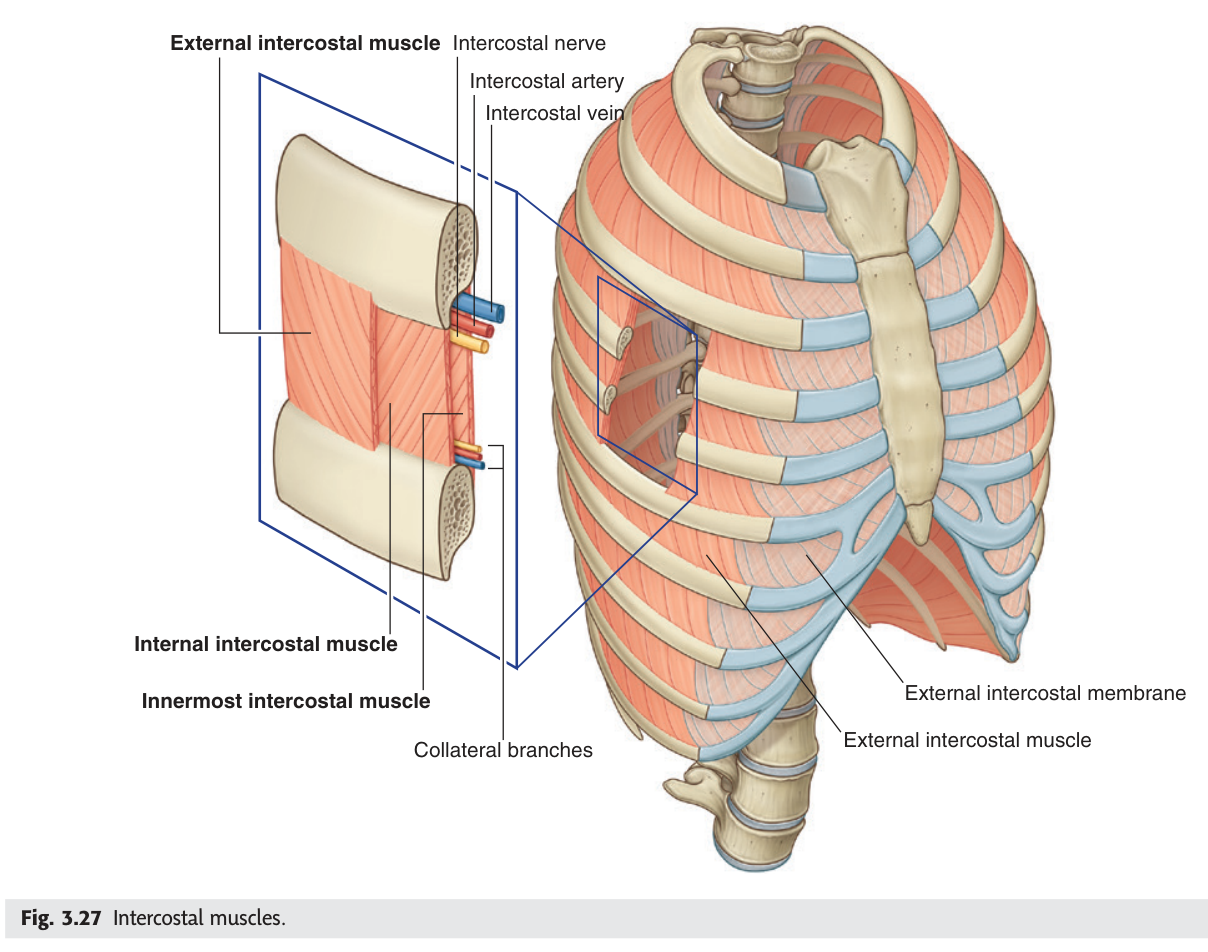
| External intercostal | Internal intercostal | |
|---|---|---|
| Fibre direction | Posterosuperior → anteroinferior | Anterosuperior → posteroinferior |
| Muscle / membrane | Muscle behind, membrane in front | Muscle in front, membrane behind |
| Function | Inspiration — elevates the ribs | Expiration — depresses the ribs |
Neither muscle is complete all the way round. The external intercostal becomes a membrane anteriorly, where the costal cartilages already hold the ribs together; the internal intercostal becomes a membrane posteriorly, close to the vertebral column where movement is least. Each muscle exists exactly where its pull is useful — the membranes are not gaps but economies.
- The three muscle layers of the intercostal space? → External · internal · innermost intercostal
- Which acts in inspiration? → External intercostal
- Where does the external intercostal become membranous? → Anteriorly
- Where does the internal intercostal become membranous? → Posteriorly
The neurovascular bundle ★★★
This is the single most examined fact in the thorax, and TMU marks it ★★★ on the slide. In the costal groove on the lower border of each rib, three structures lie in a fixed order. From above downwards: vein, artery, nerve.
From superior to inferior: intercostal Vein · intercostal Artery · intercostal Nerve.
They lie in the costal groove of the upper rib of the space, in the plane between the internal and innermost intercostal muscles.
A collateral branch of each runs along the upper border of the rib below.
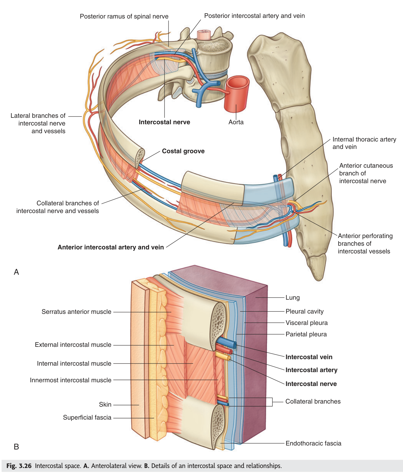
Vein, Artery, Nerve, from the top down. The nerve is lowest, which means it is the least protected by the groove and the first thing a needle meets coming from below. Everything clinical follows from that.
A needle or drain inserted along the lower border of a rib runs straight into the neurovascular bundle — bleeding from the intercostal artery, and lasting neuralgia from the nerve. So every chest drain, pleural aspiration and intercostal block is inserted immediately above the upper border of the rib below, where only the small collateral branch lies.
This is one of the few places where a single anatomical fact directly prevents a common, serious iatrogenic injury — which is exactly why examiners keep asking it.
- Order in the costal groove, top to bottom? → Vein · artery · nerve (VAN)
- In which plane does the bundle lie? → Between the internal and innermost intercostal muscles
- On which rib is the costal groove? → The lower border of the upper rib of the space
- Where is a chest drain inserted, and why? → Just above the upper border of the rib below, to avoid the bundle
The internal thoracic artery
Reflect the anterior chest wall and you find a vessel descending about 1 cm lateral to the edge of the sternum on each side, with two veins beside it. This is the internal thoracic artery, a branch of the first part of the subclavian artery, and it is the anterior half of the chest wall's blood supply.
Pericardiacophrenic artery — long and slender, accompanies the phrenic nerve between the pericardium and mediastinal pleura to the diaphragm.
Anterior intercostal arteries — supply the upper six intercostal spaces.
Superior epigastric artery — terminal branch, into the rectus sheath.
Musculophrenic artery — terminal branch, along the costal margin.
So each intercostal space has a double supply: a posterior intercostal artery from the thoracic aorta behind, and an anterior intercostal artery from the internal thoracic artery in front. They anastomose in the space, which is why the chest wall tolerates coarctation of the aorta so well.
The internal thoracic artery — surgeons usually call it the internal mammary artery — is the graft of choice for coronary bypass. Left in place proximally and swung down onto the left anterior descending artery, its patency at ten years is far better than a saphenous vein graft's. The reason is partly that it is an artery accustomed to arterial pressure, and partly that its endothelium resists atherosclerosis.
- Where does the internal thoracic artery run? → About 1 cm lateral to the sternal margin
- Its two terminal branches? → Superior epigastric and musculophrenic arteries
- Which branch accompanies the phrenic nerve? → The pericardiacophrenic artery
- How many spaces do its anterior intercostal branches supply? → The upper six
The pleura — four parts ★★
The pleura is a single continuous serous membrane folded on itself, like a fist pushed into a partly inflated balloon. The layer against the lung is the visceral pleura; the layer lining the wall is the parietal pleura; and the potential space between them is the pleural cavity, holding only a film of fluid.
The parietal pleura is named in four parts by what it lines — and TMU has asked for all four in a past paper.
1. Cupula (cervical) pleura — extends up into the neck over the apex of the lung, 2–3 cm above the medial third of the clavicle.
2. Costal pleura — lines the inner surface of the chest wall.
3. Mediastinal pleura — lines the mediastinum. Below the lung root it forms the pulmonary ligament, a redundant fold that allows the root structures to move.
4. Diaphragmatic pleura — lines the upper surface of the diaphragm.
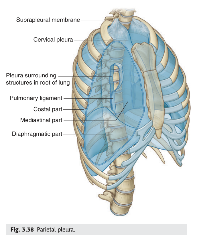
Irritation of the diaphragmatic pleura centrally is felt in the shoulder tip, because the phrenic nerve is C3–C5.
- The four parts of the parietal pleura? → Cupula (cervical) · costal · mediastinal · diaphragmatic
- How high does the cupula reach? → 2–3 cm above the medial third of the clavicle
- What is the pulmonary ligament? → A redundant fold of pleura below the lung root allowing movement
- Which pleura is pain-sensitive? → The parietal — via intercostal and phrenic nerves
Pleural recesses ★★★
The pleural cavity is larger than the lung that occupies it. In quiet breathing the lung does not reach the extreme corners of the cavity, and those unfilled corners are the pleural recesses. Two matter, and one of them is a past-paper definition.
The slit-like interval on each side where the costal pleura becomes continuous with the diaphragmatic pleura.
It is the lowest part of the pleural cavity, and the lung does not fill it even in deep inspiration.
Clinical meaning: fluid in the pleural cavity collects here first, so it is the site chosen for pleural aspiration and chest drain insertion.
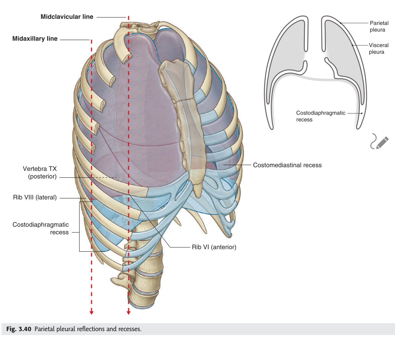
The smaller costomediastinal recess lies anteriorly, where the costal pleura meets the mediastinal pleura behind the sternum — most obvious on the left, where the heart creates the cardiac notch.
- What is the costodiaphragmatic recess? → The slit between costal and diaphragmatic pleura — the lowest part of the pleural cavity
- Why does it matter clinically? → Pleural fluid collects there first; it is the site for aspiration and chest drains
- Does the lung fill it in deep inspiration? → No
- Where is the costomediastinal recess? → Anteriorly, between costal and mediastinal pleura behind the sternum
Surface projections — the two-rib rule
TMU gives a table of the lower borders of the lungs and pleura at three vertical lines, and it looks like six numbers to memorise. It is really one rule plus one number.
| Lower border | Midclavicular line | Midaxillary line | Beside the vertebral column |
|---|---|---|---|
| Lung | 6th rib | 8th rib | 10th rib |
| Pleura | 8th rib | 10th rib | 12th rib |
The lung runs 6 · 8 · 10 at the three lines; the pleura runs 8 · 10 · 12. The pleura is always two ribs lower than the lung — and that two-rib gap is the costodiaphragmatic recess. The numbers and the recess are the same fact stated two ways.
Because the pleura extends two ribs below the lung, there is a zone where you can enter the pleural cavity without touching lung. Aspiration is classically performed in the midaxillary line around the 8th or 9th intercostal space — inside the pleural cavity, below the lung edge, and above the diaphragm. Go lower and you are in the abdomen, risking liver or spleen.
- Lower border of the lung at the three lines? → 6th · 8th · 10th rib
- Lower border of the pleura? → 8th · 10th · 12th rib
- What is the relationship between them? → The pleura is always two ribs lower
- What does that gap correspond to? → The costodiaphragmatic recess
Revision layer
The drawing homework for this lecture is "the structures going through the intercostal spaces". Everything you need for it is here.
The intercostal space, from outside in
| Layer | Note |
|---|---|
| Skin and superficial fascia | Cutaneous branches of the intercostal nerve |
| External intercostal | Fibres down and forward; membranous anteriorly; inspiration |
| Internal intercostal | Fibres down and backward; membranous posteriorly; expiration |
| Neurovascular bundle | In the costal groove: vein, artery, nerve from above down |
| Innermost intercostal | Thin, incomplete |
| Endothoracic fascia | Loose connective tissue |
| Parietal pleura | Costal part |
Blood supply of an intercostal space
| Vessel | Source | Territory |
|---|---|---|
| Posterior intercostal artery | Thoracic aorta (spaces 3–11) | Posterior part of the space |
| Anterior intercostal artery | Internal thoracic artery | Upper six spaces, anteriorly |
| Posterior intercostal veins | → azygos system | Right: azygos · Left: hemiazygos and accessory hemiazygos |
- Draw a cross-section of an intercostal space with all layers and the bundle in order
- State the VAN order and explain where a chest drain goes and why
- Name the four parts of the parietal pleura
- Define the costodiaphragmatic recess with its clinical meaning
- Give the lung and pleural lower borders at the three vertical lines