Deep Neck & Root of the Neck
The thyroid gland
The thyroid is the largest endocrine gland in the body, and it sits wrapped around the front of the trachea like a bow tie. Two lateral lobes lie against the sides of the larynx and trachea, joined across the midline by a narrow isthmus that crosses tracheal rings two to four. In about half of people a pyramidal lobe projects upward from the isthmus.
It is enclosed by the pretracheal fascia, which binds it to the larynx and trachea. That single fact explains the classic bedside test: because the gland is tethered to the larynx, a thyroid swelling moves up when the patient swallows, while a lymph node or a skin lump does not.
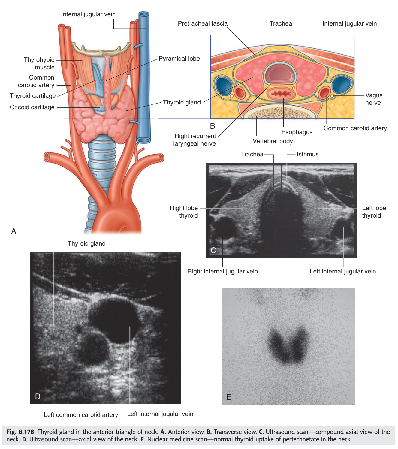
Functionally the gland makes two quite different hormones. Thyroxine sets the basal metabolic rate and drives normal growth of bone and the nervous system; calcitonin, from the parafollicular C cells, lowers blood calcium.
- What does the isthmus cross? → Tracheal rings 2–4
- What encloses the gland? → The pretracheal fascia, binding it to the larynx and trachea
- Why does a thyroid swelling move on swallowing? → It is tethered to the larynx by pretracheal fascia
- Its two hormones? → Thyroxine (metabolic rate, growth) and calcitonin (lowers blood calcium)
The arteries of the thyroid ★★★
This is the drawing homework for the lecture, and it is one of the most surgically loaded diagrams in the whole course. The gland has two arteries on each side, and — crucially — they come from different parent vessels.
Superior thyroid artery — the first branch of the external carotid artery. Descends to the upper pole.
Inferior thyroid artery — a branch of the thyrocervical trunk of the subclavian artery. Ascends to the lower pole, approaching from behind.
Thyroidea ima artery — inconstant (present in around 10%), arising from the brachiocephalic trunk or the arch of the aorta, ascending to the isthmus.
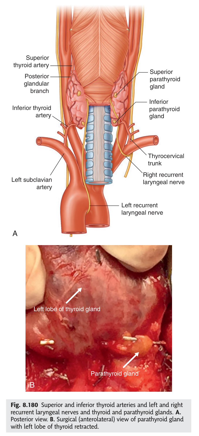
The thyroid develops as a downgrowth from the floor of the pharynx and descends through the neck. It picks up a blood supply from above early — the external carotid — and acquires a second one from below as it arrives — the subclavian, via the thyrocervical trunk. The dual supply is a fossil of that migration, and it is also why the gland survives ligation of either artery.
- Parent vessel of the superior thyroid artery? → The external carotid artery (its first branch)
- Parent vessel of the inferior thyroid artery? → The thyrocervical trunk of the subclavian artery
- Which pole does each supply? → Superior → upper pole; inferior → lower pole
- What is the thyroidea ima? → An inconstant artery to the isthmus from the brachiocephalic trunk or aortic arch
The two nerves at risk ★★★
Here is the reason this diagram is set as homework. Each thyroid artery travels with a branch of the vagus that supplies the larynx, and a surgeon tying that artery can tie the nerve with it. The two pairings are different, the consequences are different, and examiners love the distinction.
Surgical rule: ligate the superior thyroid artery close to the gland, where the nerve has already diverged.
Unilateral injury causes hoarseness with a paralysed cord. Bilateral injury is an airway emergency — both cords lie near the midline and the patient develops stridor and may need an emergency tracheostomy.
Surgical rule: ligate the inferior thyroid artery away from the gland, laterally, where the nerve is not yet closely applied.
Superior artery → external laryngeal nerve → tie it close to the gland.
Inferior artery → recurrent laryngeal nerve → tie it far from the gland.
The rules are opposites because the nerves converge on the gland from opposite directions.
The two recurrent laryngeal nerves are not symmetrical. The right hooks under the right subclavian artery; the left descends into the thorax and hooks under the arch of the aorta before returning up the neck. This is a legacy of the embryonic aortic arches being drawn down into the chest.
The clinical consequence is that a left recurrent laryngeal palsy can be caused by disease inside the chest — a bronchial carcinoma at the left hilum, an aortic arch aneurysm, or an enlarged left atrium. Unexplained hoarseness warrants a chest X-ray, and the reason is this nerve's detour.
- Nerve with the superior thyroid artery? → External branch of the superior laryngeal nerve — supplies cricothyroid
- Nerve with the inferior thyroid artery? → The recurrent laryngeal nerve
- Effect of unilateral vs bilateral recurrent laryngeal injury? → Hoarseness vs stridor and airway obstruction
- Where does the left recurrent laryngeal nerve hook? → Under the arch of the aorta
- Which artery is tied close to the gland, and why? → The superior — to spare the external laryngeal nerve
The veins of the thyroid
The veins do not mirror the arteries, and that asymmetry is the examinable point. There are three veins on each side and they drain to two different destinations.
| Vein | Drains into |
|---|---|
| Superior thyroid vein | Internal jugular vein |
| Middle thyroid vein | Internal jugular vein |
| Inferior thyroid vein | Brachiocephalic vein (left, or either) |
So the superior and middle veins go up and laterally to the internal jugular; only the inferior vein goes down, descending in front of the trachea to reach the brachiocephalic vein. The inferior veins often form a plexus in front of the trachea — which is exactly why they are a hazard during emergency tracheostomy.
- The three thyroid veins? → Superior, middle, inferior
- Where do the superior and middle drain? → The internal jugular vein
- Where does the inferior drain? → The brachiocephalic vein
- Why do the inferior veins matter in tracheostomy? → They form a plexus in front of the trachea and can bleed
The parathyroid glands
Four small glands — usually two on each side — lie on the posterior surface of the lateral lobes of the thyroid, inside its fascial sheath. They secrete parathyroid hormone, which raises blood calcium: the exact opposite of calcitonin from the thyroid next door.
Both superior and inferior parathyroids are supplied mainly by the inferior thyroid artery, which is another reason that vessel is treated with such respect during thyroid surgery.
Remove or devascularise the parathyroids during a total thyroidectomy and the patient's calcium falls within a day or two. The signs are those of neuromuscular irritability — tingling around the mouth and fingers, carpopedal spasm, and the classic Trousseau's and Chvostek's signs. Serum calcium is therefore checked routinely after thyroidectomy. Their position on the posterior surface of the gland is precisely why they are at risk.
- Where do the parathyroids lie? → On the posterior surface of the thyroid lobes, within its sheath
- What does parathyroid hormone do? → Raises blood calcium — the opposite of calcitonin
- Their main blood supply? → The inferior thyroid artery
- Complication of removing them? → Hypocalcaemia with tetany after thyroidectomy
The root of the neck
The root of the neck is the doorway between the neck and the thorax — the area above the first rib and behind the manubrium, where everything passing between head and chest is funnelled into a small space. Because the thoracic inlet slopes downward and forward, the apex of each lung and the cervical pleura rise above the first rib into the neck.
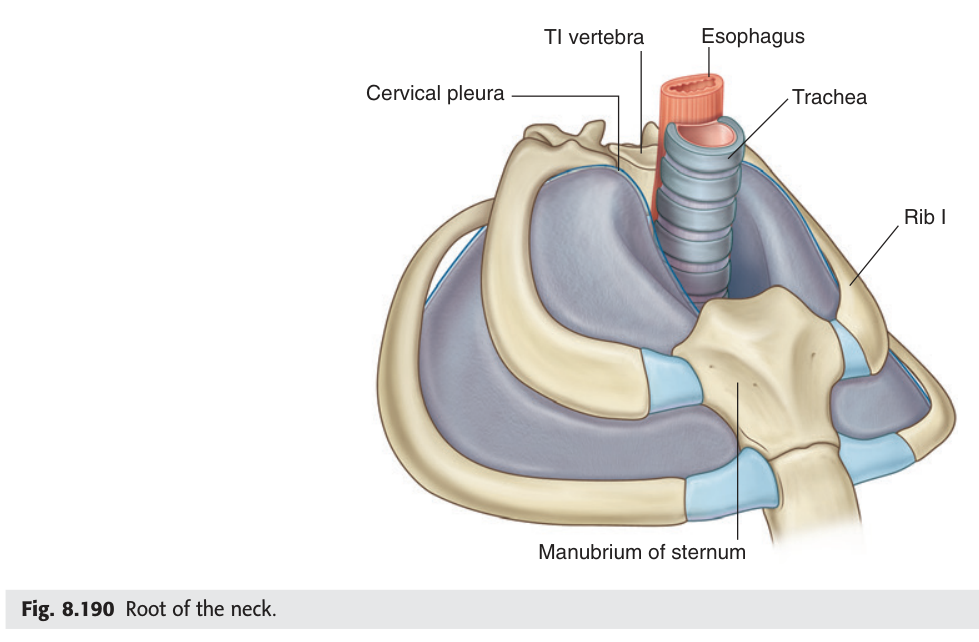
The cupula (dome) of the pleura projects into the root of the neck, up to about 2.5 cm above the medial third of the clavicle. A needle aimed at the subclavian vein that passes too deep punctures it and causes a pneumothorax — the commonest serious complication of subclavian central line insertion, and the reason a chest X-ray is taken afterwards. A stab wound at the root of the neck can do exactly the same.
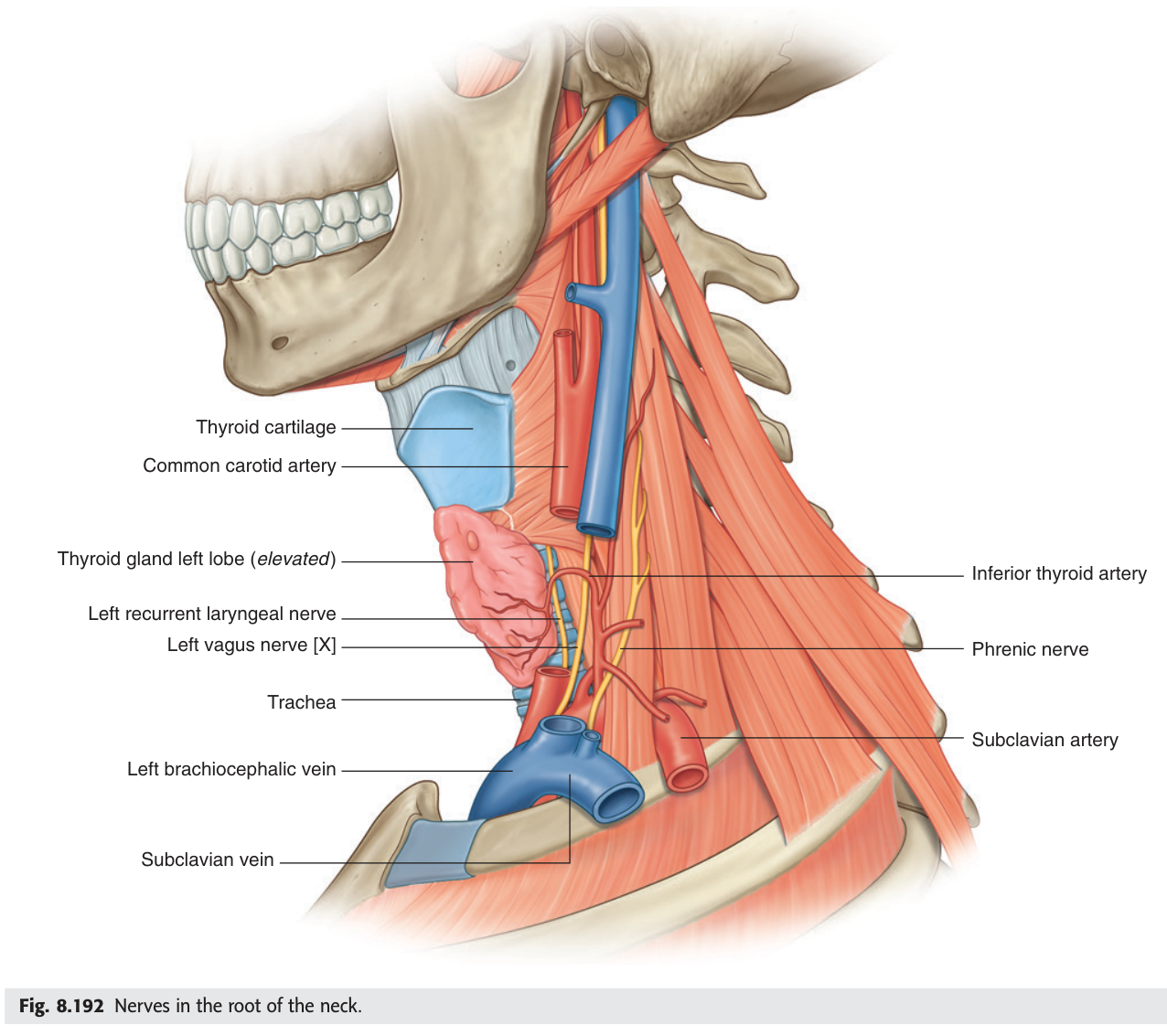
Two other structures belong here. The cervical sympathetic trunk lies on the prevertebral fascia in front of the transverse processes, with superior, middle and cervicothoracic (stellate) ganglia. And the thoracic duct arches laterally on the left, behind the carotid sheath, to reach the left venous angle.
Damage to the cervical sympathetic trunk — by an apical lung tumour (a Pancoast tumour), neck trauma or surgery — produces Horner's syndrome on the same side: a drooping eyelid (ptosis), a constricted pupil (miosis), and loss of sweating on that side of the face (anhidrosis). The eye signs point to a lesion in the neck or chest apex, not the eye itself.
- How far does the cervical pleura rise? → About 2.5 cm above the medial third of the clavicle
- Commonest serious complication of subclavian line insertion? → Pneumothorax
- Where does the thoracic duct end? → The left venous angle
- The triad of Horner's syndrome? → Ptosis, miosis, anhidrosis
The subclavian artery
The subclavian artery is divided into three parts by scalenus anterior — the same trick as the axillary artery and pectoralis minor. The first part is medial to the muscle, the second behind it, the third lateral to it, and the third part is what you found in the scalene fissure in Unit 9.
| Part | Relation to scalenus anterior | Branches |
|---|---|---|
| First | Medial | Vertebral artery · internal thoracic artery · thyrocervical trunk |
| Second | Behind | Costocervical trunk |
| Third | Lateral | Usually none (occasionally the dorsal scapular artery) |
The thyrocervical trunk from the first part is the one to remember, because it gives the inferior thyroid artery — closing the loop back to the thyroid diagram — along with the transverse cervical and suprascapular arteries that you saw crossing in front of scalenus anterior.
- What divides the subclavian artery into three parts? → Scalenus anterior
- Branches of the first part? → Vertebral · internal thoracic · thyrocervical trunk
- Which branch gives the inferior thyroid artery? → The thyrocervical trunk
- Which part lies in the scalene fissure? → The third part
Revision layer
TMU's homework for this lecture is a single diagram: the arteries of the thyroid gland and their relations with the nerves. Everything below serves that drawing.
Thyroid — arteries, nerves and the surgical rule
| Artery | Arises from | Reaches | Nerve alongside | Surgical rule |
|---|---|---|---|---|
| Superior thyroid | External carotid artery (1st branch) | Upper pole | External laryngeal nerve (to cricothyroid) | Ligate close to the gland |
| Inferior thyroid | Thyrocervical trunk of subclavian artery | Lower pole, from behind | Recurrent laryngeal nerve (all other intrinsic muscles) | Ligate away from the gland |
| Thyroidea ima (10%) | Brachiocephalic trunk or aortic arch | Isthmus | — | Beware in tracheostomy |
Thyroid venous drainage
| Vein | Destination |
|---|---|
| Superior thyroid | Internal jugular vein |
| Middle thyroid | Internal jugular vein |
| Inferior thyroid | Brachiocephalic vein |
Laryngeal nerve lesions
| Nerve | Muscle supplied | Effect of injury |
|---|---|---|
| External laryngeal | Cricothyroid (tenses the cord) | Loss of high notes; weak, tiring voice |
| Recurrent laryngeal — unilateral | All other intrinsic laryngeal muscles | Hoarseness, paralysed cord |
| Recurrent laryngeal — bilateral | As above, both sides | Stridor and airway obstruction — emergency |
- Draw the thyroid with both arteries, their parent vessels and the nerve running with each
- State the surgical rule for ligating each artery and why they differ
- Give the three thyroid veins and their destinations
- Explain why unexplained hoarseness needs a chest X-ray
- Give the three parts and branches of the subclavian artery