Sternocleidomastoid & Anterior Regions of the Neck
The four subdivisions of the anterior triangle
The anterior triangle — between the midline, the mandible and the anterior border of sternocleidomastoid — is too big to be useful on its own. The digastric muscle above and the superior belly of omohyoid below cut it into four smaller triangles, and every structure in the front of the neck is then located by naming one of them.
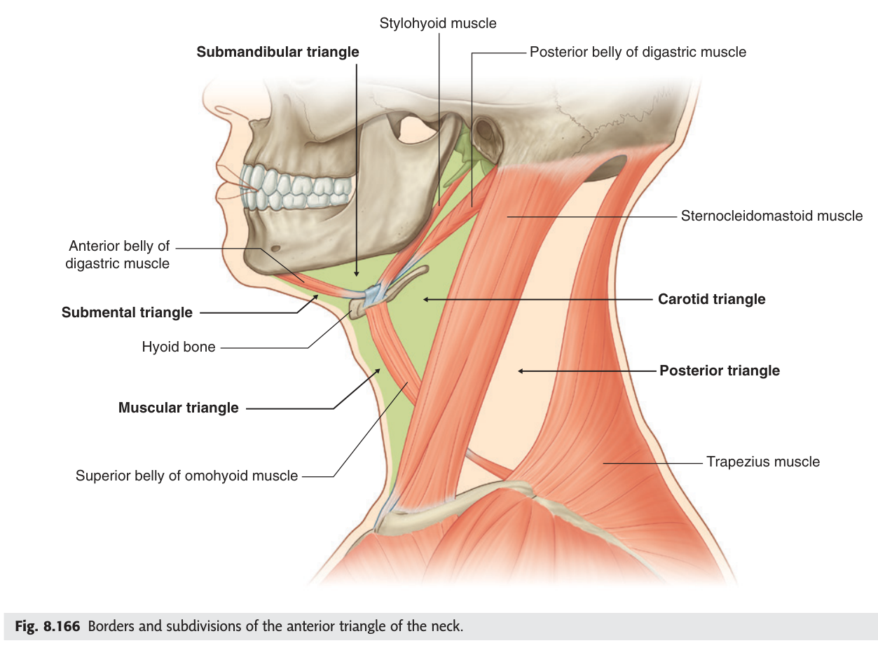
| Triangle | Boundaries | Key contents |
|---|---|---|
| Submandibular | Lower border of mandible; both bellies of digastric | Submandibular gland · facial artery and vein · hypoglossal nerve · lymph nodes |
| Submental | Anterior bellies of digastric on each side; hyoid bone below (unpaired, in the midline) | Submental lymph nodes · anterior jugular veins |
| Carotid | Superior belly of omohyoid; posterior belly of digastric; anterior border of SCM | Carotid arteries and bifurcation · internal jugular vein · vagus and hypoglossal nerves |
| Muscular | Midline; superior belly of omohyoid; anterior border of SCM | Infrahyoid muscles · thyroid and parathyroid glands · larynx and trachea |
- The four subdivisions of the anterior triangle? → Submandibular · submental · carotid · muscular
- Which is unpaired? → The submental triangle, in the midline
- Which muscles do the dividing? → Digastric above, superior belly of omohyoid below
Suprahyoid and infrahyoid muscles
The hyoid bone floats in the front of the neck, held by muscles above and below it and articulating with no other bone. The muscles above it — the suprahyoid group — pull it up and help open the mouth; those below — the infrahyoid or strap muscles — pull it down again. Between them they move the larynx up and down during swallowing and speech, which you can feel on your own throat.
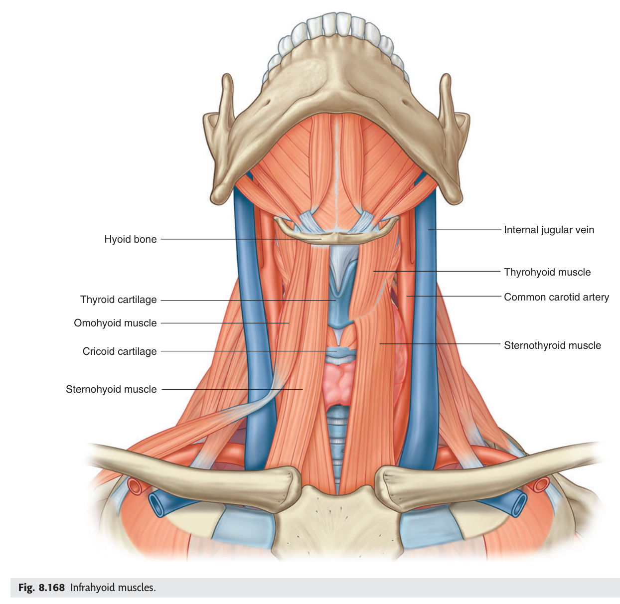
| Group | Muscles | Action |
|---|---|---|
| Suprahyoid | Digastric (two bellies) · stylohyoid · mylohyoid · geniohyoid | Elevate the hyoid; depress the mandible; elevate the floor of the mouth in swallowing |
| Infrahyoid (strap) | Sternohyoid · sternothyroid · thyrohyoid · omohyoid | Depress the hyoid and larynx after swallowing; steady the hyoid |
Two details are worth having. The digastric has two bellies joined by an intermediate tendon held to the hyoid by a fibrous loop — the anterior belly from the digastric fossa of the mandible, the posterior belly from the mastoid process. The omohyoid likewise has two bellies with an intermediate tendon, and its inferior belly is what subdivides the posterior triangle. Both are landmarks precisely because they cross regions.
- The four suprahyoid muscles? → Digastric · stylohyoid · mylohyoid · geniohyoid
- The four infrahyoid muscles? → Sternohyoid · sternothyroid · thyrohyoid · omohyoid
- What supplies the infrahyoid muscles? → The ansa cervicalis (C1–C3)
- Which two muscles have two bellies and an intermediate tendon? → Digastric and omohyoid
The carotid triangle
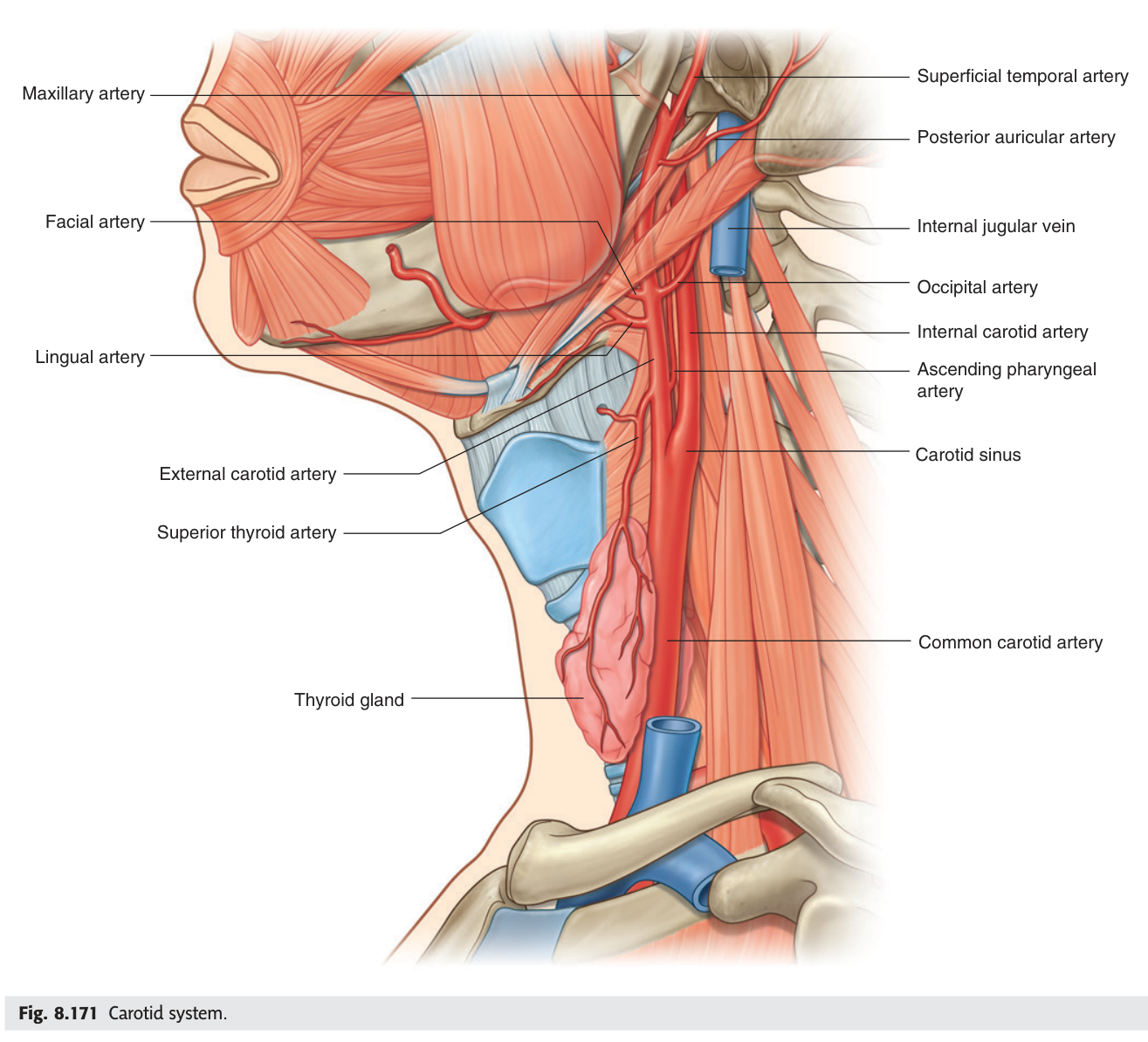
Of the four subdivisions, the carotid triangle is the one that matters most, because the great vessels of the head are exposed in it. The common carotid artery ascends within the carotid sheath and divides at the upper border of the thyroid cartilage — roughly the level of C4 — into the internal and external carotid arteries.
The distinction is simple and absolute: the internal carotid gives no branches in the neck at all and ascends to enter the skull, while the external carotid gives every branch to the face and neck. If you see a branch in the neck, it is external.
Two cranial nerves are found here in a fixed relationship. The accessory [XI] and hypoglossal [XII] nerves both descend between the internal carotid artery and the internal jugular vein. The accessory then passes backwards and downwards to sternocleidomastoid; the hypoglossal turns forwards, crossing the external carotid artery at about the level of the angle of the mandible, on its way to the tongue.
- Where does the common carotid artery divide? → At the upper border of the thyroid cartilage (~C4)
- How many branches does the internal carotid give in the neck? → None
- Which artery does the hypoglossal nerve cross? → The external carotid artery
- Boundaries of the carotid triangle? → Superior belly of omohyoid · posterior belly of digastric · anterior border of SCM
Carotid sinus and carotid body ★★
At the bifurcation sit two small structures that look almost identical and do completely different jobs. Confusing them is a guaranteed lost mark, and the way to keep them apart is to translate the names: a sinus is a dilatation, and a body is a lump of tissue.
A localised dilatation of the terminal part of the common carotid artery and the beginning of the internal carotid artery.
Function: contains baroreceptors that monitor arterial blood pressure.
Innervation: a branch of the glossopharyngeal nerve [IX].
A small ovoid mass of tissue lying in the fork of the carotid bifurcation, behind it.
Function: a chemoreceptor, responding to changes in the composition of the blood — principally oxygen, and also carbon dioxide and pH.
Innervation: branches of both the glossopharyngeal [IX] and vagus [X] nerves.
Sinus — stretch — pressure — baroreceptor.
Body — blood chemistry — chemoreceptor.
Both are at the bifurcation; the sinus is in the vessel wall, the body is behind the fork.
Pressure on the carotid sinus stimulates the baroreceptors, which reflexly slow the heart and drop the blood pressure. In susceptible people — usually elderly — a tight collar, shaving, or turning the head sharply can be enough to cause syncope. Doctors also use this deliberately: carotid sinus massage can terminate a supraventricular tachycardia. It is done on one side only, and never in a patient with a carotid bruit.
- What is the carotid sinus? → A dilatation at the carotid bifurcation containing baroreceptors
- What is the carotid body? → A chemoreceptor mass behind the bifurcation, sensing blood oxygen
- Nerve to the sinus? → Glossopharyngeal [IX]
- Nerves to the body? → Both glossopharyngeal [IX] and vagus [X]
Branches of the external carotid artery
The external carotid artery gives eight branches, and TMU's dissection asks you to trace the first three — superior thyroid, lingual and facial — which come off the front of the vessel in that order from below upwards.
| Direction from the vessel | Branches |
|---|---|
| Anterior (3) | Superior thyroid · lingual · facial |
| Posterior (2) | Occipital · posterior auricular |
| Medial (1) | Ascending pharyngeal |
| Terminal (2) | Superficial temporal · maxillary |
Superior thyroid · Ascending pharyngeal · Lingual · Facial · Occipital · Posterior auricular · Maxillary · Superficial temporal.
The important structural point beneath the mnemonic: the superior thyroid artery is the first branch, and it descends with the external laryngeal nerve — a relationship that becomes surgically critical in the next unit.
- The three anterior branches of the external carotid? → Superior thyroid · lingual · facial
- Its two terminal branches? → Superficial temporal and maxillary
- Which is the first branch, and what runs with it? → Superior thyroid artery, with the external laryngeal nerve
The venous angle ★★★
At the root of the neck on each side, the internal jugular vein coming down from the head meets the subclavian vein coming in from the arm. The angle where they join to form the brachiocephalic vein is the venous angle — and it is the destination of the entire lymphatic system of the body.
The angle formed by the union of the internal jugular vein and the subclavian vein on each side, where they join to form the brachiocephalic vein.
The thoracic duct opens into the LEFT venous angle.
The right lymphatic duct opens into the RIGHT venous angle.
Get the sides right and a whole chapter of lymphatics falls into place. The thoracic duct drains everything below the diaphragm plus the left half of the body above it — roughly three quarters of the body — into the left angle. The much smaller right lymphatic duct drains only the right head and neck, right arm and right thorax into the right angle.
Because the thoracic duct empties into the left venous angle, tumour cells travelling up it can lodge in the lymph nodes there. An enlarged, hard left supraclavicular node — Virchow's node, and the finding called Troisier's sign — is a classic indicator of abdominal malignancy, most often gastric carcinoma. A lump in the left supraclavicular fossa sends the clinician looking at the stomach, and the reason is purely anatomical.
- What forms the venous angle? → Junction of the internal jugular and subclavian veins
- Which duct opens into the left angle? → The thoracic duct
- Which opens into the right? → The right lymphatic duct
- What is Virchow's node and what does it suggest? → An enlarged left supraclavicular node suggesting abdominal (often gastric) malignancy
Revision layer
The anterior triangle — four subdivisions
| Triangle | Boundaries | Contents |
|---|---|---|
| Submandibular | Mandible above; both bellies of digastric | Submandibular gland, facial vessels, hypoglossal nerve |
| Submental | Anterior bellies of digastric; hyoid (midline, unpaired) | Submental nodes, anterior jugular veins |
| Carotid | Superior belly of omohyoid; posterior belly of digastric; anterior border of SCM | Carotid bifurcation, IJV, vagus, hypoglossal |
| Muscular | Midline; superior belly of omohyoid; anterior border of SCM | Strap muscles, thyroid, larynx, trachea |
Structures at the carotid bifurcation
| Structure | What it is | Function | Nerve |
|---|---|---|---|
| Carotid sinus | Dilatation of the vessel wall | Baroreceptor — blood pressure | Glossopharyngeal [IX] |
| Carotid body | Ovoid mass behind the fork | Chemoreceptor — blood oxygen | Glossopharyngeal [IX] + vagus [X] |
- Name the four subdivisions of the anterior triangle with boundaries and contents
- List the suprahyoid and infrahyoid muscles and their nerve supply
- Define the carotid sinus and carotid body, with function and innervation
- Give the eight branches of the external carotid artery
- Define the venous angle and state which duct enters on each side