Superficial Layer & Lateral Region of the Neck
Dividing up the neck
The neck is a short tube crammed with structures that are, individually, easy — and the only way to keep them straight is to divide the tube up first. TMU does this in two steps, and both appear in the exam.
First, the whole neck is separated from the head above and the thorax below. The superior boundary runs along the lower border of the mandible, the mastoid process, the superior nuchal line and the external occipital protuberance. The inferior boundary runs along the jugular notch of the sternum, the clavicle, the acromion, and the spine of the seventh cervical vertebra.
Second, the sternocleidomastoid is used as the great landmark. It divides each side into an anterior triangle in front of it and a posterior (lateral) triangle behind it, with the muscle itself forming a sternocleidomastoid region between the two. Almost every structure in the neck is then described by which triangle it lies in.
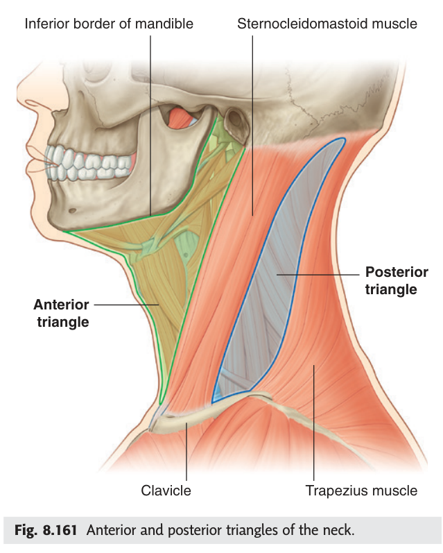
| Region | Boundaries |
|---|---|
| Anterior region | Anteriorly the median line of the neck · posteriorly the anterior border of sternocleidomastoid · superiorly the lower border of the mandible |
| Sternocleidomastoid region | Between the anterior and posterior borders of sternocleidomastoid |
| Lateral (posterior) region | Anteriorly the posterior border of sternocleidomastoid · posteriorly the anterior border of trapezius · inferiorly the middle third of the clavicle |
- Which muscle divides the neck into triangles? → Sternocleidomastoid
- Boundaries of the posterior triangle? → SCM in front, trapezius behind, middle third of clavicle below
- Origin and insertion of sternocleidomastoid? → Manubrium and clavicle → mastoid process
- What does one sternocleidomastoid do acting alone? → Tilts the head to the same side and rotates the face to the opposite side
The cervical fascia ★★★
Nothing in the neck makes sense until you understand its fascia. The neck is not a bag of loose structures — it is a set of fascial tubes sliding on one another, which is how you can swallow, turn your head and speak without everything tearing. It is also how infection travels, and where it travels is entirely determined by which tube it starts in.
Beneath the superficial fascia — which contains the platysma, the superficial veins and the cutaneous nerves — the deep cervical fascia is arranged in three layers, plus the carotid sheath.
1. Investing (superficial) layer — surrounds the whole neck like a collar. It splits to enclose two muscles and two glands: trapezius, sternocleidomastoid, the parotid and the submandibular glands. Attached above to the external occipital protuberance, superior nuchal line and mandible; below to the clavicle, manubrium, acromion and spine of the scapula.
2. Pretracheal (middle / visceral) layer — encloses the viscera: pharynx, larynx, trachea, oesophagus, thyroid and parathyroid glands. Extends from the hyoid bone down to fuse with the fibrous pericardium.
3. Prevertebral (deep) layer — surrounds the vertebral column and its muscles (prevertebral and scalene). Extends from the base of the skull to the superior mediastinum, and prolongs laterally over the subclavian vessels and brachial plexus as the axillary sheath.
A condensation receiving a contribution from all three layers, enclosing on each side:
Common and internal carotid arteries — medially
Internal jugular vein — laterally
Vagus nerve [X] — posteriorly, in the angle between the two vessels
Deep cervical lymph nodes lie along it.
For the investing layer remember it splits around two muscles and two glands — trapezius and sternocleidomastoid, parotid and submandibular. Four structures, one sentence.
For the sheath, the order is artery medial, vein lateral, nerve behind and between. The vagus hides in the groove — which is exactly why it is spared in most neck injuries and why an expanding haematoma in the sheath compresses it.
The pretracheal layer runs from the hyoid bone straight into the fibrous pericardium. That means a deep infection of the neck — from a dental abscess or a perforated oesophagus — can track down that plane into the mediastinum and cause mediastinitis, which carries a high mortality. The retropharyngeal space behind the pretracheal layer does the same. Anatomy here is not academic: it explains why neck infections are treated aggressively.
- The three layers of deep cervical fascia? → Investing · pretracheal · prevertebral
- What does the investing layer split to enclose? → Trapezius, sternocleidomastoid, parotid and submandibular glands
- Where does the pretracheal layer end below? → It fuses with the fibrous pericardium
- What does the prevertebral layer become in the limb? → The axillary sheath
- Contents of the carotid sheath? → Common/internal carotid artery medially · internal jugular vein laterally · vagus nerve posteriorly between them
Superficial veins and cutaneous nerves
In the superficial fascia, deep to platysma, run the veins you can see through the skin of the neck. The external jugular vein crosses the surface of sternocleidomastoid almost vertically, from the angle of the mandible down to the middle of the clavicle, where it pierces the deep fascia to reach the subclavian vein. The anterior jugular veins run beside the midline.
Because the external jugular vein drains into the subclavian vein with no valves worth the name, its distension reflects central venous pressure. A visibly engorged external jugular vein in a patient sitting at 45° suggests right heart failure or fluid overload. It is also a route for emergency venous access.
There is a trap: the vein is held open by the fascia it pierces, so a laceration here does not collapse — it can suck air in and cause a fatal air embolism. Wounds of the neck are pressed on immediately for that reason.
- Where does the external jugular vein run? → Vertically across sternocleidomastoid to the middle of the clavicle
- Into what does it drain? → The subclavian vein
- Why is a laceration dangerous? → The vein is held open by fascia and can admit air — risk of air embolism
The posterior (lateral) triangle
The posterior triangle is a flat, roughly triangular area you can see on any thin neck. Its floor is made of the prevertebral fascia over the scalene and other deep muscles; its roof is the investing layer. The inferior belly of omohyoid crosses its lower part and subdivides it into a large occipital triangle above and a small supraclavicular (omoclavicular) triangle below.
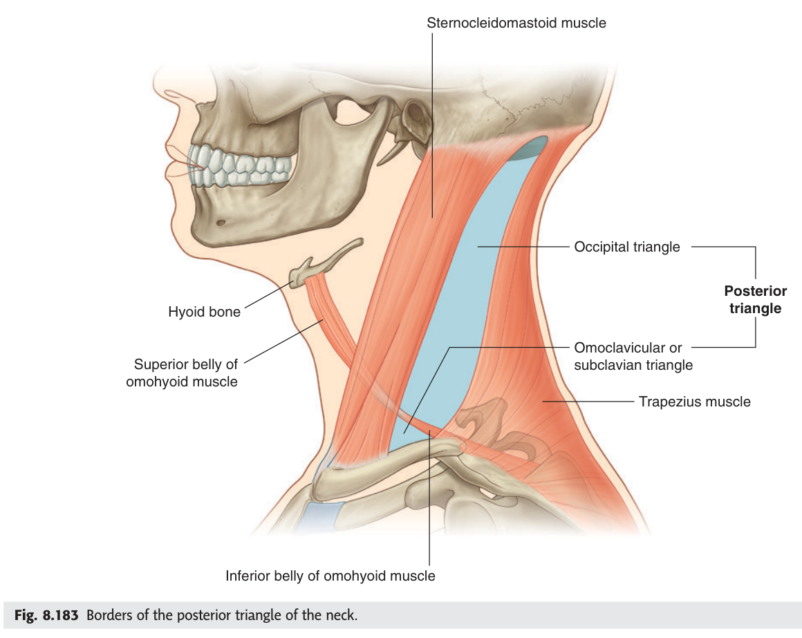
- Accessory nerve [XI] — crossing it superficially
- Cutaneous branches of the cervical plexus — four of them
- Roots and trunks of the brachial plexus — in the lower part
- Third part of the subclavian artery
- External jugular vein and superficial cervical lymph nodes
- Transverse cervical and suprascapular arteries crossing the floor
- What subdivides the posterior triangle? → The inferior belly of omohyoid
- Into what two triangles? → Occipital (above) and supraclavicular/omoclavicular (below)
- What forms its floor and roof? → Floor: prevertebral fascia over the deep muscles · Roof: investing layer
The accessory nerve ★★
The accessory nerve [XI] supplies sternocleidomastoid and trapezius. In the posterior triangle it is remarkable for being almost unprotected: it crosses the triangle lying within the investing fascia, with nothing but skin and fascia over it. That is why it is the nerve most often injured in minor neck surgery — even a lymph node biopsy.
A line joining two points:
(1) a point slightly above the middle of the posterior border of sternocleidomastoid, and
(2) a point about 5 cm above the clavicle at the anterior border of trapezius.
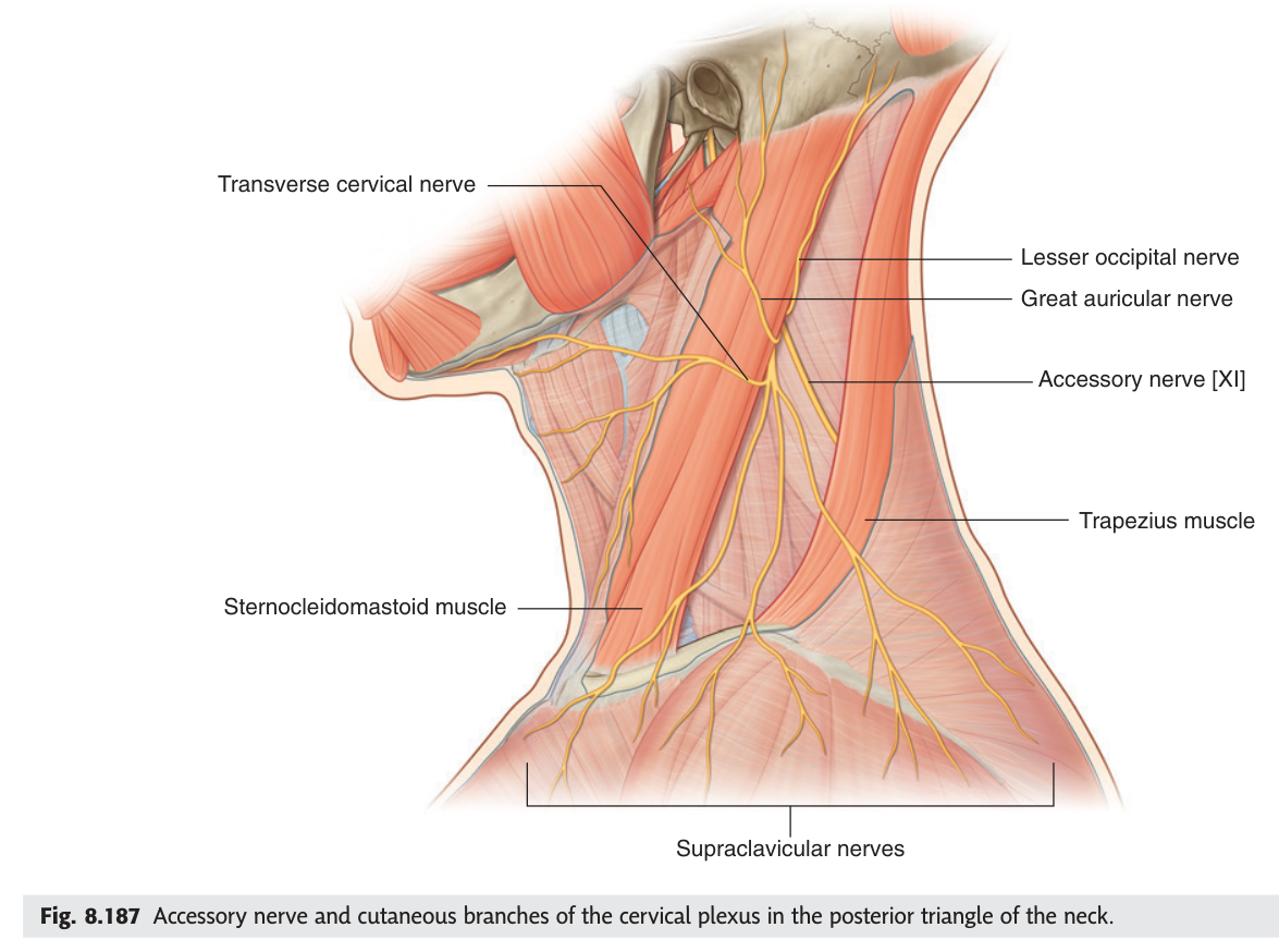
Trapezius wastes, the shoulder droops, and the patient cannot shrug against resistance or raise the arm fully above the head — because trapezius rotates the scapula upward. There is often a dull aching pain around the shoulder girdle. Because the nerve is so superficial in the posterior triangle, this is a recognised complication of node biopsy, and surgeons deliberately identify it.
- Which two muscles does the accessory nerve supply? → Sternocleidomastoid and trapezius
- Give its surface marking? → From just above the middle of the posterior border of SCM to a point 5 cm above the clavicle at the anterior border of trapezius
- Signs of injury? → Trapezius wasting, drooping shoulder, weak shrug, cannot raise the arm fully
The cervical plexus
The cervical plexus is formed from the anterior rami of C1–C4, and it gives both cutaneous and muscular branches. The four cutaneous branches all emerge from a single point — the middle of the posterior border of sternocleidomastoid, the so-called nerve point — and then fan out.
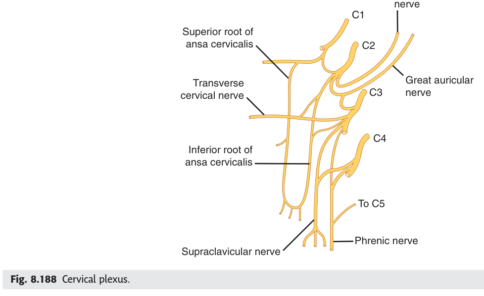
| Cutaneous branch | Course and territory |
|---|---|
| Lesser occipital | Runs up and back along the posterior border of SCM — skin behind the ear |
| Great auricular | Ascends vertically on the surface of SCM — skin over the parotid and the ear |
| Transverse cervical | Runs horizontally forwards across SCM — skin of the front of the neck |
| Supraclavicular | Descend in medial, intermediate and lateral groups — skin over the clavicle and shoulder |
- Root value of the cervical plexus? → C1–C4
- The four cutaneous branches? → Lesser occipital · great auricular · transverse cervical · supraclavicular
- Where do they emerge? → The nerve point, at the middle of the posterior border of SCM
- Root value of the phrenic nerve? → C3, C4, C5
- What does the ansa cervicalis supply? → The infrahyoid strap muscles
The scalene fissure ★★★
Deep in the lower part of the posterior triangle, the scalene muscles running from the cervical vertebrae to the first rib leave a triangular gap between them. This is the scalene fissure — Snell's and Gray's call it the interscalene triangle — and it is the doorway through which the upper limb's nerve and arterial supply leave the neck.
A triangular gap in the root of the neck.
Anteriorly: scalenus anterior. Posteriorly: scalenus medius. Inferiorly: the first rib.
Contents: the subclavian artery (third part) and the roots of the brachial plexus.
One more structure to place here: the phrenic nerve descends vertically across the front of scalenus anterior, and the transverse cervical and suprascapular arteries cross in front of the muscle running backwards. So scalenus anterior is the key: the phrenic nerve on its face, the vein in front of it, the artery and plexus behind it.
Narrowing of the scalene fissure — by a cervical rib, a fibrous band, or hypertrophied scalene muscles — compresses whatever is inside it. Compress the lower trunk of the plexus (C8, T1) and you get pain and wasting of the intrinsic hand muscles along the medial border of the forearm; compress the subclavian artery and you get a cold, pale arm with a diminished radial pulse, sometimes worse when the arm is raised. The contents of the gap predict the syndrome exactly.
- The three boundaries of the scalene fissure? → Scalenus anterior in front · scalenus medius behind · first rib below
- Its contents? → Subclavian artery (third part) and the roots of the brachial plexus
- Where is the subclavian vein? → In front of scalenus anterior — NOT in the fissure
- What lies on the front of scalenus anterior? → The phrenic nerve
- What is thoracic outlet syndrome? → Compression of the plexus or artery by narrowing of this gap
Revision layer
The fascial layers — what each encloses
| Layer | Encloses | Extends to |
|---|---|---|
| Investing (superficial) | Trapezius · sternocleidomastoid · parotid · submandibular gland | Mandible and occiput above; clavicle, manubrium, acromion below |
| Pretracheal (middle) | Pharynx · larynx · trachea · oesophagus · thyroid and parathyroids | Hyoid bone → fibrous pericardium |
| Prevertebral (deep) | Vertebral column · prevertebral and scalene muscles | Base of skull → superior mediastinum; becomes the axillary sheath |
| Carotid sheath | Common/internal carotid artery · internal jugular vein · vagus nerve | Base of skull → root of the neck |
Around scalenus anterior — the one relationship to have cold
| Position | Structure |
|---|---|
| On its anterior surface | Phrenic nerve (descending vertically) |
| In front of the muscle | Subclavian vein |
| Behind the muscle (in the fissure) | Subclavian artery and roots of the brachial plexus |
| Crossing in front | Transverse cervical and suprascapular arteries |
- Name the three fascial layers and what each encloses
- Define the carotid sheath with the position of each content
- Define the scalene fissure and state what is NOT in it
- Give the surface marking of the accessory nerve
- Name the four cutaneous branches of the cervical plexus and where they emerge