The Back of the Upper Limb
Triceps and the spiral groove ★★
The back of the arm holds one muscle. Triceps brachii has three heads — a long head from the infraglenoid tubercle of the scapula, and lateral and medial heads from the humerus — converging on the olecranon of the ulna. It extends the elbow, and the long head, crossing the shoulder, also extends and adducts the arm.
Running between the lateral and medial heads, directly on the bone, is the radial nerve with the profunda brachii (deep brachial) artery. They lie in the radial (spiral) groove — your TMU slide calls it the humeromuscular tunnel — winding from medial to lateral around the back of the shaft.
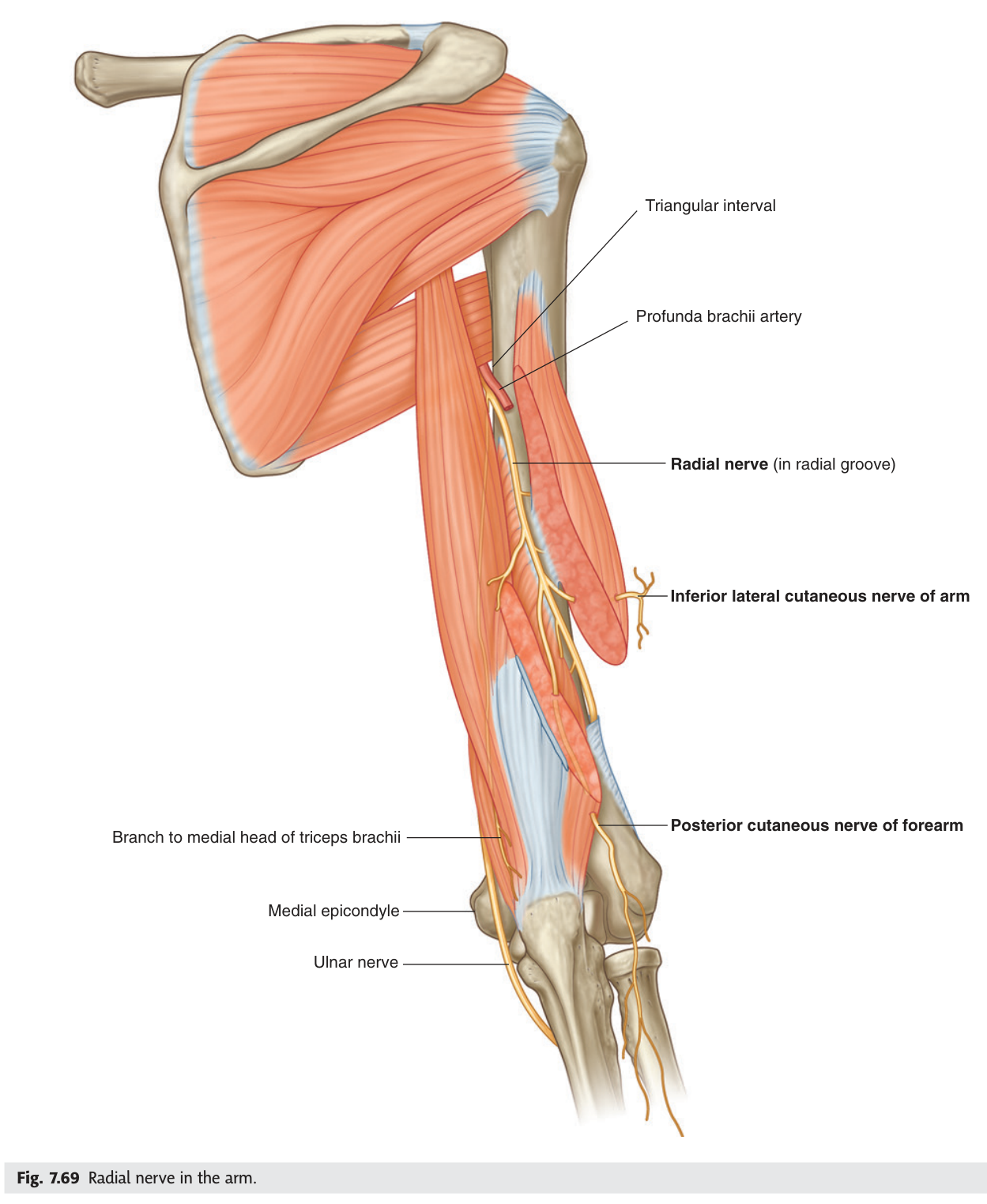
In the spiral groove the radial nerve is pressed against bare bone with no muscle cushion. A mid-shaft humeral fracture injures it, and so does prolonged pressure — an arm hung over the back of a chair while asleep or drunk, the origin of the name Saturday night palsy. The result is wrist drop: all the extensors of the wrist and fingers are paralysed, so the hand hangs limp. Note that the triceps usually escapes, because its branches leave the nerve before it enters the groove — elbow extension is preserved, and that is how you localise the lesion.
- The three heads of triceps and their nerve? → Long, lateral, medial — radial nerve
- What lies in the spiral groove? → The radial nerve and profunda brachii artery
- What is the sign of a radial nerve injury there? → Wrist drop
- Why is elbow extension preserved? → Branches to triceps arise before the nerve enters the groove
The extensors of the forearm
The posterior forearm is built in two layers, and your slide arranges both from the radial side — which is exactly how you meet them in dissection.
| Layer | Muscles, from the radial side |
|---|---|
| Superficial | Extensor carpi radialis longus · extensor carpi radialis brevis · extensor digitorum · extensor digiti minimi · extensor carpi ulnaris |
| Deep | Supinator · abductor pollicis longus · extensor pollicis brevis · extensor pollicis longus · extensor indicis |
Almost all of them arise from the lateral epicondyle by a common extensor origin — the mirror image of the flexors on the medial side — and all are supplied by the radial nerve or its deep branch.
Lateral epicondylitis (tennis elbow) is degeneration at the common extensor origin — pain on resisted wrist extension. Medial epicondylitis (golfer's elbow) is the same process at the common flexor origin — pain on resisted wrist flexion. Which epicondyle, which muscle group, which movement hurts. That is the whole distinction.
- Where do the superficial extensors arise? → Common extensor origin, lateral epicondyle
- Which nerve supplies the posterior compartment? → The radial nerve and its deep branch
- Name the deep layer from the radial side? → Supinator · abductor pollicis longus · extensor pollicis brevis · extensor pollicis longus · extensor indicis
The posterior interosseous nerve
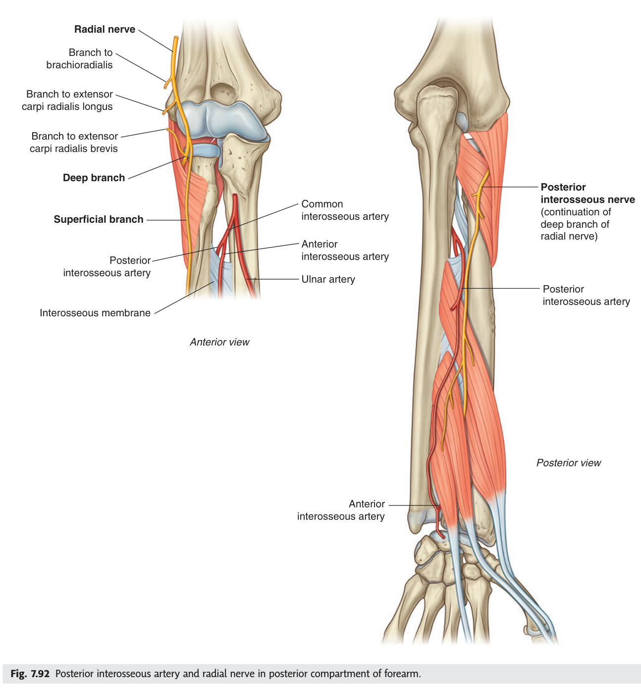
At the level of the lateral epicondyle the radial nerve divides into a superficial branch, which is purely sensory and runs down to supply skin on the back of the hand, and a deep branch, which is purely motor. The deep branch pierces supinator, winds round the neck of the radius and emerges on the back of the forearm as the posterior interosseous nerve, descending on the interosseous membrane with the posterior interosseous artery.
- Where does the radial nerve divide? → At the level of the lateral epicondyle
- Which branch is sensory, which motor? → Superficial = sensory; deep = motor
- What does the deep branch pierce, and what is it then called? → Supinator; it becomes the posterior interosseous nerve
- Key difference from a spiral groove lesion? → No sensory loss, and wrist extension is preserved
The dorsum of the hand
Turn the hand over and the superficial fascia holds the dorsal venous network — the veins used for cannulation, and the origin of both great veins of the arm. The cephalic vein begins from its radial end, the basilic vein from its ulnar end. That is the same lateral/medial pairing you learned in the leg, where the great saphenous begins medially and the small saphenous laterally.
Cutaneous supply of the dorsum is split between two nerves: the superficial branch of the radial nerve takes the lateral part, and the dorsal cutaneous branch of the ulnar nerve the medial part — a boundary running roughly down the middle finger.
- Which vein arises from the radial side of the dorsal venous network? → The cephalic vein
- And from the ulnar side? → The basilic vein
- Which two nerves supply the skin of the dorsum? → Superficial radial and dorsal branch of the ulnar
The extensor retinaculum ★★
At the back of the wrist a strong transverse fibrous band, the extensor retinaculum, holds the extensor tendons down against the bone so they do not bowstring when you extend the wrist. Septa from its deep surface divide the space into six compartments carrying nine tendons, each in a synovial sheath. TMU sets this as the drawing homework for this lecture.
1. Abductor pollicis longus and extensor pollicis brevis
2. Extensor carpi radialis longus and brevis
3. Extensor pollicis longus
4. Extensor digitorum and extensor indicis
5. Extensor digiti minimi
6. Extensor carpi ulnaris
Inflammation of the sheath of the first compartment — abductor pollicis longus and extensor pollicis brevis — causes pain at the radial side of the wrist, worse on gripping and on ulnar deviation with the thumb tucked in (Finkelstein's test). It is common in new parents repeatedly lifting an infant, and the anatomy is the diagnosis: two tendons, one tight tunnel.
- How many compartments and tendons under the extensor retinaculum? → Six compartments, nine tendons
- What is in compartment 1? → Abductor pollicis longus and extensor pollicis brevis
- What is in compartment 3? → Extensor pollicis longus
- Which compartment is inflamed in De Quervain's? → The first
The anatomical snuffbox ★★★
Extend your thumb fully and a small triangular hollow appears at the base of it, on the back of the wrist. This is the anatomical snuffbox — named because ground tobacco was once placed there to be sniffed — and it is one of the highest-yield small structures in the whole upper limb, because of what lies in its floor.
A triangular depression on the lateral part of the proximal dorsum of the hand, visible when the thumb is extended.
Lateral (anterior) border: tendons of abductor pollicis longus and extensor pollicis brevis.
Medial (posterior) border: tendon of extensor pollicis longus.
Proximal border: the distal end of the radius (styloid process).
Floor: the scaphoid and trapezium.
Contents: the radial artery crossing the floor; the superficial branch of the radial nerve and the beginning of the cephalic vein cross its roof.
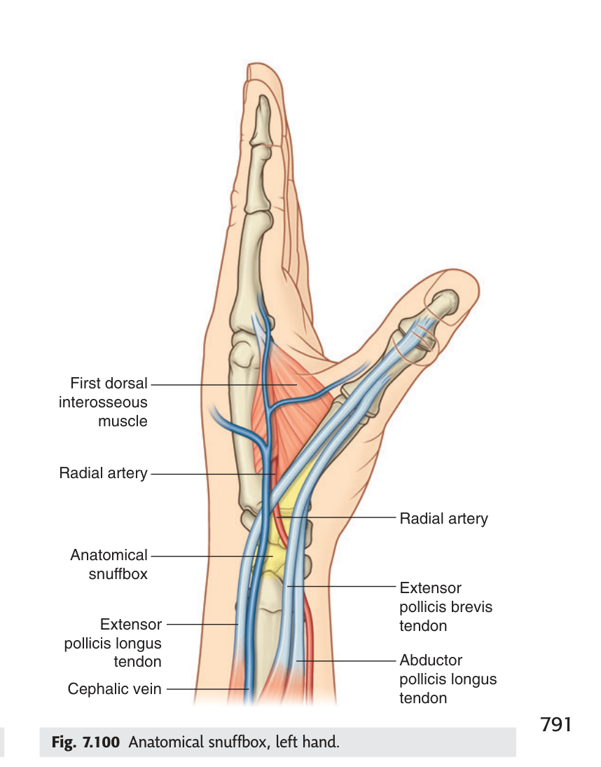
The scaphoid forms the floor. It is the most commonly fractured carpal bone, typically from a fall on the outstretched hand, and tenderness in the snuffbox is the cardinal sign.
The danger is vascular. The scaphoid's blood supply enters mainly at its distal end and runs backwards, so a fracture across the waist cuts off the proximal fragment entirely. That fragment can undergo avascular necrosis, leading to non-union and later wrist arthritis. Worse, the fracture is often invisible on the initial X-ray. This is why a patient with snuffbox tenderness and a normal film is still immobilised and re-imaged in ten to fourteen days — the clinical sign outranks the radiograph.
- The three borders of the snuffbox? → Lateral: APL and EPB tendons · Medial: EPL tendon · Proximal: radial styloid
- What forms the floor? → The scaphoid and trapezium
- What crosses the floor? → The radial artery
- What does snuffbox tenderness mean? → Scaphoid fracture until proved otherwise
- Why is a normal X-ray not reassuring? → The fracture is often occult initially; risk of avascular necrosis of the proximal fragment
Revision layer
The three nerves of the upper limb — lesion, site, sign
| Nerve | Classic site of injury | Motor loss | Sensory loss |
|---|---|---|---|
| Axillary | Surgical neck of humerus; shoulder dislocation | Deltoid — abduction lost | Regimental badge area |
| Radial | Spiral groove; mid-shaft humeral fracture | Wrist and finger extension — wrist drop | Back of hand, lateral |
| Posterior interosseous | Radial neck; piercing supinator | Finger extension only — wrist extension spared | None |
| Median | Carpal tunnel; wrist laceration | Thenar muscles — thumb abduction | Lateral 3½ fingers, sparing thenar skin |
| Ulnar | Behind medial epicondyle; Guyon's canal | Intrinsic hand muscles — claw hand | Medial 1½ fingers |
Two definitions to have word-perfect
| Structure | Borders | Contents / floor |
|---|---|---|
| Anatomical snuffbox | Lateral: APL + EPB tendons · Medial: EPL tendon · Proximal: distal radius | Floor: scaphoid and trapezium · Radial artery crosses the floor |
| Extensor retinaculum | Transverse band at the back of the wrist | Six compartments, nine tendons in synovial sheaths |
- Draw the six extensor compartments and name every tendon
- Define the anatomical snuffbox with borders, floor and contents
- Explain wrist drop, and why triceps is spared
- Distinguish a spiral groove lesion from a posterior interosseous lesion
- Explain why snuffbox tenderness is managed as a fracture despite a normal X-ray