Anterior Compartment of Arm & Forearm
The superficial veins — the ones you actually use
Before any muscle, the front of the arm gives you the three veins every clinician uses daily. The cephalic vein runs up the lateral (radial) side, the basilic vein up the medial (ulnar) side, and in front of the elbow the median cubital vein slants between them, connecting the two.
The median cubital vein is the vein of routine venepuncture, and its safety is anatomical. It lies on the bicipital aponeurosis, a flat sheet spreading medially from the biceps tendon, and that sheet is all that separates the vein from the brachial artery and median nerve underneath. The aponeurosis is the reason a needle in the right place does not reach the artery — which is also why it was once called the grace à Dieu fascia.
- Which side does the cephalic vein run on? → The lateral (radial) side
- Which side does the basilic vein run on? → The medial (ulnar) side
- What separates the median cubital vein from the brachial artery? → The bicipital aponeurosis
The anterior compartment of the arm
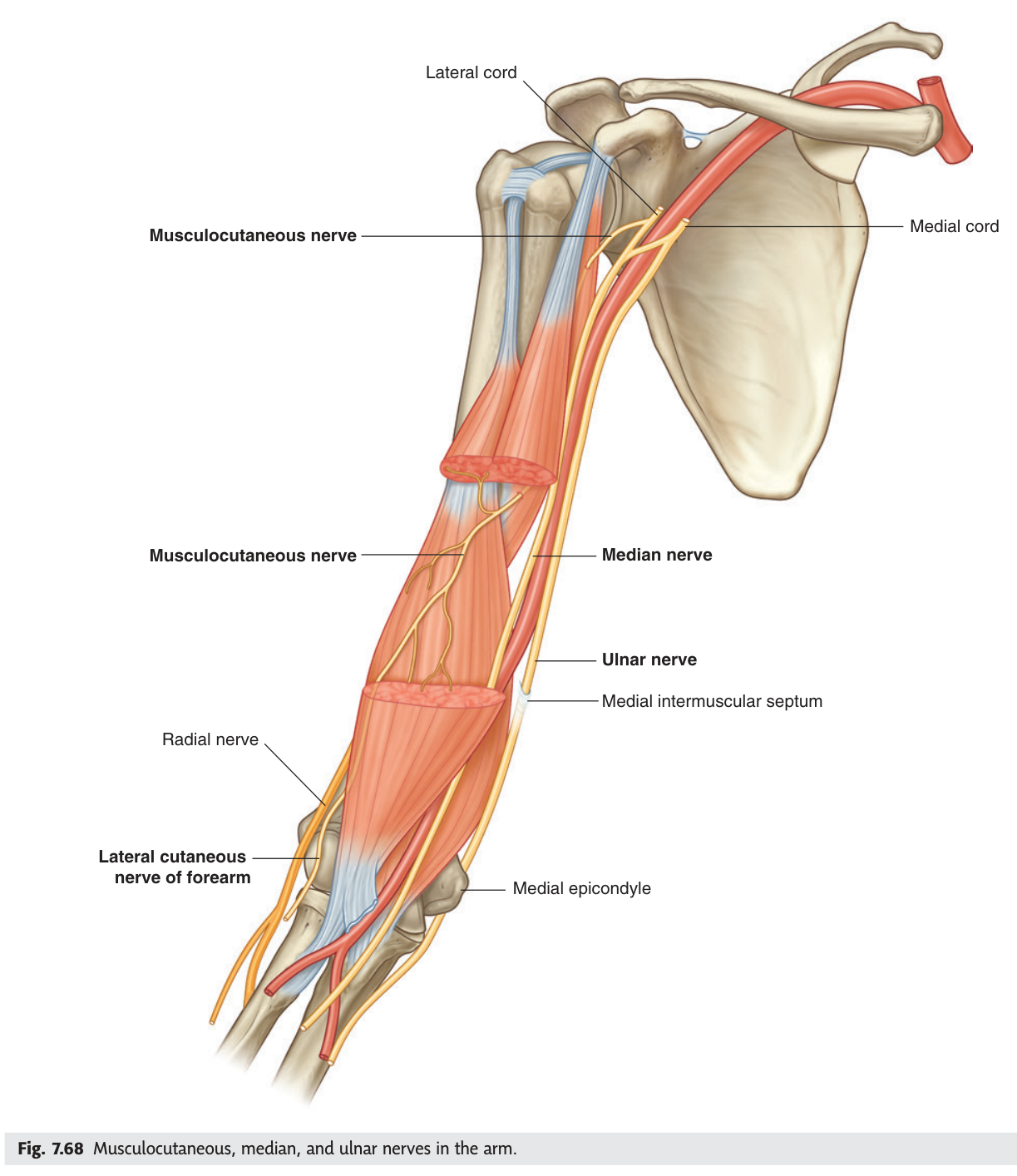
Three muscles, one nerve — this is the simplest compartment in the body. Biceps brachii, brachialis beneath it and coracobrachialis above and medially are all supplied by the musculocutaneous nerve, which pierces coracobrachialis on its way through.
Biceps has two heads with different origins and a single job that surprises students. The long head arises from the supraglenoid tubercle inside the shoulder joint, the short head from the coracoid process, and both insert on the radial tuberosity. Because it attaches to the radius, biceps is not primarily an elbow flexor at all — it is the most powerful supinator of the forearm. That is why you turn a screwdriver clockwise with your right hand, and why the muscle bulges when you do.
- The three anterior arm muscles and their nerve? → Biceps brachii, brachialis, coracobrachialis — musculocutaneous nerve
- Where does the long head of biceps arise? → Supraglenoid tubercle, inside the shoulder joint
- What is biceps's most powerful action? → Supination of the forearm
- What does the musculocutaneous nerve become distally? → The lateral cutaneous nerve of the forearm
Nerves and artery of the arm
Four structures run down the arm, and each takes a different route — which is why they get injured separately and produce different pictures. The brachial artery and median nerve travel together on the medial side, the artery gradually passing from medial to lateral. The ulnar nerve leaves them halfway down, pierces the medial intermuscular septum and passes behind the medial epicondyle — the "funny bone" groove. The radial nerve has already left the compartment entirely, spiralling round the back of the humerus.
Behind the medial epicondyle the ulnar nerve lies subcutaneously against bare bone with nothing over it. Knock it and you get the electric shock down the little finger everyone recognises. Fracture the medial epicondyle, or let the nerve be compressed there chronically, and you get cubital tunnel syndrome — numbness of the medial one and a half fingers and weakness of the intrinsic hand muscles.
- Which two structures run together down the medial arm? → Brachial artery and median nerve
- Where does the ulnar nerve cross the elbow? → Behind the medial epicondyle
- Where is the radial nerve at mid-humerus? → In the radial (spiral) groove, posteriorly
The cubital fossa ★★★
The hollow in front of your elbow is a triangle, and TMU marks it ★★★ on its own slide and sets it as a drawing homework. Its shape comes from two muscles diverging as they leave the elbow: pronator teres heading down and medially, brachioradialis heading down and laterally, with an imaginary line between the two epicondyles closing the top.
A triangular depression on the anterior aspect of the elbow.
Base (superiorly): an imaginary line between the medial and lateral epicondyles of the humerus.
Medially: lateral border of pronator teres. Laterally: medial border of brachioradialis.
Floor: brachialis and supinator. Roof: deep fascia reinforced by the bicipital aponeurosis, with the median cubital vein on it.
Contents, lateral → medial: tendon of biceps brachii · brachial artery and veins · median nerve.
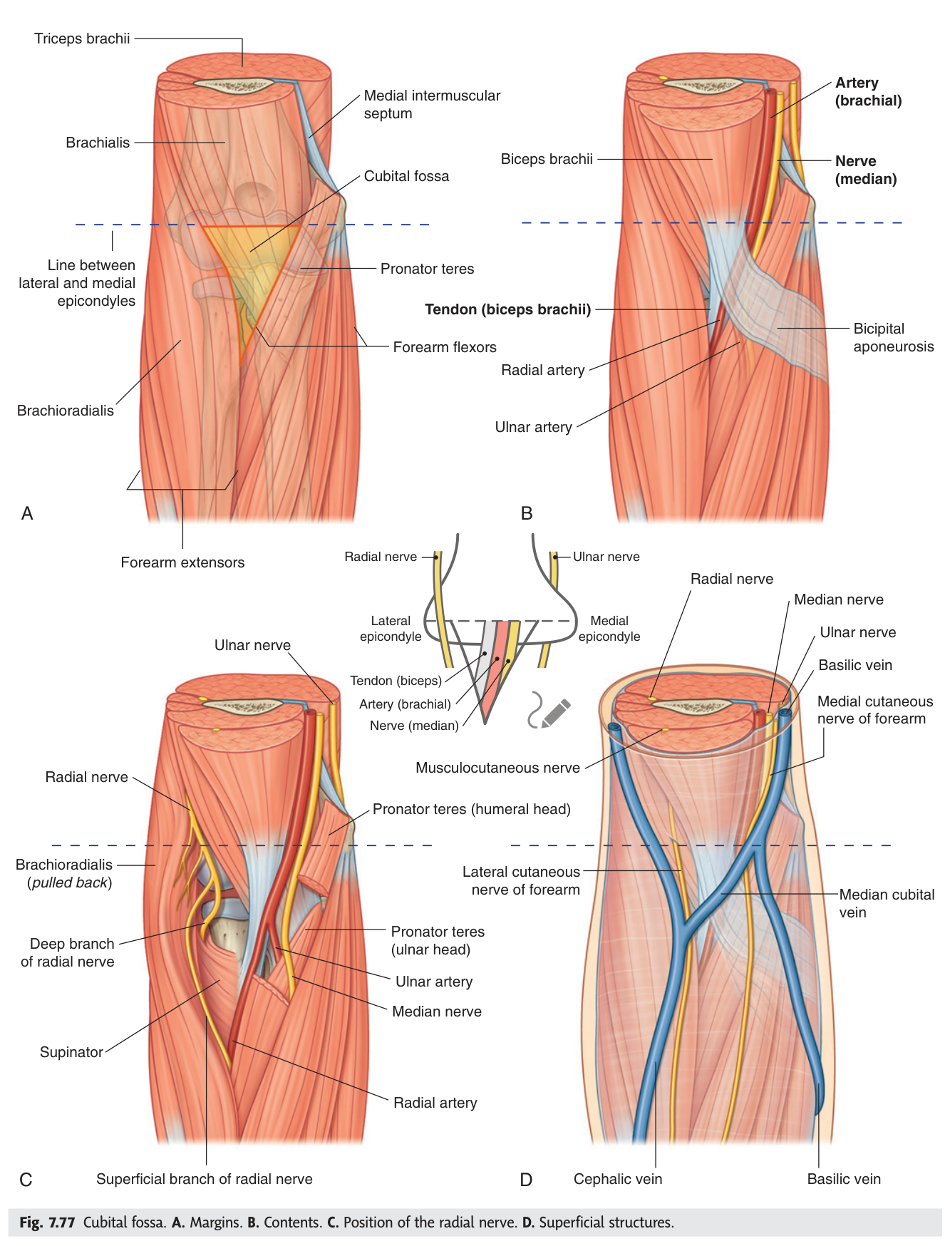
Your TMU slide labels them M · A · N from lateral to medial — Muscle tendon (biceps), Artery (brachial), Nerve (median). Three letters, correct order, and it tells you the artery is sandwiched between the tendon and the nerve.
The radial nerve is not a classical content — it lies deep and lateral, under brachioradialis, dividing into superficial and deep branches. Mention it as a relation, not as a content.
You take the blood pressure here. The stethoscope goes over the brachial artery, just medial to the biceps tendon, at the point where the pulse is felt. You also take blood from the median cubital vein on the roof, and this is where a supracondylar fracture of the humerus threatens the brachial artery — producing the ischaemic contracture of the forearm flexors known as Volkmann's contracture if it is missed.
- Three boundaries of the cubital fossa? → Interepicondylar line above · pronator teres medially · brachioradialis laterally
- Its floor and roof? → Floor: brachialis and supinator · Roof: deep fascia + bicipital aponeurosis
- Contents lateral to medial? → Biceps tendon · brachial artery · median nerve (M-A-N)
- Where do you place a stethoscope for blood pressure? → Over the brachial artery, medial to the biceps tendon
The flexor muscles of the forearm
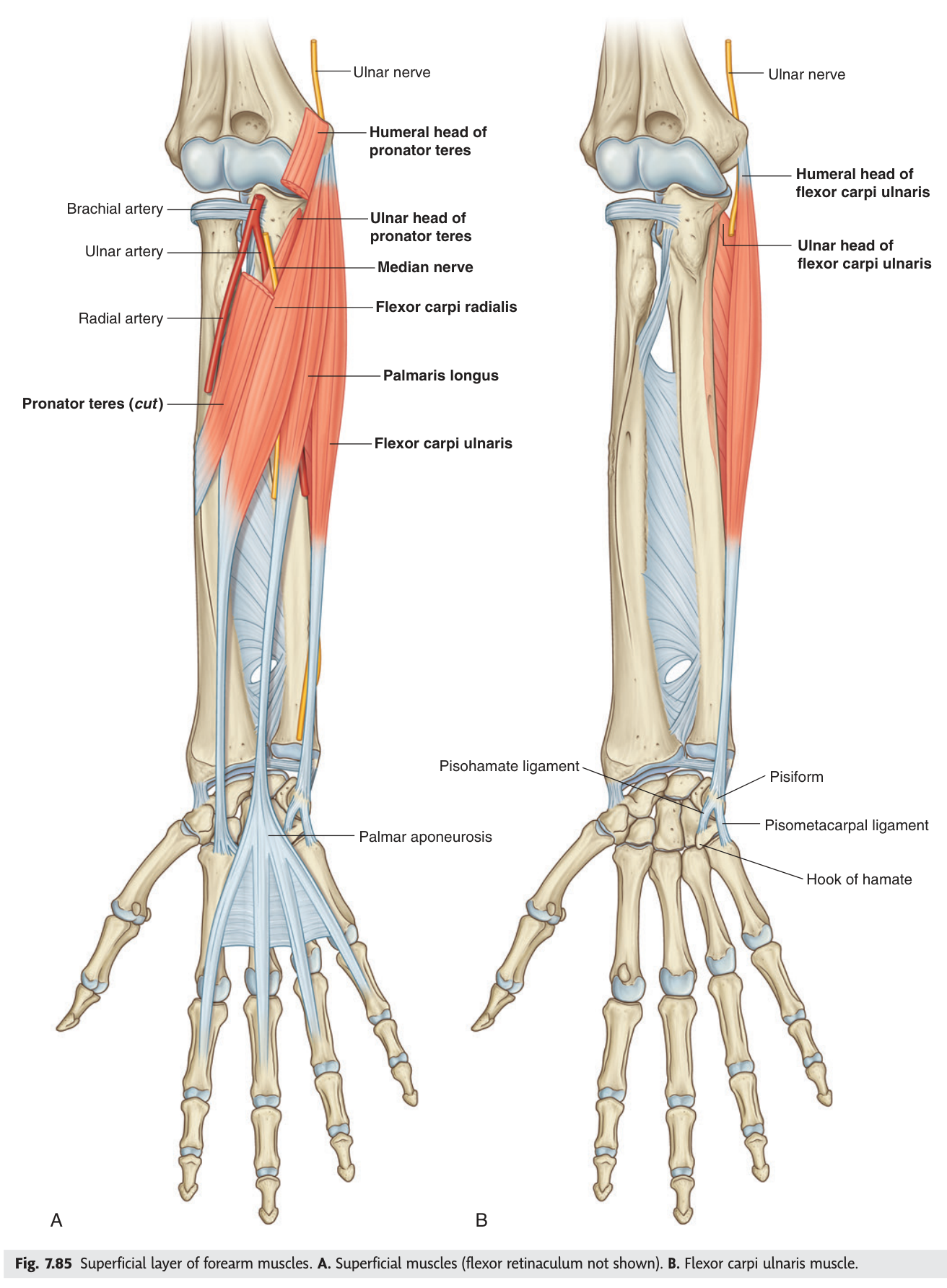
The front of the forearm holds the flexors, arranged in layers. The superficial layer — from the radial side: brachioradialis, pronator teres, flexor carpi radialis, palmaris longus, flexor carpi ulnaris — arises largely from the medial epicondyle by a common flexor origin. Deep to them lies flexor digitorum superficialis, and deeper still flexor pollicis longus, flexor digitorum profundus and pronator quadratus.
The nerve supply has one rule and one exception, and the exception is the exam question. The median nerve supplies everything in the anterior forearm except flexor carpi ulnaris and the medial half of flexor digitorum profundus, which take the ulnar nerve. One and a half muscles to the ulnar nerve; all the rest to the median.
- Where do the superficial flexors arise? → Common flexor origin on the medial epicondyle
- Which nerve supplies most of the anterior forearm? → The median nerve
- The two exceptions? → Flexor carpi ulnaris and the medial half of flexor digitorum profundus — ulnar nerve
- Name the deep layer? → Flexor pollicis longus · flexor digitorum profundus · pronator quadratus
The radial and ulnar arteries
The brachial artery ends in the cubital fossa, at about the level of the neck of the radius, by dividing into the radial and ulnar arteries. The radial artery runs down the lateral side, relatively superficial, and ends as the deep palmar arch. The ulnar artery runs down the medial side beneath the flexors and ends as the superficial palmar arch.
The ulnar artery gives off the common interosseous artery near its origin, which promptly divides into anterior and posterior interosseous arteries — one on each side of the interosseous membrane, supplying the deep flexors and the extensors respectively.
The radial artery is palpated at the wrist lateral to the tendon of flexor carpi radialis, where it lies on bone with only skin and fascia over it. Because the two arteries anastomose through the palmar arches, one can usually supply the whole hand alone — which is what makes it safe to cannulate or harvest the radial artery. Allen's test checks this before either procedure: compress both arteries, let the hand blanch, release the ulnar alone and watch the colour return.
- Where does the brachial artery divide? → In the cubital fossa, at the level of the neck of the radius
- Which artery forms the superficial palmar arch? → The ulnar artery
- Which forms the deep palmar arch? → The radial artery
- What does Allen's test check? → That the palmar arches can supply the hand from one artery alone
The front of the wrist ★★★
This is the second drawing homework of the lecture, and it is pure applied anatomy: at the wrist, eight structures lie side by side in a fixed order just under the skin. Knowing that order is what lets you take a radial pulse, find the median nerve for a block, or avoid cutting something important when repairing a laceration.
1. Tendon of brachioradialis
2. Radial artery
3. Tendon of flexor carpi radialis
4. Median nerve
5. Tendon of palmaris longus
6. Ulnar artery
7. Ulnar nerve
8. Tendon of flexor carpi ulnaris
The two arteries sit at positions 2 and 6, each just medial to a tendon you can see. The two nerves sit at 4 and 7. The median nerve hides between the tendons of palmaris longus and flexor carpi radialis — clench your fist and flex the wrist and those two tendons stand up; the nerve is in the gap between them. That is exactly where a median nerve block is placed, and exactly what a wrist laceration divides.
- The eight structures, lateral to medial? → Brachioradialis tendon · radial artery · FCR tendon · median nerve · palmaris longus tendon · ulnar artery · ulnar nerve · FCU tendon
- Between which two tendons does the median nerve lie? → Palmaris longus and flexor carpi radialis
- Where is the radial pulse felt? → Lateral to the tendon of flexor carpi radialis
The carpal tunnel
Deep to those surface structures, the tendons entering the hand pass through a rigid tunnel. The floor and sides are the carpal bones, arched into a concavity; the roof is the tough flexor retinaculum stretched across it. Nine tendons and one nerve share the space: the four tendons of flexor digitorum superficialis, four of flexor digitorum profundus, the tendon of flexor pollicis longus — and the median nerve.
Note what is not in the tunnel. The ulnar nerve and artery pass superficial to the retinaculum, in Guyon's canal, and are therefore spared in carpal tunnel syndrome. Palmaris longus is also superficial to it. That distinction is the whole basis of the clinical picture.
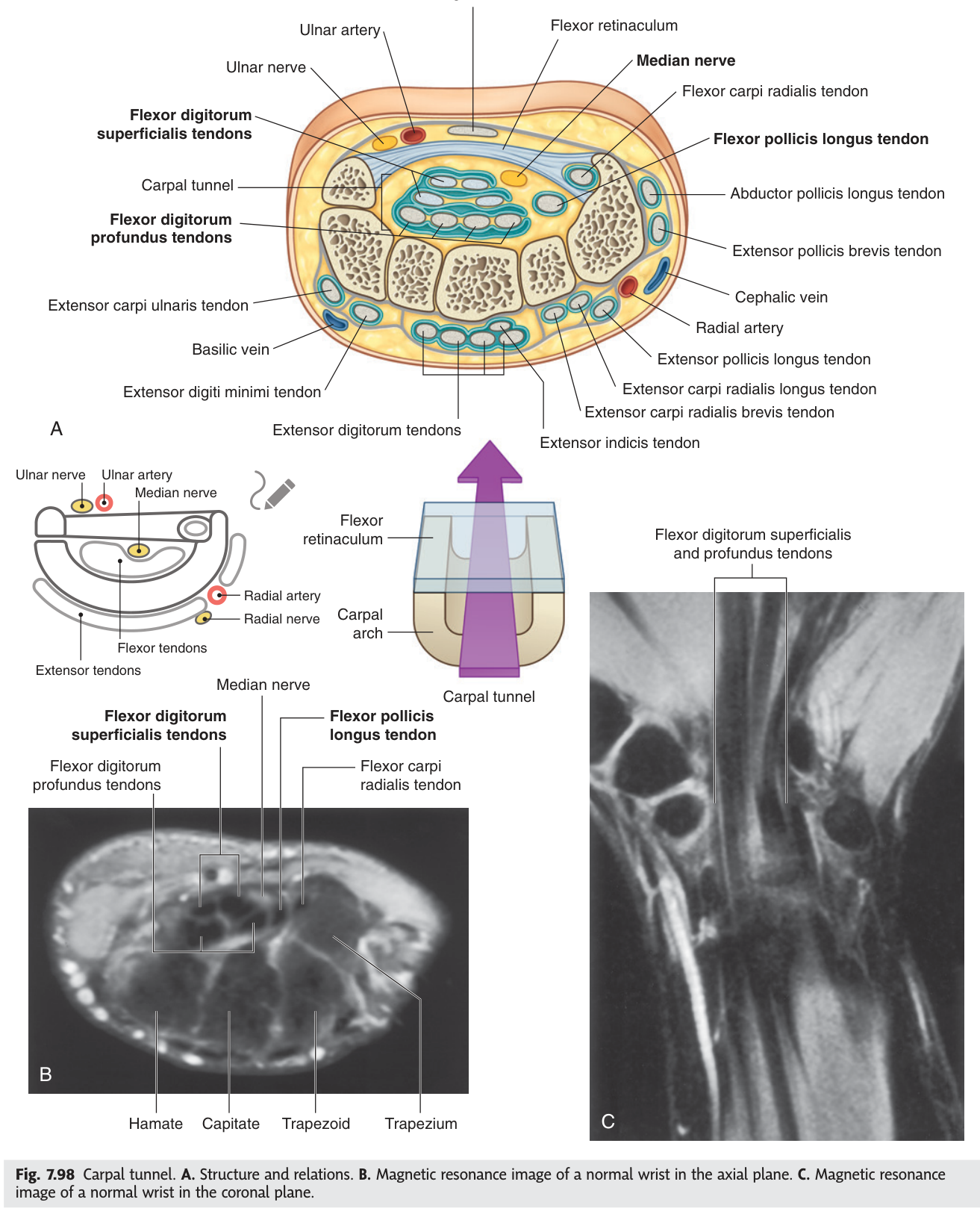
The tunnel cannot expand. Anything that raises the pressure inside it — tenosynovitis, pregnancy, hypothyroidism, rheumatoid arthritis, repetitive use — compresses the median nerve, which is the softest structure present and lies right under the retinaculum. The result is tingling and numbness in the lateral three and a half fingers, classically worse at night, and in time wasting of the thenar eminence with weak thumb abduction.
Two anatomical points decide the diagnosis. First, sensation over the thenar eminence itself is spared, because the palmar cutaneous branch of the median nerve leaves the nerve before the tunnel and passes over the retinaculum. Second, the little finger is normal, because it is ulnar territory. A patient with numbness of the whole hand does not have carpal tunnel syndrome.
- What forms the roof of the carpal tunnel? → The flexor retinaculum
- How many tendons pass through, and which nerve? → Nine tendons; the median nerve
- Which nerve and artery are NOT in the tunnel? → The ulnar nerve and artery — superficial to the retinaculum
- Why is the thenar skin spared? → The palmar cutaneous branch leaves before the tunnel and crosses over the retinaculum
Revision layer
Compartments of the arm and forearm
| Compartment | Muscles | Nerve | Artery |
|---|---|---|---|
| Arm — anterior | Biceps brachii · brachialis · coracobrachialis | Musculocutaneous n. | Brachial a. |
| Arm — posterior | Triceps brachii | Radial n. | Profunda brachii a. |
| Forearm — anterior | Superficial: brachioradialis*, pronator teres, FCR, palmaris longus, FCU · FDS · Deep: FPL, FDP, pronator quadratus | Median n. except FCU and medial half of FDP (ulnar n.) | Radial and ulnar aa. |
*Brachioradialis lies in the anterior forearm by position but is supplied by the radial nerve — another compartment exception worth a mark.
The two set-pieces
| Structure | Boundaries | Contents |
|---|---|---|
| Cubital fossa | Base: interepicondylar line · Medial: pronator teres · Lateral: brachioradialis · Floor: brachialis + supinator · Roof: deep fascia + bicipital aponeurosis | Biceps tendon · brachial artery · median nerve (lateral → medial) |
| Front of wrist | — | Lateral → medial: brachioradialis tendon · radial artery · FCR tendon · median nerve · palmaris longus tendon · ulnar artery · ulnar nerve · FCU tendon |
- Draw the cubital fossa with all boundaries, floor, roof and three contents
- List the eight structures at the front of the wrist in order
- Give the anterior forearm nerve supply including both exceptions
- Name the nine tendons and one nerve in the carpal tunnel
- Explain why thenar sensation is spared in carpal tunnel syndrome