The Axillary Fossa
What the axilla actually is
The armpit is not a hole. It is a pyramidal space between the upper arm and the chest wall, and everything travelling between the neck and the upper limb passes through it. Abduct the arm and the pyramid opens; drop the arm and it closes around its contents.
Its shape is worth fixing before anything else. The apex points upward and medially toward the neck, bounded by the clavicle, the first rib and the upper border of the scapula — that gap is the doorway from the neck. The base is the skin of the armpit that you can see. Between them are four walls, and TMU asks you to name all four.
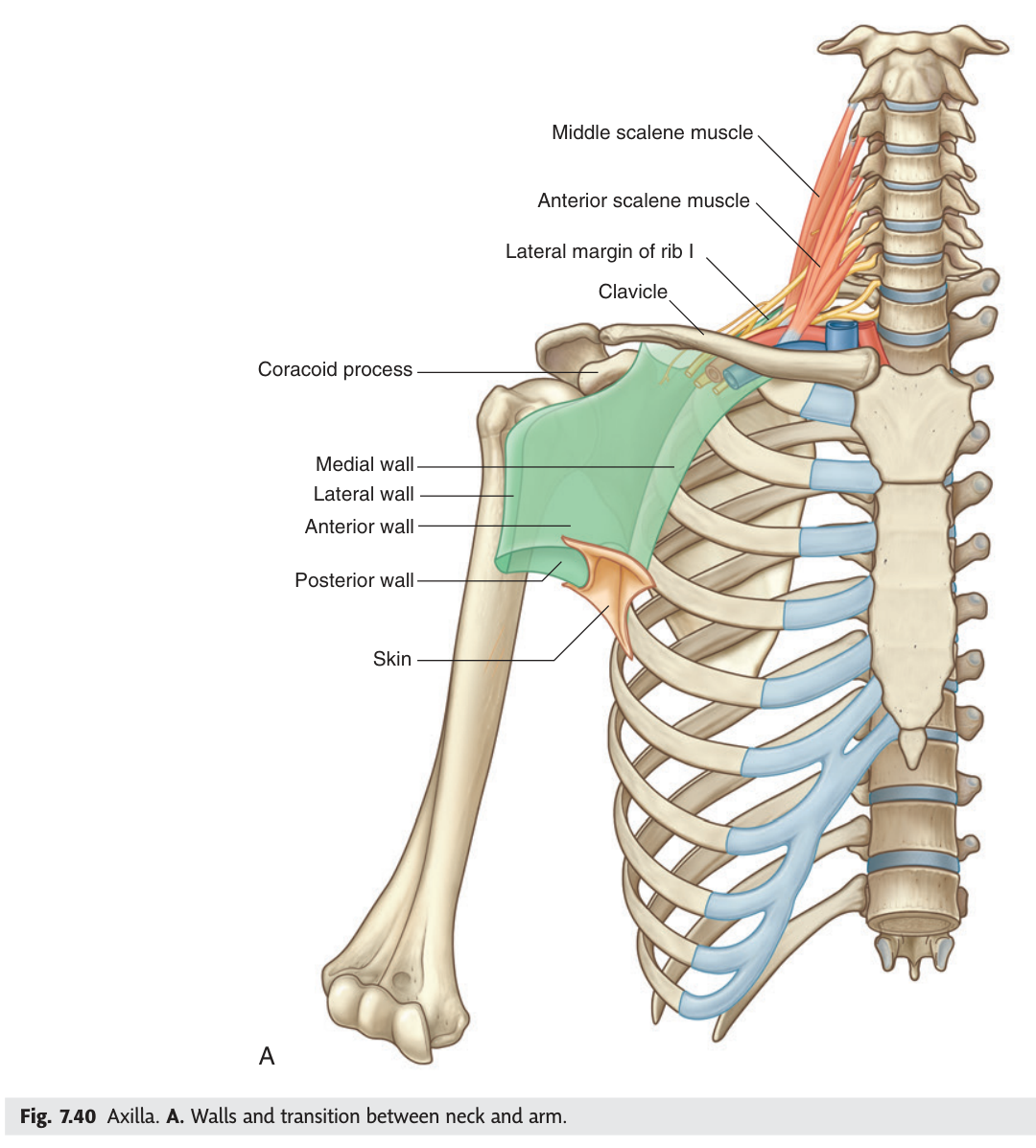
- What shape is the axilla? → A pyramid
- What bounds its apex? → Clavicle, first rib, upper border of the scapula
- What is its base? → The skin of the armpit
The four walls ★★★
Each wall is defined by what forms it, and each one has a reason to be remembered. The anterior wall is what you pinch between your fingers at the front of the armpit; the posterior wall is what you pinch at the back.
Anterior wall: pectoralis major and pectoralis minor (with subclavius).
Posterior wall: subscapularis, teres major and latissimus dorsi.
Medial wall: the upper part of serratus anterior over the first four ribs and their intercostal muscles.
Lateral wall: the intertubercular (bicipital) groove of the humerus, with coracobrachialis and the short head of biceps.
Apex: clavicle, first rib, upper border of scapula. Base: skin and axillary fascia.
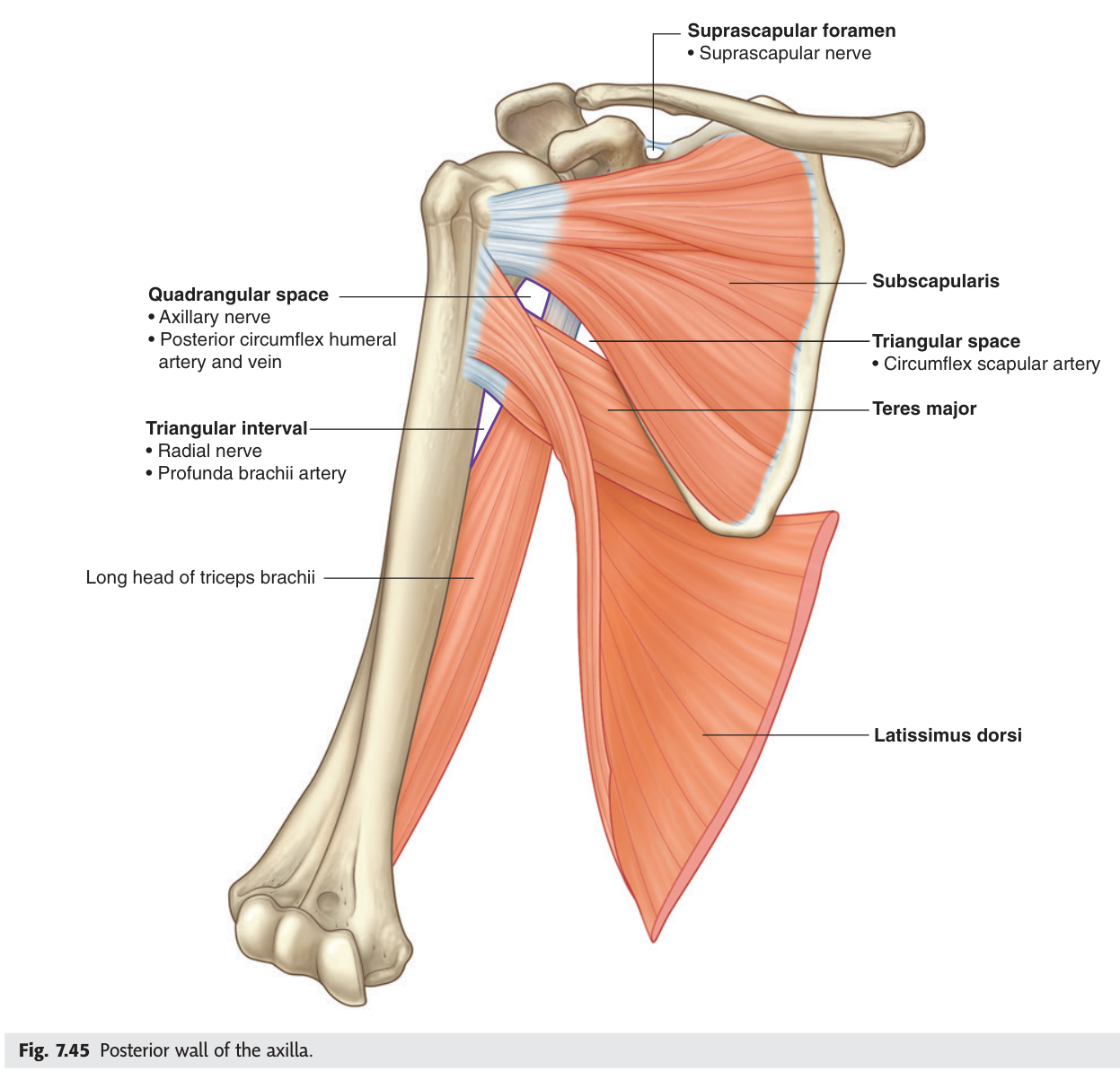
Front = the pecs. Back = the three muscles reaching from scapula and trunk to the humerus (subscapularis, teres major, latissimus dorsi). Medial = the chest wall itself, ribs plus serratus anterior. Lateral = the bone, in its groove. Nothing else could plausibly be there.
- Anterior wall? → Pectoralis major and minor
- Posterior wall? → Subscapularis, teres major, latissimus dorsi
- Medial wall? → Serratus anterior over the upper four ribs and intercostals
- Lateral wall? → Intertubercular groove of the humerus with coracobrachialis
What is inside
Inside the pyramid, wrapped together in the axillary sheath — a downward prolongation of the prevertebral fascia of the neck — lie the axillary artery, the axillary vein, and the cords and branches of the brachial plexus. Around them sit the axillary lymph nodes and a quantity of fat.
Because a single fascial tube encloses the artery and the whole plexus, an anaesthetist can block the entire upper limb with one injection. The distal sheath is compressed with finger pressure, local anaesthetic is injected proximally, and the solution is massaged along the tube. The position of the sheath is confirmed by feeling the pulsations of the third part of the axillary artery — the artery is the guide to the nerves.
- Axillary artery and its branches
- Axillary vein and its tributaries
- Cords and branches of the brachial plexus
- Axillary lymph nodes — five groups
- Axillary sheath and fat
- The tendon of the long head of biceps, crossing the space
- What is the axillary sheath derived from? → The prevertebral layer of deep cervical fascia
- Name the four main contents? → Axillary artery, axillary vein, brachial plexus cords and branches, axillary lymph nodes
- How is the sheath located during a block? → By the pulsation of the third part of the axillary artery
The axillary artery ★★
The subclavian artery becomes the axillary artery at the outer border of the first rib, and becomes the brachial artery at the lower border of teres major. Between those two points it is divided into three parts — and the divider is pectoralis minor.
The scheme is beautifully regular and worth learning as a rule rather than a list: the first part is proximal to pectoralis minor and has one branch; the second part lies behind the muscle and has two; the third part is distal to it and has three. One, two, three.
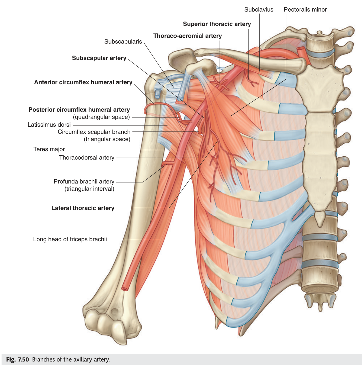
| Part | Relation to pectoralis minor | Branches |
|---|---|---|
| First | Proximal (above) | Superior (supreme) thoracic artery |
| Second | Behind | Thoracoacromial artery · lateral thoracic artery |
| Third | Distal (below) | Subscapular artery (→ circumflex scapular + thoracodorsal) · anterior circumflex humeral · posterior circumflex humeral |
Count the branches by the part number. The subscapular artery is the largest branch of the whole vessel and immediately splits again — the circumflex scapular heading back through the triangular space, the thoracodorsal running down with its nerve to latissimus dorsi.
- What divides the axillary artery into three parts? → Pectoralis minor
- Where does it begin and end? → Outer border of the first rib → lower border of teres major
- Branches of the third part? → Subscapular, anterior circumflex humeral, posterior circumflex humeral
- Which branch passes through the quadrangular space? → Posterior circumflex humeral artery
The brachial plexus ★★★
The brachial plexus is the wiring loom of the upper limb, and it looks impossible until you see that it is built in five tidy stages: roots → trunks → divisions → cords → branches. Five roots (C5–T1) merge into three trunks, each trunk splits into an anterior and a posterior division, and the divisions regroup into three cords named for their position around the axillary artery — lateral, medial and posterior.
That naming is the key to the whole thing. The cords are named by where they sit relative to the second part of the axillary artery, so if you can draw the artery you can place the cords, and if you can place the cords the branches follow.
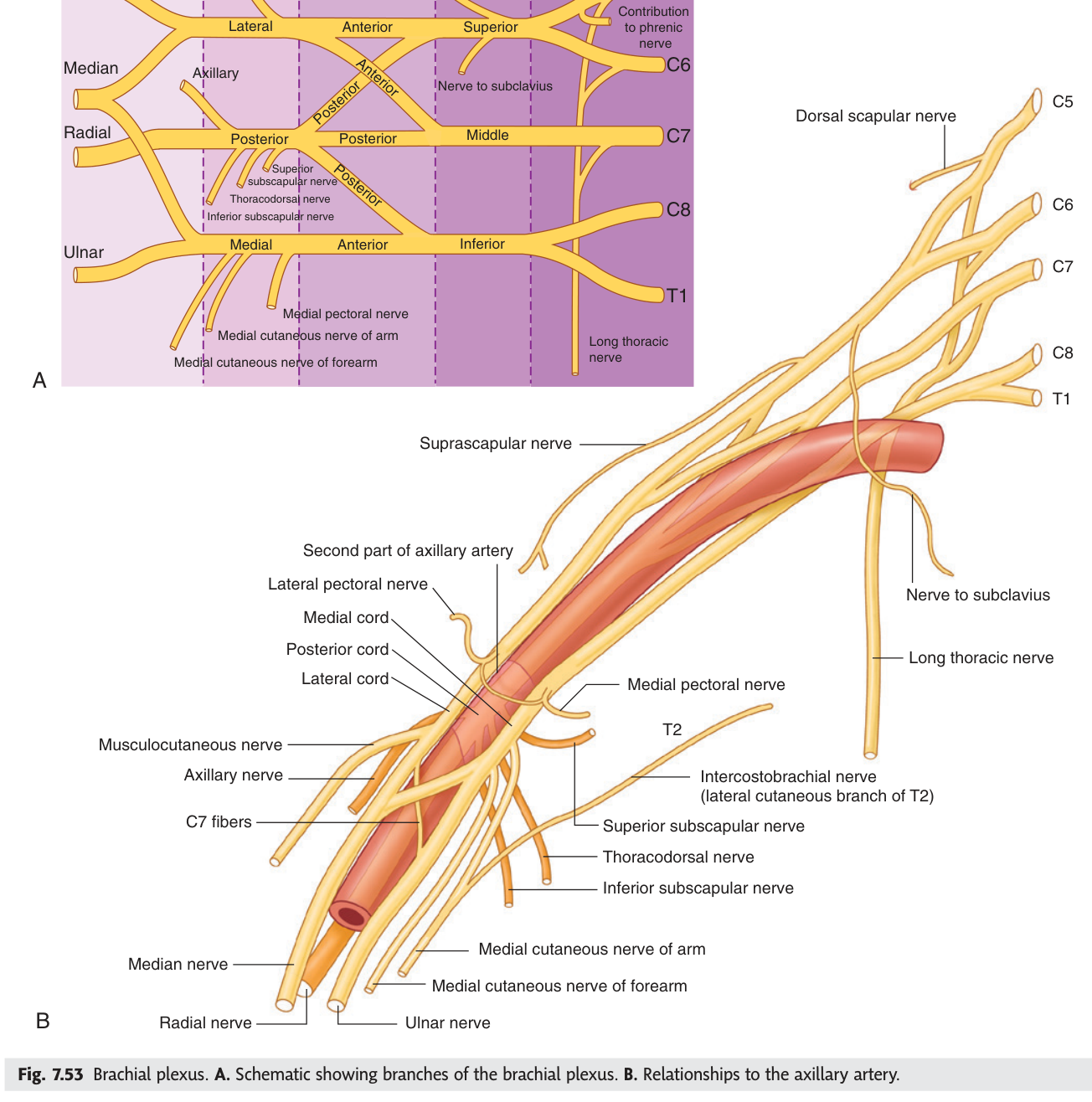
Lateral cord: musculocutaneous nerve · lateral root of the median nerve · lateral pectoral nerve.
Medial cord: ulnar nerve · medial root of the median nerve · medial pectoral nerve · medial cutaneous nerves of the arm and forearm.
Posterior cord: radial nerve · axillary nerve · thoracodorsal nerve · upper and lower subscapular nerves.
From the roots: long thoracic nerve (C5–7) · dorsal scapular nerve. From the upper trunk: suprascapular nerve · nerve to subclavius.
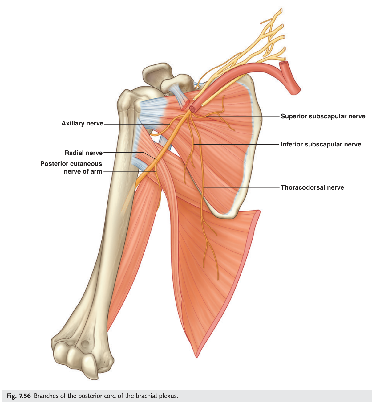
The median nerve is the one branch formed by both the lateral and medial cords — its two roots straddle the axillary artery like a letter M. Find that M in a dissection and you have identified the lateral cord to its left and the medial cord to its right, with the ulnar nerve running off medially and the musculocutaneous laterally.
- The five stages of the plexus? → Roots → trunks → divisions → cords → branches
- Why are the cords named lateral, medial and posterior? → By their position around the second part of the axillary artery
- Branches of the posterior cord? → Radial · axillary · thoracodorsal · upper and lower subscapular
- Which nerve is formed from two cords? → The median nerve — lateral and medial roots
- Which branch arises from the roots themselves? → The long thoracic nerve
Two nerves that get injured here
Two nerves in this region have signature injuries, and both appear in exams because the sign is visible across a room.
The long thoracic nerve arises directly from the roots C5, C6 and C7 and descends on the surface of serratus anterior, which it supplies. Serratus anterior holds the medial border of the scapula flat against the chest wall. Damage the nerve — in axillary surgery, a mastectomy with node clearance, or a blow to the side of the chest — and the scapula lifts away from the ribs when the patient pushes against a wall. That is a winged scapula, and the patient also cannot raise the arm fully above the head.
The thoracodorsal nerve from the posterior cord supplies latissimus dorsi. It runs down the posterior wall of the axilla with the thoracodorsal artery, which is exactly why it is at risk during axillary lymph node clearance for breast cancer — and why the surgeon deliberately identifies and preserves both.
- Root value of the long thoracic nerve? → C5, C6, C7 — directly from the roots
- Which muscle does it supply and what is the sign of injury? → Serratus anterior; winged scapula
- Which nerve supplies latissimus dorsi? → The thoracodorsal nerve, from the posterior cord
- Why do both matter in breast surgery? → Both run in the axilla and are at risk during node clearance
The axillary lymph nodes
Around twenty to thirty lymph nodes sit in the fat of the axilla, and they matter far beyond the limb: they receive most of the lymph from the breast, and their status determines the staging and treatment of breast cancer. They are described in five groups — pectoral (anterior), subscapular (posterior), humeral (lateral), central and apical — draining ultimately to the apical group and thence to the subclavian lymph trunk.
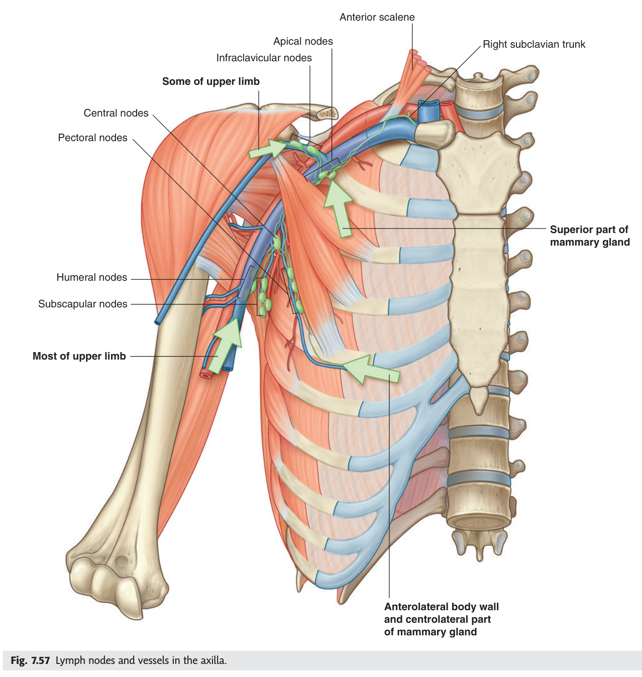
Because breast lymph reaches the pectoral group first, surgeons inject dye or tracer into the breast and remove only the first node it reaches — the sentinel node. If it is clear, the rest can usually be left. That single anatomical insight spared a generation of women full axillary clearance and the lymphoedema — chronic, disfiguring arm swelling from destroyed lymphatic drainage — that so often followed it.
- Name the five groups? → Pectoral · subscapular · humeral · central · apical
- Which group receives most breast lymph first? → The pectoral (anterior) group
- Where does the apical group drain? → The subclavian lymph trunk
- What is the consequence of clearing all of them? → Lymphoedema of the arm
Revision layer
TMU sets two summaries as homework for this lecture: the walls and contents of the axilla, and the branches of the brachial plexus and axillary artery. Both are below.
The axilla in one table
| Formed by | |
|---|---|
| Apex | Clavicle · first rib · upper border of scapula |
| Base | Skin and axillary fascia |
| Anterior wall | Pectoralis major and minor, subclavius |
| Posterior wall | Subscapularis · teres major · latissimus dorsi |
| Medial wall | Upper serratus anterior over ribs 1–4 and intercostals |
| Lateral wall | Intertubercular groove of humerus, coracobrachialis, short head of biceps |
| Contents | Axillary artery and vein · brachial plexus cords and branches · axillary lymph nodes · axillary sheath and fat |
Nerve → muscle, for the branches you must know
| Nerve | From | Supplies | Injury sign |
|---|---|---|---|
| Long thoracic | Roots C5–7 | Serratus anterior | Winged scapula |
| Thoracodorsal | Posterior cord | Latissimus dorsi | Weak adduction/extension |
| Axillary | Posterior cord | Deltoid, teres minor | Square shoulder |
| Radial | Posterior cord | Posterior arm and forearm | Wrist drop |
| Musculocutaneous | Lateral cord | Anterior arm (3 muscles) | Weak elbow flexion |
| Median | Lateral + medial cords | Most forearm flexors, thenar | Hand of benediction |
| Ulnar | Medial cord | Intrinsic hand muscles | Claw hand |
- Name the four walls, apex and base of the axilla with their contents
- Give the three parts of the axillary artery and all six branches
- Draw the brachial plexus from roots to cords and list branches by cord
- Explain winged scapula from the anatomy of the long thoracic nerve
- Name the five axillary node groups and explain sentinel node biopsy