Scapular Region & Superficial Thoracic Wall
Opening the back
The upper limb is not bolted to the trunk. It hangs from it, slung by muscle, with only one small bony joint — the sternoclavicular — connecting the whole arm to the axial skeleton. That single fact explains both the extraordinary mobility of the shoulder and its habit of dislocating: almost everything holding it in place is soft tissue, and soft tissue can be torn.
This unit is the first pass at the back and shoulder, and it exists mainly to reach two small intermuscular gaps — the quadrangular and triangular spaces — through which nerves and vessels escape from the axilla to the back of the arm. TMU sets both as a drawing question.
- Skin incisions from the external occipital protuberance to the median sacral crest
- Laterally to the acromion and along the midaxillary line
- Reflect trapezius and latissimus dorsi — the two superficial muscles of the back
- Then levator scapulae and the rhomboid muscles beneath them
- Finally the thoracolumbar fascia and the erector spinae in its sheath
The accessory nerve [XI] is found on the deep surface of trapezius — it supplies trapezius and sternocleidomastoid, and it is the nerve at risk in posterior triangle surgery.
It is not padding. The posterior and middle layers enclose erector spinae in a sheath; the anterior layer lies in front of quadratus lumborum. Latissimus dorsi and transversus abdominis pull on it from the sides, so tightening the abdominal wall tightens the fascia and splints the lumbar spine. This is the anatomy behind bracing before you lift.
- How is the upper limb attached to the axial skeleton? → Only at the sternoclavicular joint — the rest is muscular
- Which nerve supplies trapezius? → The accessory nerve [XI]
- What lies inside the sheath of the thoracolumbar fascia? → Erector spinae
Muscles of the shoulder
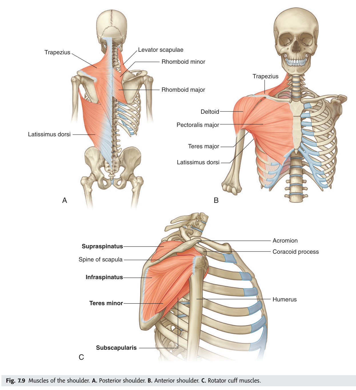
Six muscles run from the scapula to the humerus, and between them they can move the arm in almost any direction. Deltoid caps the shoulder and abducts it. Behind the scapula, supraspinatus sits above its spine and infraspinatus below; teres minor and teres major run from the lateral border toward the humerus. On the costal surface, hidden against the ribs, lies subscapularis.
Learn them by action group rather than as a list. Deltoid and supraspinatus abduct. Infraspinatus and teres minor rotate laterally. Teres major and subscapularis rotate medially and adduct. Two abduct, two turn out, two turn in — and the nerve supply follows the same grouping, which is why the table below is easier than it looks.
- Which two muscles abduct the shoulder? → Deltoid and supraspinatus
- Which two rotate it laterally? → Infraspinatus and teres minor
- Which two rotate it medially and adduct? → Teres major and subscapularis
- Nerve to deltoid and teres minor? → Axillary nerve
- Nerve to supraspinatus and infraspinatus? → Suprascapular nerve
The rotator cuff
Four of those six muscles do something beyond moving the arm: their tendons blend with the capsule of the shoulder joint and hold the head of the humerus pressed into the very shallow glenoid cavity. That is the rotator cuff — supraspinatus, infraspinatus, teres minor and subscapularis. Remember them as SITS.
The cuff is a compromise. A deep socket would be stable but stiff; the shoulder trades bony stability for range and asks muscle to make up the difference. When those muscles fail, the joint has almost nothing left.
Supraspinatus runs through a narrow tunnel between the head of the humerus below and the acromion above. Every abduction drags the tendon through that gap, and its blood supply is poorest close to its insertion. Decades of that produce degeneration, impingement and eventually rupture — the commonest rotator cuff tear by a wide margin. The patient cannot initiate abduction, though they can often hold the arm up once someone lifts it there for them, because deltoid can continue what supraspinatus had to start.
SITS — Supraspinatus · Infraspinatus · Teres minor · Subscapularis. Note who is absent: teres major is not part of the cuff, and neither is deltoid.
- The four rotator cuff muscles? → Supraspinatus · infraspinatus · teres minor · subscapularis (SITS)
- Which shoulder muscle is NOT in the cuff despite the name? → Teres major
- Why does supraspinatus tear most often? → It passes through a narrow subacromial gap and has poor blood supply near its insertion
The quadrangular space ★★★
Behind the shoulder, the muscles running to the humerus cross the long head of triceps and leave gaps between them. The largest is the quadrangular space, and it sits immediately below the shoulder joint — which is the entire reason it matters clinically.
An intermuscular space immediately below the glenohumeral joint.
Above: subscapularis and teres minor, with the capsule of the shoulder joint.
Below: teres major.
Medially: long head of triceps brachii.
Laterally: surgical neck of the humerus.
Contents: the axillary nerve and the posterior circumflex humeral artery and vein.
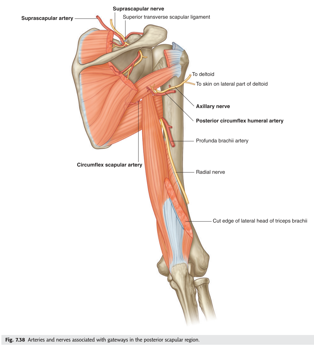
Picture the long head of triceps running vertically and the two teres muscles running horizontally toward the humerus. They make a cross. Teres minor above, teres major below — minor is the smaller and sits higher, which matches the alphabet and the anatomy at once. Triceps closes the medial side, the humerus closes the lateral side. Four walls, no memorising.
- The four boundaries of the quadrangular space? → Above: subscapularis/teres minor + capsule · Below: teres major · Medial: long head of triceps · Lateral: surgical neck of humerus
- Its two contents? → Axillary nerve and posterior circumflex humeral vessels
- Where does it lie relative to the shoulder joint? → Immediately below it
The triangular space ★★
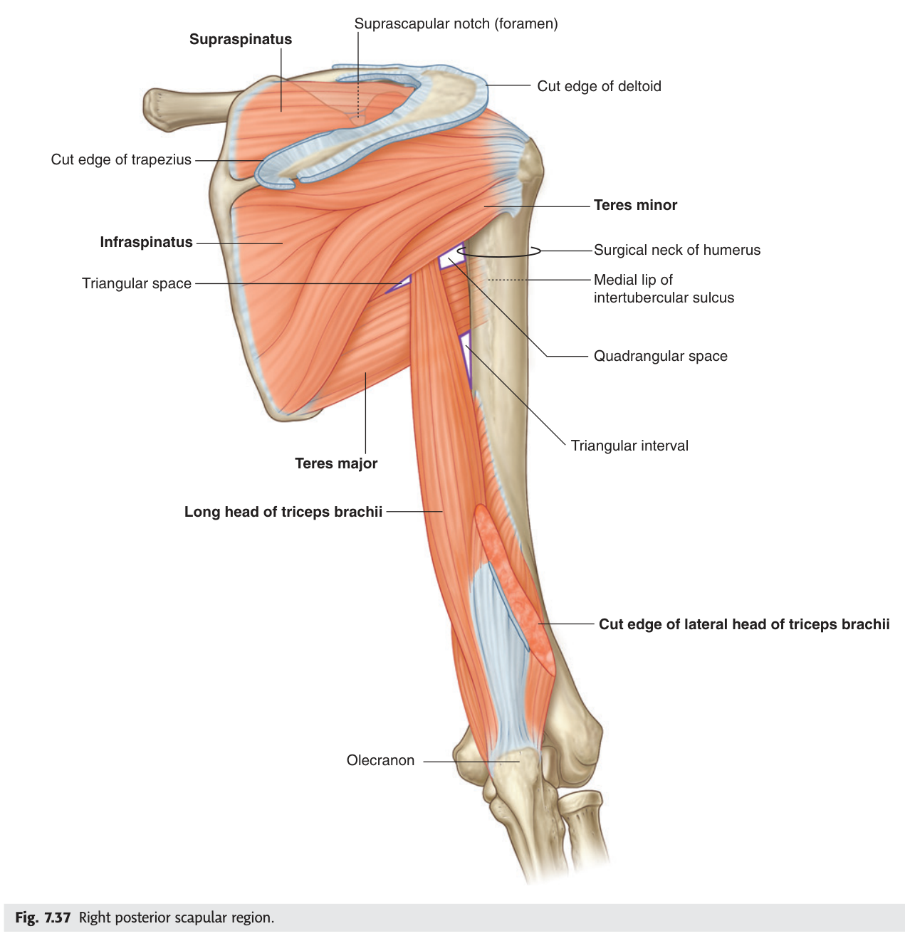
Medial to the long head of triceps is a smaller gap — the triangular space — with only three walls and only one thing passing through it. It is the easier of the pair, and it is examined precisely because students learn the quadrangular space and forget this one.
A smaller intermuscular space medial to the quadrangular space.
Above: teres minor. Below: teres major. Laterally: long head of triceps brachii.
Content: the circumflex scapular artery, a branch of the subscapular artery, which contributes to the scapular anastomosis.
| Gap | Boundaries | Contents |
|---|---|---|
| Quadrangular space | Above: subscapularis + teres minor + capsule · Below: teres major · Medial: long head of triceps · Lateral: surgical neck of humerus | Axillary nerve · posterior circumflex humeral vessels |
| Triangular space | Above: teres minor · Below: teres major · Lateral: long head of triceps | Circumflex scapular artery |
| Triangular interval | Above: teres major · Medial: long head of triceps · Lateral: shaft of humerus | Radial nerve · profunda brachii artery |
- Boundaries of the triangular space? → Teres minor above, teres major below, long head of triceps laterally
- Its content? → Circumflex scapular artery
- What passes through the triangular interval? → Radial nerve and profunda brachii artery
The axillary nerve
The axillary nerve arises from the posterior cord of the brachial plexus (C5, C6), passes backwards through the quadrangular space with the posterior circumflex humeral artery, and wraps around the surgical neck of the humerus. It supplies deltoid and teres minor, plus the skin over the lower deltoid.
Everything clinical about this nerve follows from one relationship: it is pressed against bare bone at the surgical neck, directly below a joint that dislocates more often than any other in the body.
A fracture of the surgical neck of the humerus, or an anterior dislocation of the shoulder, injures the axillary nerve. Deltoid is paralysed and wastes, so the normal rounded contour of the shoulder flattens — the "square shoulder" your slide names. Abduction is lost beyond the first few degrees, and there is a patch of numbness over the lower deltoid, the so-called regimental badge area. Testing that patch of skin is the standard bedside check after any shoulder dislocation, before reduction is attempted.
- Origin of the axillary nerve? → Posterior cord of the brachial plexus (C5, C6)
- Which two muscles does it supply? → Deltoid and teres minor
- Where is it injured? → Surgical neck of the humerus — fracture or shoulder dislocation
- Two clinical signs? → Square shoulder (deltoid wasting) and numbness over the regimental badge area
The superficial layer of the thoracic wall
Turning the cadaver over, the front of the chest is covered by pectoralis major, with pectoralis minor beneath it and serratus anterior wrapping around the ribs. In a female cadaver the breast is preserved on the skin as a landmark before removal, since it lies in the superficial fascia over pectoralis major.
The one structure worth chasing here is the cephalic vein. It ascends the lateral side of the arm, then runs in the deltopectoral groove between deltoid and pectoralis major, and finally pierces the deep fascia to drain into the axillary vein. That groove is a reliable surface landmark and a standard route for pacemaker leads and central venous access.
- Where does the cephalic vein run before it dives deep? → The deltopectoral groove
- Into what does it drain? → The axillary vein
- Which layer contains the breast? → Superficial fascia over pectoralis major
Revision layer
The prose was to make it stick. This is the table that earns the marks — TMU sets "summarise the scapular muscles and their function and nerve supply" as homework for this lecture.
Scapular muscles — function and nerve supply
| Muscle | Action | Nerve |
|---|---|---|
| Deltoid | Abducts the shoulder | Axillary nerve |
| Supraspinatus | Initiates abduction | Suprascapular nerve |
| Infraspinatus | Lateral rotation | Suprascapular nerve |
| Teres minor | Lateral rotation | Axillary nerve |
| Teres major | Adduction, medial rotation, extension | Lower subscapular nerve |
| Subscapularis | Adduction, medial rotation | Upper and lower subscapular nerves |
TMU's slide writes "infrascapular nerve" for the last two — use subscapular nerve, the term Gray's and Snell's use.
- Draw both spaces with all boundaries and contents labelled
- Distinguish the triangular space from the triangular interval
- Give all six scapular muscles with action and nerve
- Name the rotator cuff and explain why supraspinatus tears first
- Explain square shoulder from the course of the axillary nerve