The Leg & Dorsum of the Foot
Three compartments, three nerves, three jobs
The leg is the cleanest region in the whole course, because it obeys one rule almost without exception: each compartment has one nerve, one artery and one function. Learn the rule and you can reconstruct most of the detail rather than memorising it.
The tibia and fibula are joined along their length by the interosseous membrane, and two intermuscular septa run from the fibula out to the deep fascia. Those three sheets divide the leg into anterior, lateral and posterior compartments. The anterior compartment dorsiflexes (lifts the foot), the lateral everts (turns the sole outward), and the posterior plantarflexes (pushes off). Each is served by the nerve that runs inside it.
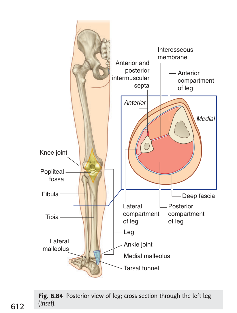
Anterior — deep fibular nerve — anterior tibial artery — dorsiflexion
Lateral — superficial fibular nerve — no artery of its own (fed by branches from the
fibular artery) — eversion
Posterior — tibial nerve — posterior tibial artery — plantarflexion
Now recall the common fibular nerve from Unit 3: it splits at the neck of the fibula into deep and superficial branches. That is why a single injury at the fibular neck knocks out both dorsiflexion and eversion — it takes out two compartments at once, and that is exactly what foot drop is.
- What forms the compartment walls? → Interosseous membrane and the anterior and posterior intermuscular septa
- Nerve and action of the anterior compartment? → Deep fibular nerve; dorsiflexion
- Nerve and action of the lateral compartment? → Superficial fibular nerve; eversion
- Nerve and action of the posterior compartment? → Tibial nerve; plantarflexion
Posterior compartment — the superficial layer
The calf you can see and grip is the superficial group: gastrocnemius, soleus and the slender plantaris. Gastrocnemius and soleus together are called the triceps surae — three heads, since gastrocnemius has two and soleus one — and they converge into the calcaneal (Achilles) tendon, the thickest tendon in the body.
The two muscles are not interchangeable. Gastrocnemius arises above the knee from the femoral condyles, so it crosses two joints and can only generate full power when the knee is straight — it is the sprinting and jumping muscle. Soleus arises below the knee from the tibia and fibula, crosses only the ankle, and works regardless of knee position — it is the standing and walking muscle. You can demonstrate this on yourself: rise onto your toes with the knee straight and you feel gastrocnemius; do it sitting with the knee bent and the work shifts to soleus.
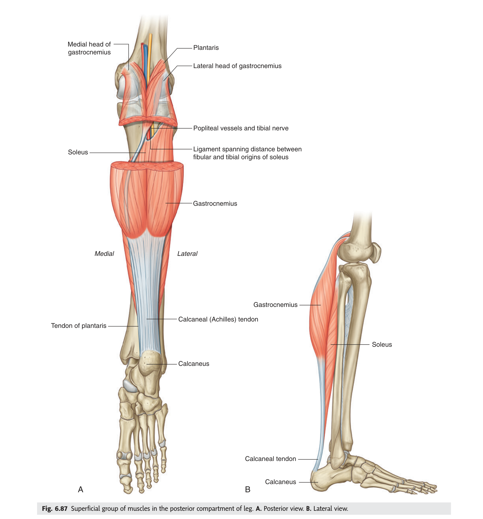
Soleus is riddled with venous sinuses. Every time it contracts during walking it squeezes blood up the deep veins against gravity, which is why the calf is called the "peripheral heart". Immobilise a patient — a long flight, a plaster cast, a hospital bed — and the pump stops, blood stagnates in those sinuses, and a deep vein thrombosis begins. The anatomy of this one muscle is the reason early mobilisation is standard care after surgery.
- What is the triceps surae? → Gastrocnemius (2 heads) + soleus (1 head), inserting via the calcaneal tendon
- Why does gastrocnemius weaken when the knee bends? → It arises above the knee, so knee flexion slackens it
- Nerve supply of the posterior compartment? → Tibial nerve
- How does soleus relate to DVT? → Its venous sinuses depend on muscular pumping; immobility causes stasis
Posterior compartment — the deep layer
Cut soleus and reflect it and you expose four deep muscles: popliteus at the top, and then three long muscles whose tendons will travel into the sole — flexor digitorum longus, tibialis posterior and flexor hallucis longus.
Their arrangement in the leg is worth fixing now, because it changes at the ankle and that change is examinable. In the leg, going from medial to lateral, the order is flexor digitorum longus (medial) → tibialis posterior (middle) → flexor hallucis longus (lateral). Behind the medial malleolus, however, tibialis posterior slips in front of flexor digitorum longus and becomes the most anterior of the two. Watch for that in the next section.
Popliteus deserves a sentence of its own. It is the "key" that unlocks the knee: with the foot on the ground it laterally rotates the femur on the tibia by a few degrees, releasing the fully-extended, locked knee so that flexion can begin.
- Name the four deep posterior muscles? → Popliteus, flexor digitorum longus, tibialis posterior, flexor hallucis longus
- Their medial-to-lateral order in the leg? → FDL (medial) · tibialis posterior (middle) · FHL (lateral)
- What is popliteus's special function? → It unlocks the fully extended knee to allow flexion
Arteries of the leg
Everything descends from the popliteal artery, which divides at the lower border of popliteus. The anterior tibial artery immediately does something distinctive: it passes forward through the aperture in the upper interosseous membrane, close to the neck of the fibula, to enter the anterior compartment. Nothing else takes that route, which is why the anterior compartment has its own dedicated supply.
The posterior tibial artery continues down between the superficial and deep muscles of the calf, giving off the fibular (peroneal) artery near its origin. At the ankle it passes through the malleolar canal and ends by dividing into the medial and lateral plantar arteries for the sole.
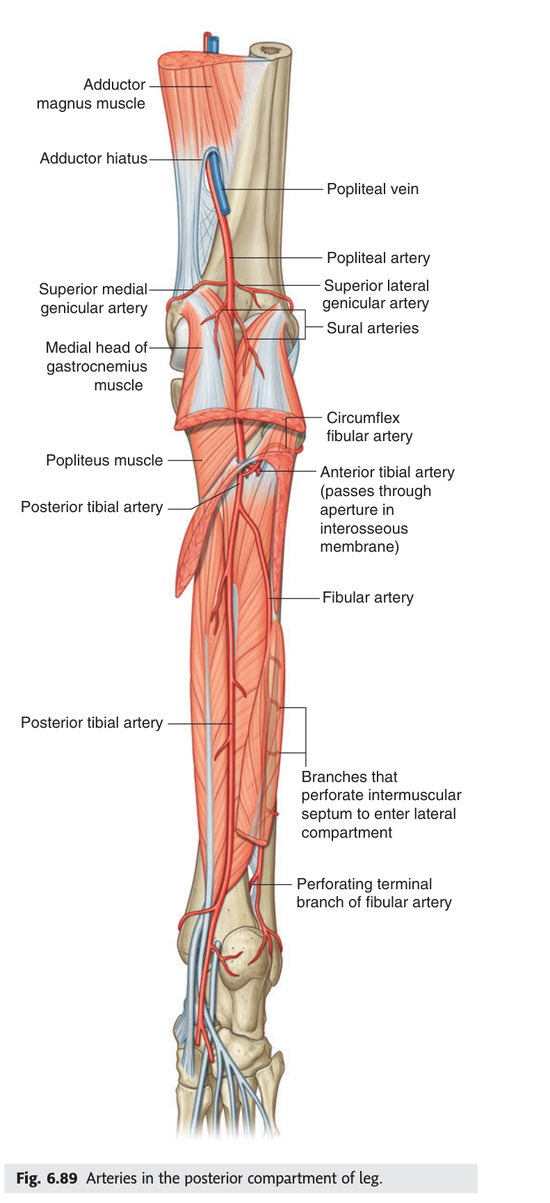
Vascular assessment of the foot uses three points, and each is an anatomical landmark you now know: the popliteal pulse behind the knee, the posterior tibial pulse midway between the medial malleolus and the heel (in the malleolar canal), and the dorsalis pedis pulse on the dorsum of the foot. Absent posterior tibial and dorsalis pedis pulses in a diabetic patient is the finding that predicts a non-healing foot ulcer.
- How does the anterior tibial artery reach its compartment? → Through the aperture in the upper interosseous membrane
- Which artery supplies the lateral compartment? → It has no artery of its own — branches from the fibular artery perforate the septum
- How does the posterior tibial artery end? → Dividing into medial and lateral plantar arteries
- Where is the posterior tibial pulse felt? → Midway between the medial malleolus and the heel
The malleolar canal ★★★
This is the set-piece of the unit — TMU's homework for this lecture is literally "draw a picture of the malleolar canal and its contents". It is the gateway through which everything from the back of the leg reaches the sole of the foot.
Your TMU slides call this the malleolar canal. Gray's and most English-language textbooks call the same structure the tarsal tunnel. They are the same space. Use "malleolar canal" in the TMU exam, but recognise "tarsal tunnel" when you read Gray's or meet the clinical syndrome.
An osteofibrous canal on the posteromedial side of the ankle.
Formed by: the medial surface of the calcaneus, the medial malleolus, and the overlying
flexor retinaculum.
Contents, from anterior to posterior:
tendon of tibialis posterior · tendon of flexor digitorum longus · posterior tibial artery and
veins · tibial nerve · tendon of flexor hallucis longus.
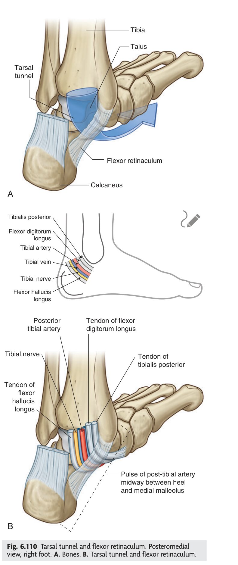
Tom, Dick And Very Nervous Harry
Tibialis posterior · flexor Digitorum longus ·
Artery (posterior tibial) · Vein · Nerve (tibial) ·
flexor Hallucis longus.
Start at the medial malleolus and work backwards towards the heel.
Compression of the tibial nerve under the flexor retinaculum causes burning pain, tingling and numbness in the sole of the foot, typically worse at night and after standing. It is the ankle's equivalent of carpal tunnel syndrome, and the anatomy explains the distribution exactly: the tibial nerve is the sole supply of the plantar surface, so the symptoms stop at the edge of the sole and spare the dorsum.
- What three structures form the malleolar canal? → Medial surface of calcaneus, medial malleolus, flexor retinaculum
- Its five contents, anterior to posterior? → Tibialis posterior tendon · FDL tendon · posterior tibial artery and veins · tibial nerve · FHL tendon
- What is the English-textbook name? → The tarsal tunnel
- Where are the symptoms of tarsal tunnel syndrome felt? → The sole, sparing the dorsum
The lateral compartment
The smallest compartment holds just two muscles — fibularis (peroneus) longus and fibularis (peroneus) brevis — supplied by the superficial fibular nerve. Both evert the foot, and both tendons pass behind the lateral malleolus, the mirror image of the malleolar canal on the other side of the ankle.
Fibularis longus does something remarkable. Its tendon crosses under the sole of the foot from lateral to medial to attach to the medial cuneiform and the first metatarsal — the same bones tibialis anterior attaches to from the other direction. The two tendons form a sling under the foot that supports the transverse arch. This is why the muscle is both an evertor and an important arch support.
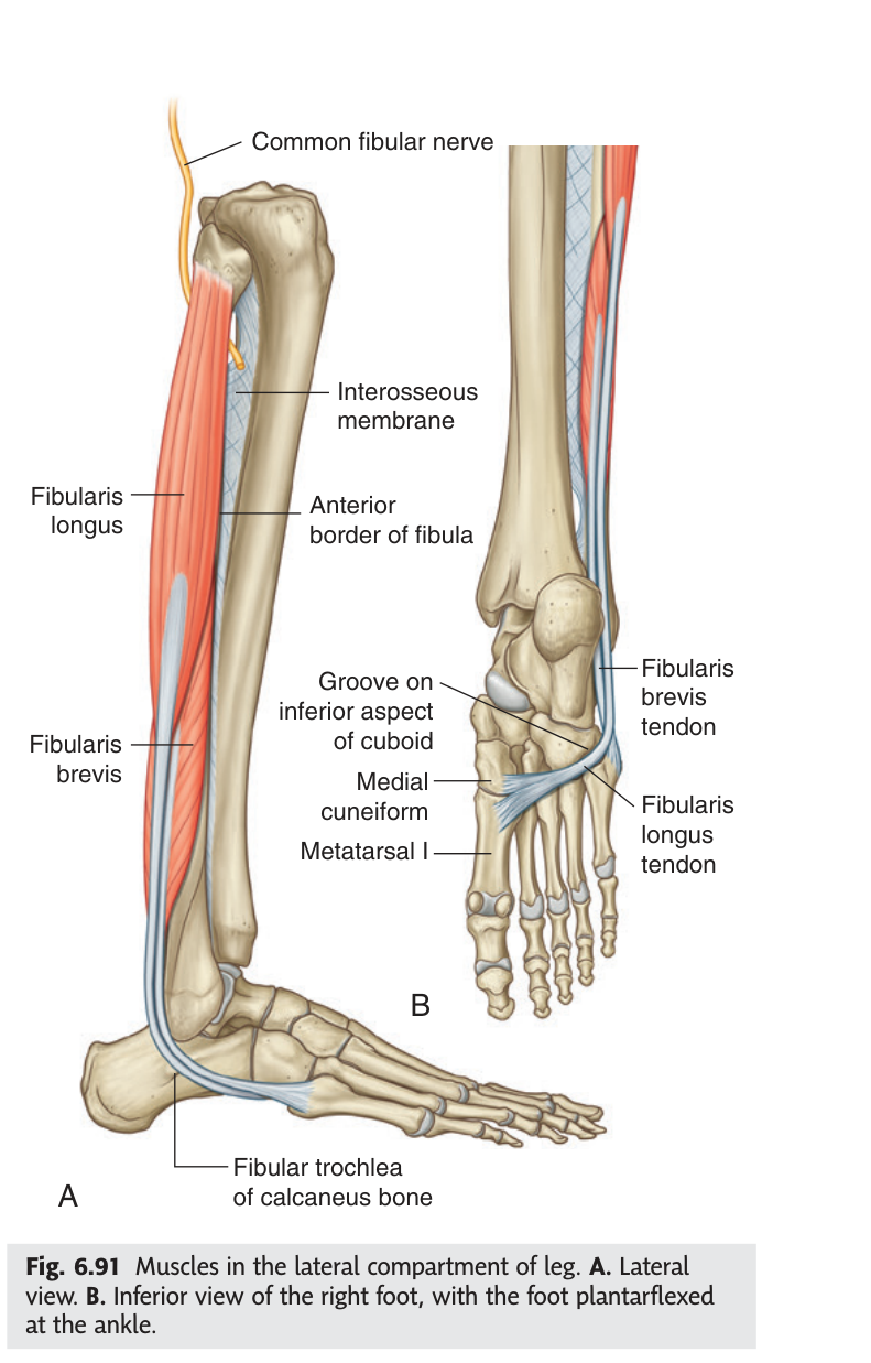
The superficial fibular nerve descends between the two muscles, supplies them both, and then pierces the deep fascia in the distal third of the leg to become cutaneous, supplying most of the skin of the dorsum of the foot.
- The two lateral compartment muscles and their nerve? → Fibularis longus and brevis; superficial fibular nerve
- Where do their tendons pass? → Behind the lateral malleolus
- Where does fibularis longus insert, and why is that odd? → Medial cuneiform and first metatarsal — it crosses the sole from lateral to medial
- Where does the superficial fibular nerve become cutaneous? → Distal one-third of the leg
The anterior compartment
Three muscles lift the foot: tibialis anterior medially, extensor hallucis longus in the middle, and extensor digitorum longus laterally. That medial-to-lateral order is worth learning as a sentence, because it is the same order in which their tendons cross the ankle. All three are supplied by the deep fibular nerve and accompanied by the anterior tibial artery.
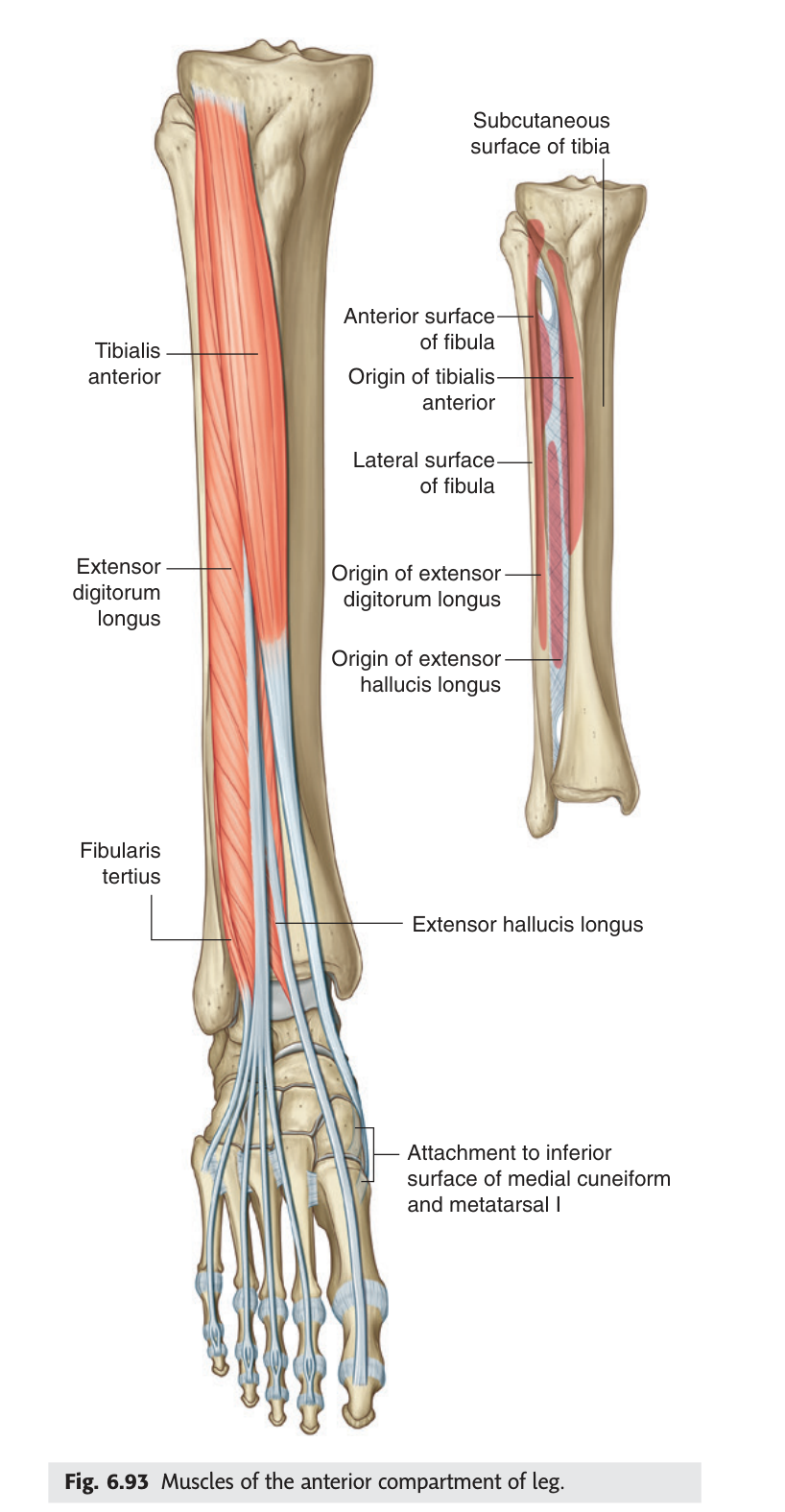
This is the compartment that most often goes wrong, and the reason is structural: it is the tightest, bounded by bone on two sides, interosseous membrane behind and unyielding deep fascia in front. A tibial fracture or crush injury raises the pressure inside, veins collapse first, pressure rises further, and finally arterial inflow fails. The warning sign is pain out of proportion to the injury, worse on passive stretch of the toes — and crucially, the dorsalis pedis pulse may still be present. Waiting for a pulse to disappear before operating means waiting until the muscle is dead. The treatment is emergency fasciotomy.
- The three anterior muscles, medial to lateral? → Tibialis anterior · extensor hallucis longus · extensor digitorum longus
- Their nerve and artery? → Deep fibular nerve; anterior tibial artery
- Earliest sign of compartment syndrome? → Pain out of proportion, worse on passive stretch
- Why is a present pulse falsely reassuring? → Arterial inflow fails last; muscle necrosis begins before the pulse goes
The dorsum of the foot
As the anterior tibial artery crosses the ankle joint it changes name and becomes the dorsalis pedis artery — the vessel you palpate to assess the circulation of the foot. Finding it is pure applied anatomy: feel lateral to the tendon of extensor hallucis longus, on the line between the two malleoli, and you are on it. Ask the patient to lift the big toe and the tendon stands up, giving you the landmark.
The deep fibular nerve accompanies the artery onto the dorsum, and its cutaneous territory is a famous exam answer: it supplies only the small web of skin between the first and second toes. Everything else on the dorsum is superficial fibular nerve territory. That tiny patch is the reliable way to test deep fibular nerve function clinically.
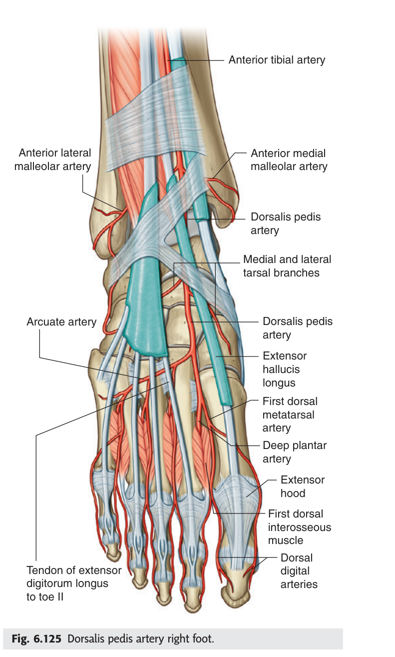
- What does the anterior tibial artery become? → The dorsalis pedis artery, at the ankle
- Where do you palpate it? → Lateral to the tendon of extensor hallucis longus
- Cutaneous territory of the deep fibular nerve? → The web space between the first and second toes only
- Who supplies the rest of the dorsum? → The superficial fibular nerve
Revision layer
The three compartments — the master table
| Compartment | Muscles | Nerve | Artery | Action |
|---|---|---|---|---|
| Anterior | Tibialis anterior; extensor hallucis longus; extensor digitorum longus (± fibularis tertius) | Deep fibular n. | Anterior tibial a. | Dorsiflexion |
| Lateral | Fibularis longus; fibularis brevis | Superficial fibular n. | Branches of fibular a. | Eversion |
| Posterior superficial |
Gastrocnemius; soleus; plantaris | Tibial n. | Posterior tibial a. | Plantarflexion |
| Posterior deep |
Popliteus; flexor digitorum longus; tibialis posterior; flexor hallucis longus | Tibial n. | Posterior tibial a. | Plantarflexion, inversion, toe flexion |
Malleolar canal — the drawing
| # | Structure (anterior → posterior) | Mnemonic |
|---|---|---|
| 1 | Tendon of tibialis posterior | Tom |
| 2 | Tendon of flexor digitorum longus | Dick |
| 3 | Posterior tibial artery and veins | And Very |
| 4 | Tibial nerve | Nervous |
| 5 | Tendon of flexor hallucis longus | Harry |
Where names change (continued from Unit 3)
| Vessel | Becomes | At |
|---|---|---|
| Anterior tibial artery | Dorsalis pedis artery | The ankle joint |
| Posterior tibial artery | Medial + lateral plantar arteries | Beyond the malleolar canal |
| Common fibular nerve | Superficial + deep fibular nerves | Neck of the fibula |
Nerve lesions of the leg — one table, three answers
| Nerve injured | Motor loss | Sensory loss |
|---|---|---|
| Common fibular (at fibular neck) | Dorsiflexion and eversion — foot drop | Dorsum of foot + lateral leg |
| Deep fibular only | Dorsiflexion | Web space between toes 1 and 2 |
| Superficial fibular only | Eversion | Most of the dorsum of the foot |
| Tibial (in malleolar canal) | Toe flexion, sole intrinsics | Sole of the foot |
- Draw the malleolar canal in cross-section with all five contents labelled in order
- Give the three compartments with muscles, nerve, artery and action
- Trace the popliteal artery to the sole of the foot, naming every change of name
- State where you palpate all three pulses below the knee
- Distinguish deep from superficial fibular nerve lesions on motor and sensory grounds