Back of the Lower Limb & Popliteal Fossa
The small saphenous vein
Turn the cadaver over and the first thing in the superficial fascia is the small saphenous vein — the mirror image of the great saphenous vein you met in Unit 1. Where the great saphenous begins medially and passes in front of the medial malleolus, the small saphenous begins on the lateral side of the dorsal venous arch and passes behind the lateral malleolus. Learn them as a pair and neither can be confused.
From there it ascends up the middle of the back of the calf, pierces the deep fascia, and drains into the popliteal vein inside the popliteal fossa. That termination is the clinically important part: incompetence at this junction is a common cause of varicose veins on the back of the calf, and it is why a duplex scan of varicose veins always examines the popliteal fossa as well as the groin.
Great — medial arch → in front of medial malleolus → up the
medial side → femoral vein in the groin.
Small — lateral arch → behind the lateral malleolus → up the back of
the calf → popliteal vein behind the knee.
Alongside it in the superficial fascia lies the posterior cutaneous nerve of the thigh, running centrally down the back of the thigh — the same nerve you traced out of the infrapiriform foramen in Unit 2. It is purely sensory, supplying the skin over the back of the thigh.
- Where does the small saphenous vein begin? → Lateral end of the dorsal venous arch of the foot
- What is its relation to the lateral malleolus? → It passes behind it
- Where does it end? → The popliteal vein, in the popliteal fossa
- Which cutaneous nerve accompanies it in the thigh? → Posterior cutaneous nerve of the thigh
The hamstrings
Three muscles fill the back of the thigh: biceps femoris laterally, and semitendinosus and semimembranosus medially. What defines a true hamstring is not position but attachment — each arises from the ischial tuberosity, crosses both the hip and the knee, and is supplied by the sciatic nerve. That triple test is why the short head of biceps femoris, which arises from the linea aspera and crosses only the knee, is strictly not a hamstring at all.
Because they cross two joints, the hamstrings extend the hip and flex the knee at the same time, and they are stretched to their limit when the hip is flexed and the knee simultaneously straightened — a sprinter's leading leg, precisely. That biomechanical fact is the whole explanation for why hamstring tears happen in sprinting rather than in slow running.
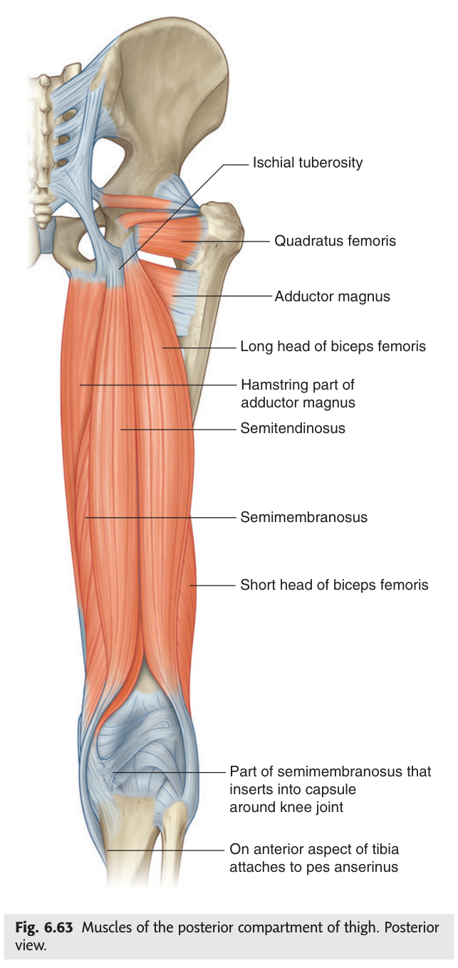
Semitendinosus has a long, cord-like tendon for its lower half — you can roll it between your fingers. Semimembranosus lies deep to it and is a broad, flat membrane at its upper end. The names describe exactly what you feel: tendinosus = tendon, membranosus = membrane.
- Name the three posterior thigh muscles? → Biceps femoris, semitendinosus, semimembranosus
- What three features define a true hamstring? → Origin from ischial tuberosity, crosses hip and knee, supplied by sciatic nerve
- Which part fails the test, and why? → Short head of biceps femoris — arises from linea aspera, crosses only the knee
- Their combined action? → Extend the hip and flex the knee
The sciatic nerve in the thigh
The sciatic nerve descends through the posterior compartment giving muscular branches to all three hamstrings and to the hamstring part of adductor magnus, plus articular branches to the hip and knee joints. It then does something you must be able to state precisely: it ends at the superior angle of the popliteal fossa by dividing into the tibial nerve and the common fibular (peroneal) nerve.
Notice that this division is the reason the popliteal fossa contains two nerves rather than one. Everything downstream in the leg and foot — which muscles are paralysed by which injury — follows from which of these two branches is involved.
- Where does the sciatic nerve divide? → At the superior angle of the popliteal fossa
- Into what? → Tibial nerve and common fibular (peroneal) nerve
- Which hamstring takes the common fibular division? → Short head of biceps femoris
The popliteal fossa — boundaries ★★★
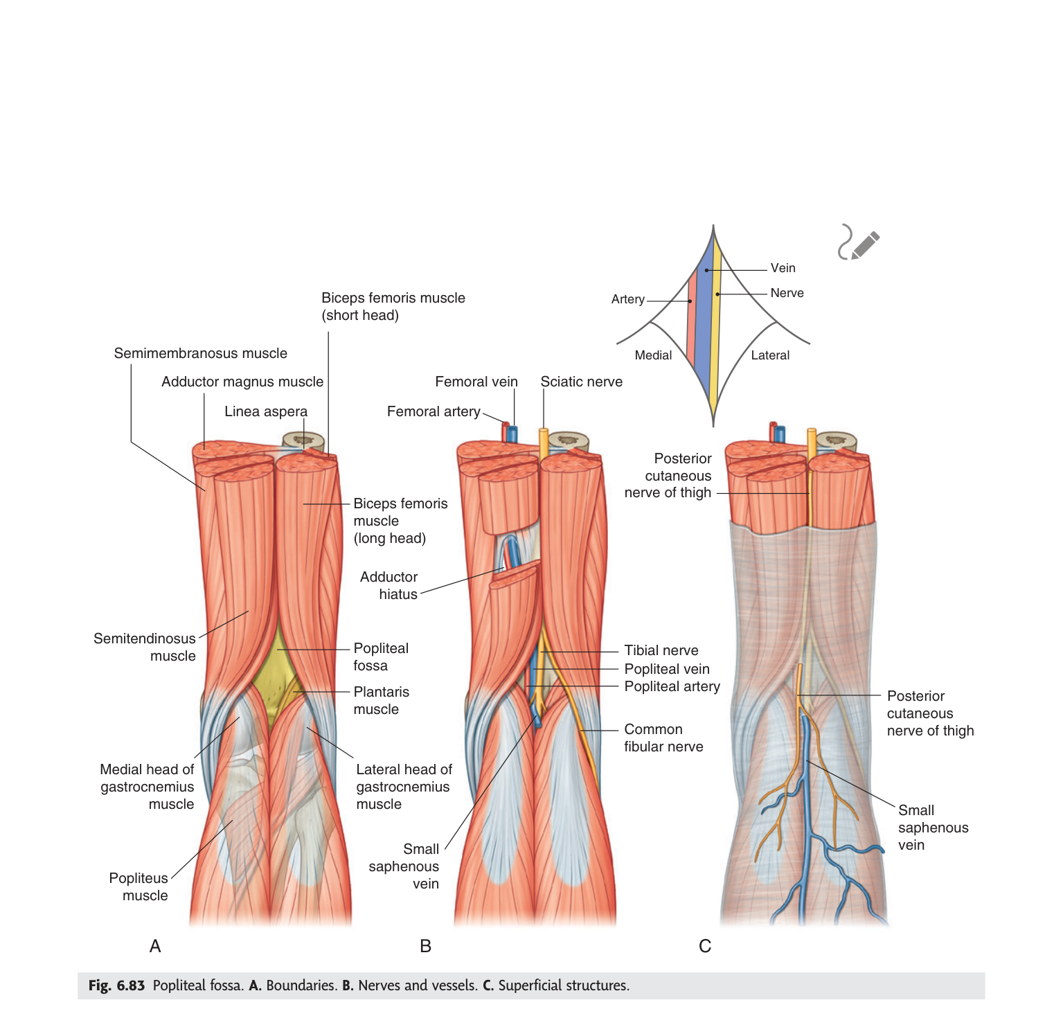
The hollow behind your knee is a diamond, and the shape comes from simple geometry: two muscles diverging from above and two converging from below. Above, the hamstrings split as they approach the knee — biceps femoris heading laterally to the fibula, semitendinosus and semimembranosus heading medially to the tibia. Below, the two heads of gastrocnemius rise from the femoral condyles and converge. Four muscle edges, four sides, one diamond.
TMU asked for exactly this in the 2021–22 paper, worth six marks. Give all four borders plus the floor and roof and you will not lose any of them.
A diamond-shaped space behind the knee joint, between the muscles of the posterior compartments of
the thigh and leg.
Superolateral: biceps femoris.
Superomedial: semitendinosus and semimembranosus.
Inferolateral: lateral head of gastrocnemius (with plantaris).
Inferomedial: medial head of gastrocnemius.
Floor: capsule of the knee joint and adjacent femur and tibia above, popliteus muscle
below.
Roof: deep fascia, continuous above with the fascia lata and below with the deep
fascia of the leg.
- What shape is the fossa and why? → A diamond — two hamstrings diverging above, two gastrocnemius heads converging below
- Superolateral border? → Biceps femoris
- Superomedial border? → Semitendinosus and semimembranosus
- The two inferior borders? → Lateral and medial heads of gastrocnemius
- The floor? → Knee joint capsule and adjacent bone, then popliteus below
The popliteal fossa — contents ★★★
The contents are easy if you get the direction right, and the classic mistake is to give them in the wrong one. Read them from superficial to deep, which is the order in which your scalpel meets them:
- Tibial nerve and common fibular nerve — most superficial
- Popliteal vein — in the middle
- Popliteal artery — deepest, lying directly on the knee joint capsule
Why that order? Because the nerves arrive from behind, descending in the plane just under the deep fascia, while the artery arrives from in front, coming through the adductor hiatus on the bone side of the limb. The vein, formed by the union of the anterior and posterior tibial veins, sits between the two. So the layering is not arbitrary — it records where each structure came from.
Superficial → deep: Nerve, Vein, Artery — "NVA".
Medial → lateral (as in Gray's inset): Artery, Vein, Nerve.
Same three structures, two different axes. Always say which axis you are answering on — the examiner
is testing whether you understand the space in three dimensions, not whether you memorised a string.
The popliteal artery is the deepest structure in the fossa, lying against the joint capsule with nerve and vein piled on top of it. That is exactly why it is the hardest peripheral pulse to palpate: you must flex the knee to relax the roof of the fascia and press firmly, with the fingers of both hands, against the back of the tibia. Its depth also explains why a popliteal aneurysm can grow large before it is noticed, and why it presents by compressing the tibial nerve or thrombosing rather than by being seen.
- Contents from superficial to deep? → Tibial and common fibular nerves → popliteal vein → popliteal artery
- Which structure lies deepest, and against what? → The popliteal artery, on the knee joint capsule
- Why is the popliteal pulse difficult? → The artery is deepest, under the vein and nerves
- What else is in the fossa? → Popliteal lymph nodes (1–2) and fat
The popliteal artery
The popliteal artery is a short vessel with a clearly defined beginning and end, and both are named by the structures it passes. It begins at the adductor hiatus, where it continues from the femoral artery, and it ends at the lower border of popliteus, where it divides into the anterior and posterior tibial arteries. Between those two points it gives off genicular branches — superior, middle and inferior — and muscular branches.
The genicular branches matter more than their dull name suggests. They form an anastomotic network around the knee that can maintain some blood flow to the leg if the main vessel is slowly occluded. "Slowly" is the operative word: this collateral circulation takes time to develop, so it protects against gradual atherosclerotic narrowing but not against sudden traumatic transection.
The artery is tethered at both ends — proximally at the adductor hiatus and distally by the fibrous arch of soleus. A posterior knee dislocation stretches the segment between those fixed points, and the vessel tears. This is why every knee dislocation is treated as a vascular emergency with mandatory assessment of distal pulses: the joint may reduce easily and look fine while the limb is ischaemic.
- Where does the popliteal artery begin? → At the adductor hiatus, continuing the femoral artery
- Where and into what does it end? → At the lower border of popliteus, into anterior and posterior tibial arteries
- Its named branches? → Superior, middle and inferior genicular arteries, plus muscular branches
- How is the popliteal vein formed? → Union of the anterior and posterior tibial veins near the lower border of popliteus
The common fibular nerve — the vulnerable one
The two nerves leave the fossa very differently, and the difference decides which one gets injured. The tibial nerve runs straight down the middle and dives deep to the gastrocnemius, staying protected in the calf. The common fibular nerve takes the risky route: it follows the tendon of biceps femoris to the lateral side, passes superficial to the lateral head of gastrocnemius, and then winds around the neck of the fibula, where it lies almost directly under the skin against bare bone. There it divides into the superficial and deep fibular nerves.
At the neck of the fibula the common fibular nerve is the most commonly injured nerve in the lower limb. A fibular neck fracture, a tight plaster cast, prolonged leg-crossing or lying anaesthetised with the leg against a hard edge will all compress it. The result is foot drop: paralysis of the anterior compartment (dorsiflexors, via the deep fibular nerve) and the lateral compartment (evertors, via the superficial fibular nerve), so the foot hangs down and inverted, and the patient lifts the knee high to clear the toes — a high-stepping gait.
- How does the common fibular nerve leave the fossa? → Along the biceps femoris tendon, superficial to the lateral head of gastrocnemius
- Where is it most vulnerable? → Winding around the neck of the fibula
- Into what does it divide? → Superficial and deep fibular nerves
- What deficit results from injury? → Foot drop — loss of dorsiflexion and eversion
Revision layer
Popliteal fossa at a glance
| Structure | |
|---|---|
| Superolateral | Biceps femoris |
| Superomedial | Semitendinosus and semimembranosus |
| Inferolateral | Lateral head of gastrocnemius (+ plantaris) |
| Inferomedial | Medial head of gastrocnemius |
| Floor | Knee joint capsule, femur and tibia; popliteus inferiorly |
| Roof | Deep fascia (continuous with fascia lata above, deep fascia of leg below) |
| Contents (superficial→deep) | Tibial n. and common fibular n. → popliteal vein → popliteal artery; plus popliteal lymph nodes and fat |
Posterior thigh muscles
| Muscle | Nerve | Action |
|---|---|---|
| Biceps femoris — long head | Tibial division of sciatic n. | Extends hip, flexes and laterally rotates knee |
| Biceps femoris — short head | Common fibular division of sciatic n. | Flexes and laterally rotates knee |
| Semitendinosus | Tibial division of sciatic n. | Extends hip, flexes and medially rotates knee |
| Semimembranosus | Tibial division of sciatic n. | Extends hip, flexes and medially rotates knee |
Where names change
| Vessel / nerve | Becomes | At |
|---|---|---|
| Femoral artery | Popliteal artery | Adductor hiatus |
| Popliteal artery | Anterior + posterior tibial arteries | Lower border of popliteus |
| Sciatic nerve | Tibial + common fibular nerves | Superior angle of the popliteal fossa |
| Common fibular nerve | Superficial + deep fibular nerves | Neck of the fibula |
- Draw the popliteal fossa: four borders, floor, roof, three contents in depth order
- Trace the small saphenous vein from foot to termination
- Name the three hamstrings with their exact nerve supply, including the short-head exception
- State where the popliteal artery begins and ends and what it becomes
- Explain foot drop from the anatomy of the common fibular nerve