Regulation of Gene Expression
Why regulation is required ★★★
Regulated expression of genes is required for development, differentiation and adaptation.
Gene expression is:
1 · regulated during ontogeny and differentiation of the organism and its cellular components
2 · needed so the organism can adapt to its environment and conserve energy and nutrients
3 · needed to respond to complex environmental challenges
Transcription level · gene amplification · gene rearrangement · post-transcriptional modifications · RNA stabilization.
The process works through the interaction of specific binding regulatory proteins with various regions of DNA, which have a positive or negative effect on transcription, and can result in tissue-specific gene expression. It is influenced by hormones, heavy metals and chemicals.
Expression could in principle be controlled anywhere between the gene and the working protein — and it is, at every level in that list. But transcription is where the decisive control sits, and the reason is economy.
Consider the cost of controlling at the last possible step. To regulate by degrading a finished protein, the cell must first pay for transcription, processing, export and translation — four high-energy phosphate bonds per residue (Unit 25) — and then throw the product away. Blocking at the first step wastes nothing.
Post-transcriptional control has a different virtue: speed. A stored mRNA can be translated in seconds; a gene that must be transcribed, spliced and exported takes minutes to hours. So the cell uses transcription for durable decisions — which cell type to be, which nutrient to metabolise — and post-transcriptional mechanisms for urgent ones. The level of control matches the timescale of the decision.
Type A response — expression rises while the inducer is present and falls when it is withdrawn; the lac operon is the classic example.
Type B response — expression rises and remains elevated even after the inducer is removed.
Type C response — occurs during the development of differentiated function in a tissue or organ, and is essentially irreversible.
- The three reasons regulation is required? → Development, differentiation and adaptation
- Name three levels of control → Transcription, gene amplification, gene rearrangement, post-transcriptional modification, RNA stabilisation
- Which response type is the lac operon? → Type A
- Which type accompanies differentiation? → Type C
Positive and negative regulation ★★★
Negative regulation: the regulatory element is present and expression is decreased. The protein responsible is a repressor.
Positive regulation: the regulatory element is present and expression is increased. That mediating positive regulation is a positive regulator or ACTIVATOR.
Derepression is the removal of a repressor — and note the logic that follows: a double negative has the effect of acting as a positive.
It is easy to read “a double negative acts as a positive” as a piece of wordplay. It is in fact a design principle, and it explains why so many biological switches are built from repressors.
A gene under negative control is off by default and needs no continuous input to stay off. Turning it on requires only that the repressor be removed — and removal can be triggered by the substrate itself, as lactose does in §4. The signal that a pathway is needed is the arrival of the very molecule the pathway acts on.
By contrast, a gene under positive control needs an activator present and maintained. That costs something continuously, but it gives finer graded control.
The lac operon uses both at once, and that is exactly why it is the textbook example: one that acts positively (the cAMP-CRP complex) and one that acts negatively (the LacI repressor). Two independent signals, one gene.
Inducible genes have relatively low basal rates of transcription and are switched up when needed.
Housekeeping genes are expressed constantly, at a roughly constant level, because their products are always needed.
A constitutive mutation is one that causes a normally inducible gene to be expressed all the time — for example, a mutation that destroys the repressor or its binding site.
- What is a repressor? → A protein mediating negative regulation
- What is an activator? → A protein mediating positive regulation
- What is derepression? → Removal of a repressor — a double negative acting as a positive
- Inducible vs housekeeping genes? → Inducible have low basal transcription; housekeeping are always expressed
- What is a constitutive mutation? → One causing an inducible gene to be expressed permanently
Operons and polycistronic mRNA ★★★
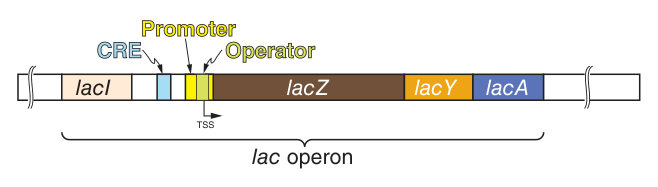
An operon can be regulated by a single promoter or regulatory region. It is a cluster of genes transcribed together as one unit.
A cistron is the genetic unit encoding one polypeptide — the “one cistron, one subunit” concept, which refines the older “one gene, one enzyme”.
Polycistronic mRNA is transcribed into one large mRNA molecule that contains multiple independent translation start (AUG) and stop (UAA) codons for each cistron. The result is coordinate expression — all the genes rise and fall together.
Polycistronic mRNA works only if a ribosome can start translating in the middle of a message. Bacteria can: their ribosomes bind an internal sequence upstream of each AUG.
Eukaryotic ribosomes cannot. As Unit 25 established, the 5′ cap directs the 40S ribosomal subunit to the 5′ end of the mRNA, which then scans forward to the first suitable AUG. There is only one entry point, so there can be only one protein per message.
The cap-dependent scanning mechanism is precisely what makes operons impossible in eukaryotes. One feature of initiation determines an entire architecture of gene organisation — and it is why eukaryotes must coordinate related genes by shared regulatory elements scattered across the genome instead of by physical clustering.
- Define an operon → A cluster of genes regulated by a single promoter or regulatory region
- What is a cistron? → The genetic unit encoding one polypeptide
- What is polycistronic mRNA? → One transcript containing independent start and stop codons for several cistrons
- Why can eukaryotes not use operons? → Cap-dependent scanning allows only one initiation site per mRNA
The lac operon ★★★
Analysis of lactose metabolism in E. coli led to the operon model — the foundation of everything in this unit, and the reason Harper's says prokaryotes provide models for the study of gene expression in mammalian cells.
| Gene | Product |
|---|---|
| lacZ | β-galactosidase, which cleaves lactose |
| lacY | A permease — the permeation of lactose into the cell |
| lacA | A thiogalactoside transacetylase |
| lacI | The lac operon repressor protein — note that it lies OUTSIDE the operon and has its own promoter |
The lac repressor has four identical subunits of molecular weight 38,000 and a high affinity for the operator locus.
The operator locus is a region of double-stranded DNA 27 base pairs long, with a twofold rotational symmetry and an inverted palindrome. LacI repressor binding covers 17 base pairs, and only two subunits of the repressor appear to bind.
This is not decoration — it is a consequence of the protein's shape, and the same logic recurs throughout Unit 26.
The repressor is a multimer with twofold rotational symmetry: rotate it 180° and it looks the same. A symmetric protein must bind a symmetric site. An inverted palindrome — reading the same on both strands — is exactly a DNA sequence with twofold rotational symmetry.
Once you see this, you can predict it elsewhere. The λ operator sites in §6 are 17-bp elements bound by dimeric proteins. The leucine zipper and many helix-turn-helix proteins in §9 bind as dimers to palindromic sites. Protein symmetry predicts DNA symmetry — and a palindrome in a regulatory region is a strong hint that a dimer binds there.
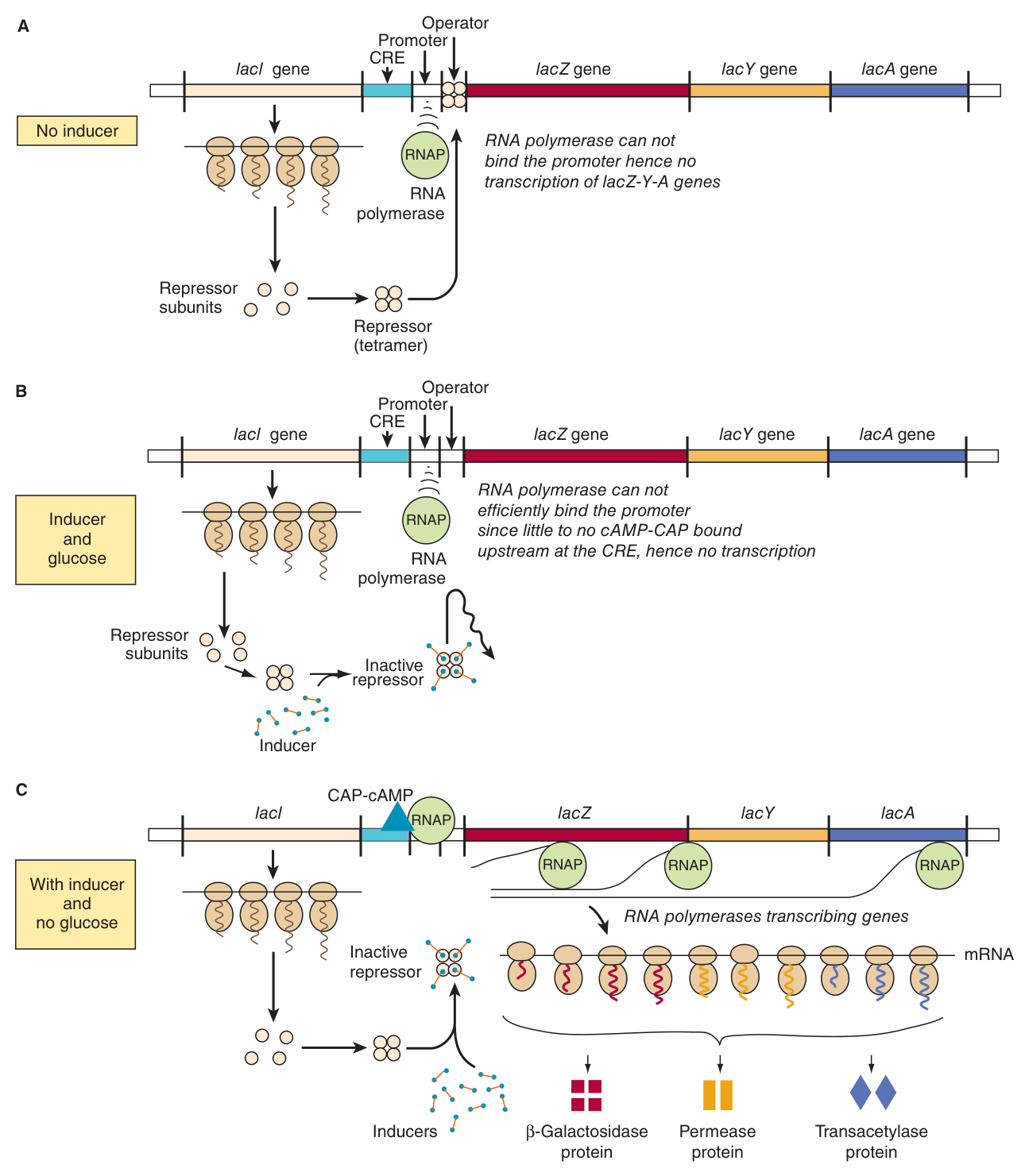
Addition of lactose or of a gratuitous inducer such as IPTG causes the inducer to bind the repressor, which then releases the operator: an inducer DEREPRESSES the lac operon.
The kinetics are fast: at high concentrations of lactose with no or very low glucose, lac-specific mRNAs are fully induced within 5–6 minutes, and β-galactosidase protein is maximal within 10 minutes. This is the type A response.
Translation of the polycistronic mRNA can occur even before transcription is completed — the prokaryotic coupling of Unit 24.
- The three structural genes of the lac operon, and their products? → lacZ β-galactosidase, lacY permease, lacA thiogalactoside transacetylase
- What does lacI encode? → The lac operon repressor protein
- Structure of the repressor? → Four identical subunits of MW 38,000
- Size and character of the operator? → 27 bp of dsDNA, twofold rotational symmetry, an inverted palindrome
- How much does the repressor cover, and with how many subunits? → 17 bp, using only two subunits
- How fast is induction? → mRNA fully induced within 5–6 minutes, protein maximal within 10
Catabolite repression ★★★
When exposed to both lactose and glucose as sources of carbon, bacteria first metabolize the glucose and then temporarily stop growing, until the genes of the lac operon become induced and they gain the ability to metabolize lactose as a usable energy source.
This is CATABOLITE REPRESSION, and it is mediated by a catabolite gene activator protein (CAP) in conjunction with cAMP. This protein is also referred to as the cAMP regulatory protein (CRP).
When glucose or glycerol is present in concentrations sufficient for growth, the bacteria will lack sufficient cAMP to bind to CAP, because the glucose inhibits adenylyl cyclase, the enzyme that converts ATP to cAMP.
Without cAMP, CAP cannot bind DNA and cannot activate transcription. The CAP-cAMP regulator acts as a POSITIVE regulator.
So the operon is controlled by two regulatory proteins: one that acts positively (the cAMP-CRP complex) and one that acts negatively (the LacI repressor). Maximal activity requires lactose present AND glucose absent.
| Lactose | Glucose | cAMP | LacI repressor | Transcription |
|---|---|---|---|---|
| Absent | Present | Low | Bound to operator | Off |
| Absent | Absent | High | Bound to operator | Off |
| Present | Present | Low | Released | Very low — no CAP-cAMP activation |
| Present | Absent | High | Released | MAXIMAL — repressor off AND activator on |
Ask what each signal actually reports.
The LacI repressor answers: is there any lactose to eat? Releasing it when lactose appears is obviously sensible — there is no point making β-galactosidase with no substrate.
But that alone would be wasteful. Lactose might be present while glucose, a better fuel, is also available. So CAP-cAMP answers a second question: is there anything better? Glucose inhibits adenylyl cyclase, so cAMP is low precisely when glucose is plentiful, and the activator is absent.
Only when the answer is “lactose yes, glucose no” does the cell commit. That is an AND gate built from one negative and one positive regulator — the simplest possible logic circuit, assembled from two proteins.
Note also that cAMP is doing here exactly what it does in Unit 12: reporting the absence of glucose. In the hepatocyte, glucagon raises cAMP when blood glucose falls and glycogen breakdown begins. In both cases cAMP is the “glucose is scarce” signal — the same messenger, the same meaning, three billion years apart.
- What is catabolite repression? → Suppression of the lac operon by glucose
- Which protein mediates it, and with what? → Catabolite gene activator protein (CAP), also called CRP, with cAMP
- How does glucose lower cAMP? → It inhibits adenylyl cyclase
- Is CAP-cAMP positive or negative? → Positive
- When is transcription maximal? → Lactose present AND glucose absent
The lambda switch ★★
The genetic switch of bacteriophage lambda chooses between the lysogenic pathway — the virus integrates and lies dormant as a prophage — and the lytic pathway — the virus replicates and destroys the host.
Exposure of its lysogenic bacterial host to DNA-damaging agents causes the dormant bacteriophage to become “induced”, switching from the prophage state to a lytic infection.
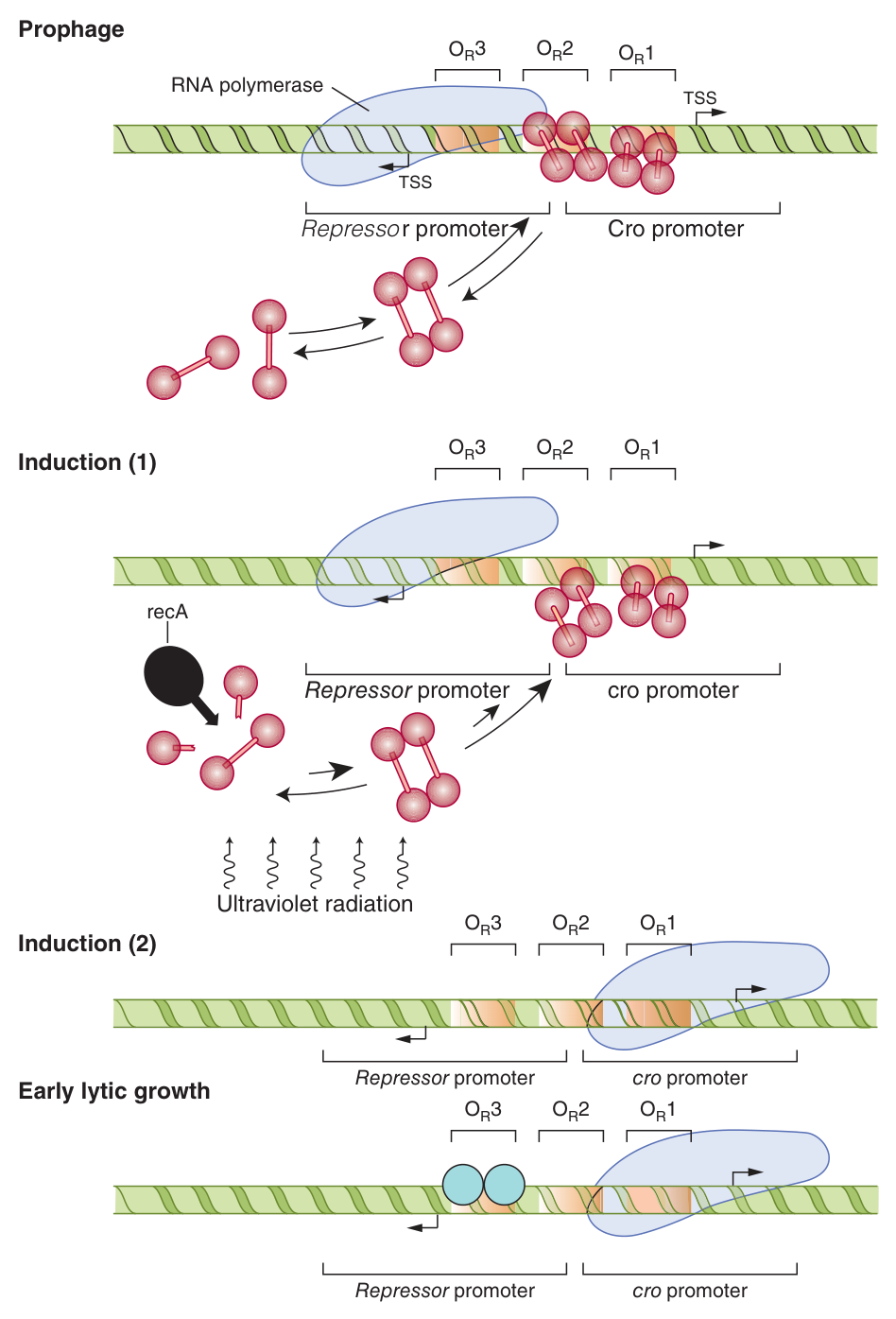
The right operator (OR) lies in the DNA region between the cro and repressor genes, and is subdivided into three discrete, evenly spaced, 17-bp cis-active DNA elements whose nucleotide sequences are similar but not identical. Each may be bound by either cI or Cro proteins, and the relative affinities of cI and Cro for each of the sites varies.
There are two promoter sequences: one directing rightward transcription of cro and other distal genes, the other leftward transcription of the repressor gene.
The repressor protein (cI) is 236 amino acids, 27 kDa. The Cro protein is 66 amino acids, 9 kDa, a single domain mediating both operator binding and dimerization.
When the repressor gene is on, the cro gene is off — the prophage state; when the cro gene is on, the repressor gene is off — lytic growth.
In a lysogenic bacterium the repressor's affinities run OR1 > OR2 > OR3. Binding of repressor to OR2 both blocks cro transcription and activates its own gene.
Ultraviolet light strikes the lysogenic host bacterium → fragments of single-stranded DNA are generated → these activate a specific protease coded by a bacterial gene (recA) → the protease hydrolyzes the repressor protein → dissociation of the repressor molecules from OR2 and OR1 → RNA polymerase immediately has access to the rightward promoter.
Cro protein then binds the operator region as a dimer, expression of other lambda genes begins as part of the lytic cycle, and later Cro comes to occupy OR1, reducing expression and effecting the final stages of the lytic cycle.
The switch looks elaborate for a virus. But read the trigger: it is DNA damage in the host, detected through recA, the bacterium's own damage-response protein.
A prophage sitting quietly in a healthy bacterium is in an excellent position — it is copied free of charge every time the cell divides. But a bacterium whose DNA is being shredded by ultraviolet light is about to die, and a prophage would die with it. The virus is eavesdropping on the host's distress signal and abandoning ship.
That is why the switch is worth its complexity: it is not a random coin-toss but a conditional decision, made on the best available evidence about the host's prospects. And it is bistable — when the repressor gene is on the cro gene is off, and vice versa — so there is no unstable intermediate state. Two mutually repressing genes make a memory device, and the same architecture underlies differentiation decisions in our own cells.
- The two lambda pathways? → Lysogenic (prophage) and lytic
- Structure of the right operator? → Three evenly spaced 17-bp elements, similar but not identical
- The two competing proteins? → cI repressor (236 aa, 27 kDa) and Cro (66 aa, 9 kDa)
- What triggers induction? → UV light → single-stranded DNA fragments → activation of the recA protease → repressor hydrolysis
- Repressor affinity order? → O_R1 > O_R2 > O_R3
Chromatin remodelling ★★★
Chromatin remodeling is an important aspect of eukaryotic gene expression. Chromatin structure provides an additional level of control of gene transcription — one that prokaryotes, having no nucleosomes, do not possess.
Acetylation and deacetylation occur on lysine residues in the amino terminal tails of histone molecules.
Acetylation reduces the positive charge of these tails, which decreases the binding affinity of histone for the negatively charged DNA, causing disruption of nucleosomal structure. That gives access of transcription factors to cognate regulatory DNA elements and enhances binding of the basal transcription machinery to the promoter.
Histone deacetylation would have the opposite effect.
Methylation of deoxycytidine residues in the sequence 5′-mCpG-3′ silences genes.
In mouse liver, only the UNmethylated ribosomal genes can be expressed, and many animal viruses are not transcribed when their DNA is methylated.
Remodelling proceeds through multiple protein-binding DNA elements and the binding of transcription factors to these elements, which can disrupt the structure of the nucleosome, prevent its re-formation, or recruit further remodelling activities.
As Unit 24 put it: promoter accessibility, and hence PIC formation, is often modulated by nucleosomes, and nucleosome eviction by chromatin-active coregulators facilitates PIC formation and transcription.
A bacterium has roughly 4,000 genes and needs, at any moment, most of them. A human cell has about 20,000 and needs, in any given tissue, a minority — and it must keep the rest reliably and durably silent, through every cell division, for the lifetime of the organism.
Repressor proteins are not adequate for that. A repressor must be continuously present at every site it silences, and it is diluted at every division. Chromatin solves the problem structurally: package the gene away and it is off by default, with no ongoing cost.
And crucially, the packaging is heritable. Methylation patterns are copied to the daughter strand after replication, and modified histones are redistributed to both daughter chromatids. A liver cell's daughters are liver cells. That is the whole mechanism of differential expression — and it is why this level of control belongs to eukaryotes alone.
- Where does histone acetylation occur? → On lysine residues in the amino-terminal tails
- Why does it activate transcription? → It reduces the positive charge, lowering histone affinity for DNA and disrupting the nucleosome
- What does deacetylation do? → The opposite — it silences
- Which DNA modification silences genes? → Methylation of deoxycytidine in 5′-mCpG-3′
- Give the evidence → In mouse liver only unmethylated ribosomal genes are expressed; methylated viral DNA is not transcribed
Enhancers and the enhanceosome ★★
Certain DNA elements enhance or repress transcription of eukaryotic genes. The classic example is the simian virus 40 (SV40) enhancer.
Enhancer binding proteins interact with a plethora of other transcription proteins, and unlike promoter elements, enhancers work at great distance — 1000 to 10⁵ bp — and in either orientation.
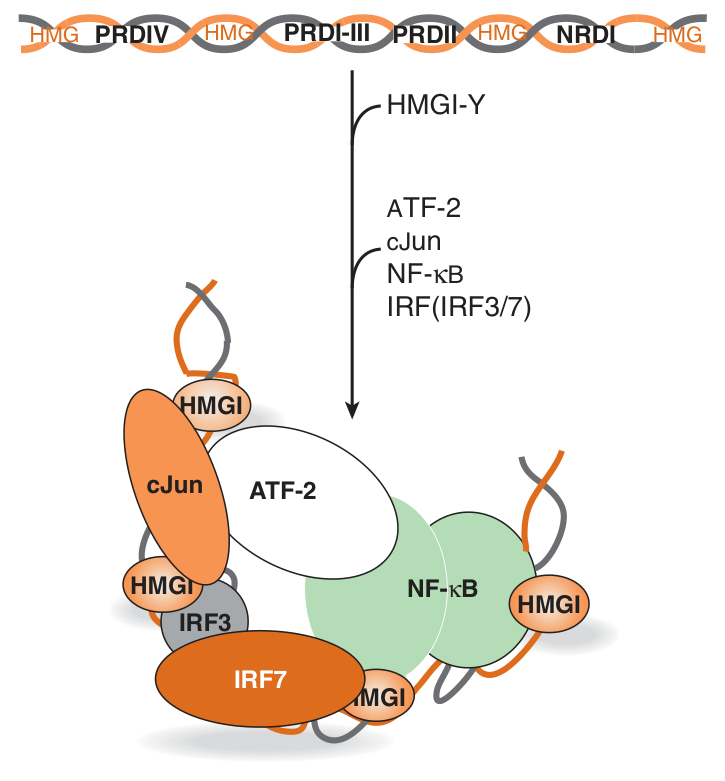
The β-interferon gene is induced upon viral infection of mammalian cells. Its enhancer element is located between nucleotides −110 and −45 and contains four distinct clustered cis elements, bound by four trans factors:
1 · NF-κB
2 · a member of the IRF (interferon regulatory factor) family
3 · the heterodimeric leucine zipper factor ATF-2/c-Jun
4 · HMG I(Y), a ubiquitous architectural transcription factor
HMG I(Y) induces a significant bend in the DNA, and along with the three trans factors, by inducing a series of critically spaced DNA bends, brings about the formation of a unique, stereospecific, three-dimensional structure in which all four factors are active — the ENHANCEOSOME.
HMG I(Y) is the interesting member of that quartet, because it does not activate transcription itself. All it does is bend the DNA.
But bending is what makes the other three work. Spread along a straight helix, the four sites are too far apart for their proteins to touch. Bend the DNA at critically spaced points and they are brought into contact, forming a surface that no one of them could present alone. The complex, not any component, is the activator.
Two consequences follow, and both are exam-worthy. First, this is why the assembly is stereospecific: the spacing of the elements matters as much as their sequence, because spacing determines geometry. Second, it makes the enhanceosome a coincidence detector — it fires only when all four signals are present at once. An interferon response should not be triggered by any single stimulus, and this architecture guarantees it is not.
This also explains how enhancers act from thousands of base pairs away: the intervening DNA simply loops out, bringing distant elements to the promoter.
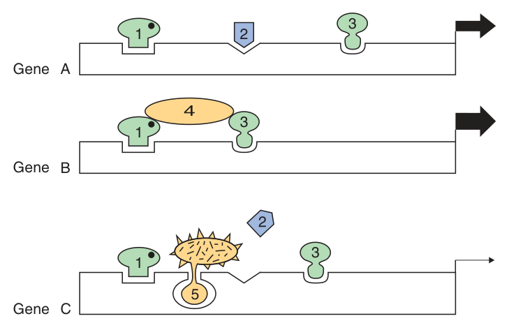
Combinations of DNA elements and associated proteins provide diversity in responses: the same factor may activate one gene and, in different company, inhibit another — gene C is inactivated by the combination of factors 1, 5 and 3, because factor 5 precludes the essential binding of factor 2 to DNA.
Tissue-specific expression may result from the action of enhancers or repressors.
Reporter genes are used to define enhancers and other regulatory elements: a DNA fragment of 2 kb of 5′-flanking DNA and its cognate promoter is ligated to a reporter, transfected, and the reporter activity detected; addition of one or more hormones then reveals functional hormone response elements (HRE).
Transcription domains can be defined by locus control regions and insulators.
- How far can an enhancer act? → 1000 to 10⁵ bp, in either orientation
- Where is the β-interferon enhancer? → Between nucleotides −110 and −45
- Name the four factors of the enhanceosome → NF-κB, an IRF family member, ATF-2/c-Jun, and HMG I(Y)
- What does HMG I(Y) do? → Induces a significant bend in the DNA — an architectural factor
- How are enhancers mapped experimentally? → With reporter gene constructs
DNA-binding motifs ★★★
Several motifs mediate the binding of regulatory proteins to DNA: the helix-turn-helix, the zinc finger, and the leucine zipper.
1 · Binding must be of high affinity to the specific site and of low affinity to other DNA.
2 · Binding is usually by a dimer or higher multimer, to a site with matching symmetry.
3 · The protein-DNA interactions are maintained by hydrogen bonds and van der Waals forces.
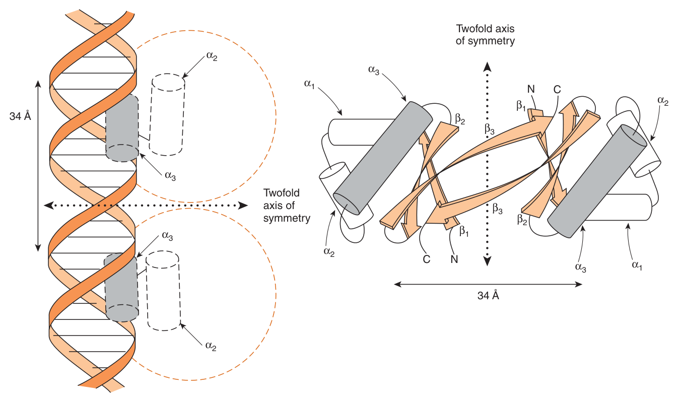
Illustrated by the Cro protein and its binding to DNA.
The Cro monomer consists of three antiparallel β sheets (β1–β3) and three α-helices (α1–α3). The α3 and α2 helices are held at about 90 degrees to each other by a turn of four amino acids, and the α3 helix of Cro is the DNA recognition surface.
The distance between comparable points on the two DNA α-helices is 34 Å — exactly one turn of B-DNA, so a dimer places one recognition helix in each of two successive major grooves.
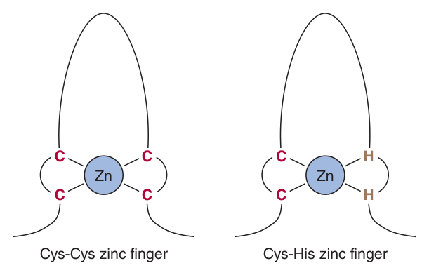
The zinc finger motif — a small domain folded around a zinc ion, which stabilises the structure so that a short α-helix can enter the major groove. Zinc fingers occur in tandem arrays, each finger reading a few base pairs.
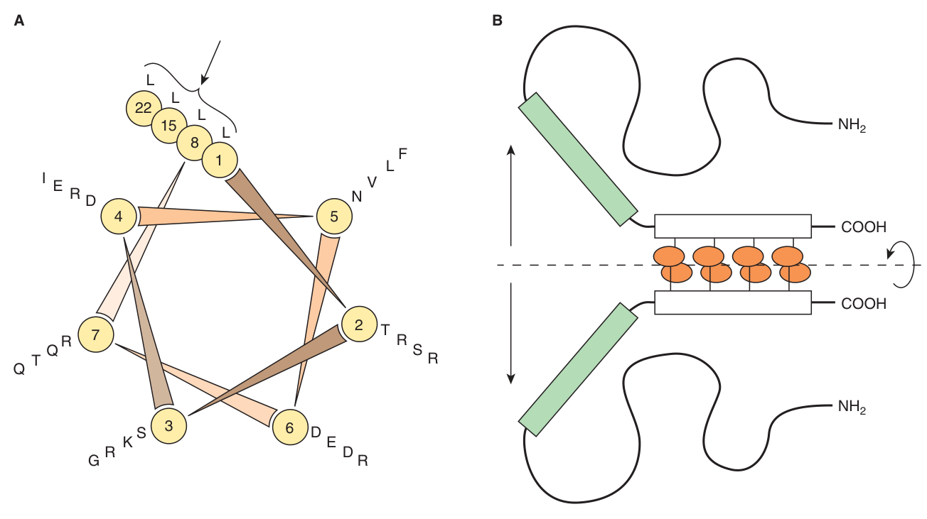
The leucine zipper motif — a helical wheel consisting of seven spokes that correspond to the seven amino acids of each turn, in which leucine residues occur at every seventh position. The leucines line up along one face of the helix and interdigitate with those of a partner helix — hence “leucine zippers”. The zipper mediates dimerisation; an adjacent basic region does the DNA binding.
Three structurally unrelated folds, arrived at independently — and all three do the same thing: they present a short α-helix to the major groove of DNA.
That convergence is not coincidence. Unit 23 explained the constraint: the bases are on the inside of the helix, so a protein must read the sequence from outside, and the edges of the base pairs are exposed at the floor of the major groove. The groove is about the width of an α-helix. There is essentially one way to solve the problem, and evolution has found it repeatedly.
Note too the recurring role of dimerisation. The leucine zipper does nothing but hold two helices together; Cro's single domain mediates both operator binding and dimerization; the lac repressor binds as a pair of subunits to a palindrome. A dimer doubles the number of contacts and therefore squares the specificity — which is exactly what principle 1 demands: high affinity to the specific site and low affinity to other DNA.
And dimerisation buys something else. Heterodimers — such as the ATF-2/c-Jun pair in the enhanceosome — let a limited set of proteins generate a combinatorial variety of specificities. Two hundred factors can make far more than two hundred decisions.
- Name the three DNA-binding motifs → Helix-turn-helix, zinc finger, leucine zipper
- Which helix of Cro recognises DNA? → α3, held at ~90° to α2 by a four-amino-acid turn
- Distance between the two recognition helices? → 34 Å — one turn of B-DNA
- What defines a leucine zipper? → Leucine at every seventh position, on a seven-spoke helical wheel
- What does the zipper itself do? → Mediates dimerisation; an adjacent basic region binds DNA
- What forces hold protein to DNA? → Hydrogen bonds and van der Waals forces
Prokaryotes versus eukaryotes ★★★
| Prokaryote | Eukaryote | |
|---|---|---|
| Gene organisation | Operons — polycistronic mRNA, coordinate expression | One gene, one mRNA; coordination by shared regulatory elements |
| Chromatin | None | Nucleosomes — an additional level of control |
| DNA methylation | Restriction/modification only | 5′-mCpG-3′ silences genes; heritable |
| Distance of control | Adjacent operator and promoter | Enhancers at 1000–10⁵ bp, in either orientation |
| Transcription and translation | Coupled — translation begins before transcription ends | Separated by the nuclear membrane |
| Post-transcriptional control | Limited | Splicing, alternative promoters, RNA editing, miRNA, stability |
| Typical response | Rapid, reversible adaptation to nutrients | Durable, heritable differentiation |
A bacterium regulates to adapt; a eukaryote regulates to differentiate.
Everything in the table follows from that difference of purpose. A bacterium must respond to a sugar appearing in its medium within 5–6 minutes and reverse the decision just as fast, so it uses freely diffusible repressors and activators binding adjacent sites — cheap, quick, and completely reversible.
A liver cell must keep the muscle genes off permanently, through every division, for eighty years. That demands mechanisms that are structural and heritable — chromatin packaging and DNA methylation — rather than dependent on the continuous presence of a protein.
And yet Harper's insists that prokaryotes provide models for the study of gene expression in mammalian cells, and it is right. The lac operon's vocabulary — promoter, operator, repressor, activator, induction, cis and trans — is the vocabulary of the whole field. The eukaryotic cell did not replace that logic; it added layers on top of it.
- Which organisms use operons, and why can the other not? → Prokaryotes; eukaryotic cap-dependent scanning allows only one initiation site per mRNA
- Which level of control is unique to eukaryotes? → Chromatin structure
- Which control is heritable through cell division? → DNA methylation and chromatin modification
- State the organising contrast → Bacteria regulate to adapt; eukaryotes regulate to differentiate
Revision layer
The lac operon in one table — the most examinable thing in this unit
| Component | What it is | What it does |
|---|---|---|
| lacZ | Structural gene | β-galactosidase |
| lacY | Structural gene | Permease — permeation of lactose into the cell |
| lacA | Structural gene | Thiogalactoside transacetylase |
| lacI | Regulatory gene, outside the operon | Encodes the repressor — four identical subunits, MW 38,000 |
| Operator | cis DNA element | 27 bp, twofold rotational symmetry, inverted palindrome; repressor covers 17 bp |
| Inducer | Lactose, or gratuitous inducer IPTG | Derepresses the operon |
| CAP / CRP | trans protein | POSITIVE regulator, active only with cAMP |
| Glucose | Metabolite | Inhibits adenylyl cyclase, lowering cAMP — catabolite repression |
Numbers to have ready
| Quantity | Value |
|---|---|
| lac repressor | Four identical subunits, MW 38,000 |
| Operator | 27 bp; repressor binding covers 17 bp, using two subunits |
| lac induction kinetics | mRNA full within 5–6 min; β-galactosidase maximal within 10 min |
| λ right operator | Three evenly spaced 17-bp elements |
| λ cI repressor | 236 amino acids, 27 kDa |
| λ Cro protein | 66 amino acids, 9 kDa, single domain |
| β-interferon enhancer | Nucleotides −110 to −45, four clustered cis elements |
| Cro recognition helices | 34 Å apart — one turn of B-DNA |
| Leucine zipper | Leucine at every seventh position |
| Reporter construct | 2 kb of 5′-flanking DNA plus its cognate promoter |
Terms that are commonly confused
| This one | Not this one | |
|---|---|---|
| cis vs trans | cis — a DNA sequence, acting only on the molecule it is part of | trans — a diffusible protein, acting on any copy |
| Operator vs promoter | Operator — where the repressor binds | Promoter — where RNA polymerase binds (Unit 24) |
| Repressor vs activator | Repressor — negative regulation | Activator — positive regulation, e.g. CAP-cAMP |
| Induction vs derepression | Derepression — a repressor is removed; a double negative | Direct activation — an activator is supplied |
| Inducible vs constitutive | Inducible — low basal rate, raised on demand | Constitutive — expressed always; a constitutive mutation makes an inducible gene permanent |
| lacI vs operator mutation | lacI — affects a diffusible protein, acts in trans | Operator — a DNA site, acts only in cis |
“Z-Y-A: Zap it, Yank it in, Acetylate it.” lacZ = β-galactosidase (zaps the lactose), lacY = permease (yanks it in), lacA = transacetylase.
“cAMP means glucose is gone.” True in the bacterium — glucose inhibits adenylyl cyclase, so CAP has no partner — and true in the hepatocyte, where glucagon raises cAMP as blood glucose falls. The same messenger with the same meaning, three billion years apart.
- Why is regulated gene expression required? → For development, differentiation and adaptation
- Define an operon → A cluster of genes regulated by a single promoter or regulatory region
- What is polycistronic mRNA? → One transcript with independent start and stop codons for several cistrons
- The three lac structural genes and their products? → lacZ β-galactosidase, lacY permease, lacA transacetylase
- Describe the lac operator → 27 bp of dsDNA with twofold rotational symmetry — an inverted palindrome
- What is catabolite repression, and its mechanism? → Glucose suppresses the lac operon by inhibiting adenylyl cyclase, so cAMP is low and CAP cannot activate
- When is lac transcription maximal? → Lactose present AND glucose absent — repressor off and activator on
- How does histone acetylation activate a gene? → It neutralises the positive charge of the histone tails, loosening their grip on DNA
- Which DNA modification silences genes heritably? → Methylation of deoxycytidine in 5′-mCpG-3′
- Name the three DNA-binding motifs → Helix-turn-helix, zinc finger, leucine zipper