Protein Synthesis & the Genetic Code
Why the code is a triplet ★★★
The argument is arithmetical, and an examiner may well ask you to reproduce it. It is worth being able to derive rather than recite.
There are twenty different amino acids, so the code needs at least 20 distinct codons. But there are only four different nucleotides in mRNA.
Codons consisting of two nucleotides each could provide for only 16 (4²) specific codons — not enough.
Codons of three nucleotides could provide 64 (4³) specific codons — more than enough.
Therefore each codon consists of a sequence of three nucleotides; that is, it is a TRIPLET code.
Three of the 64 possible codons do not code for specific amino acids; these have been termed NONSENSE codons. These nonsense codons are utilized in the cell as termination signals; they specify where the polymerization of amino acids into a protein molecule is to stop.
The remaining 61 codons code for the 20 naturally occurring amino acids.
The three stop codons are UAA, UAG and UGA. One codon — AUG — codes for start and also for methionine.
A code with exactly 20 codons would be maximally efficient and catastrophically fragile: every single-base change would alter the protein.
With 61 coding codons for 20 amino acids, most amino acids have several. That surplus buys two things. First, silent mutations become possible — many base changes produce no change in the protein at all. Second, the assignments are arranged so that codons differing in one base often specify chemically similar amino acids, so even a mutation that does change the residue often changes it to something comparable.
The redundancy is a shock absorber. It is the reason Harper's can list haemoglobin variants at the same position — Hb Milwaukee (glutamic acid), Hb Bristol (aspartic acid), Hb Sydney (alanine) — of which some mutations have no apparent effect at all.
- Why must the code be a triplet? → 4² = 16 is too few for 20 amino acids; 4³ = 64 is enough
- How many codons code for amino acids? → 61
- How many are nonsense codons, and what do they do? → 3 — UAA, UAG, UGA — they signal termination
- Which codon is both start and an amino acid? → AUG, which also codes for methionine
The five features of the code ⭐
| Feature | What it means |
|---|---|
| DEGENERATE | Multiple codons decode the same amino acid. Six different codons — UCU, UCC, UCA, UCG, AGU and AGC — all specify serine; other amino acids, such as methionine and tryptophan, have a single codon |
| UNAMBIGUOUS | For any specific codon, only a single amino acid is specified. The distinction between ambiguity and degeneracy is an important concept |
| NONOVERLAPPING | Each nucleotide belongs to one codon only; the message is read in successive triplets |
| NOT PUNCTUATED | Once the reading is commenced at a specific codon, there is no punctuation between codons |
| UNIVERSAL | The genetic code is universal, with noted exceptions — mitochondria require only 22 tRNA molecules, whereas the cytoplasmic translation system possesses a full complement of 31 tRNA species |
“Degeneracy” in the genetic code means that MULTIPLE CODONS DECODE THE SAME AMINO ACID.
Some amino acids are encoded by several codons — six for serine — while methionine and tryptophan have a single codon each.
In general, the third nucleotide in a codon is less important than the first two in determining the specific amino acid to be incorporated, and it is there that the degeneracy mostly resides — see §3.
These two words describe opposite directions of the same mapping, and candidates lose marks by blurring them.
Degenerate: several codons → one amino acid. Reading backwards, from protein to nucleic acid, is uncertain — you cannot deduce the mRNA sequence from a protein sequence.
Ambiguous: one codon → several amino acids. That would make reading forwards uncertain — and it would be catastrophic, since the ribosome could not know which amino acid to insert. With rare exceptions, the genetic code is unambiguous.
So: the code is degenerate but unambiguous. Translation is reliable; back-translation is not. If you can say that in one sentence, the definition mark is secure.
The unambiguous but degenerate code can be explained in molecular terms.
Each tRNA molecule contains a specific sequence, complementary to a codon, which is termed its ANTICODON. For a given codon in the mRNA, only a single species of tRNA molecule possesses the proper anticodon. Since each tRNA molecule can be charged with only one specific amino acid, each codon therefore specifies only one amino acid.
However, some tRNA molecules can utilize the anticodon to recognize more than one codon — which is where the degeneracy comes from.
- Define degeneracy → Multiple codons decode the same amino acid
- Which amino acid has six codons? → Serine — UCU, UCC, UCA, UCG, AGU, AGC
- Which two have only one? → Methionine and tryptophan
- Degenerate vs ambiguous? → Degenerate = several codons per amino acid; ambiguous = several amino acids per codon, which the code is NOT
- The five features? → Degenerate, unambiguous, nonoverlapping, not punctuated, universal
- How many tRNAs do mitochondria need? → 22, against 31 in the cytoplasm
The anticodon and wobble ⭐
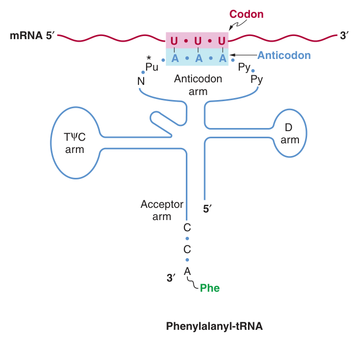
A specific sequence in a tRNA molecule, complementary to a codon.
The anticodon region (arm) consists of seven nucleotides, and it recognizes the three-letter codon in mRNA. The sequence read from the 3′ to 5′ direction in that anticodon loop consists of a variable base (N) — modified purine (Pu*) — XYZ (the anticodon) — pyrimidine (Py) — pyrimidine (Py) — 5′.
Note that this direction of reading the anticodon is 3′→5′, whereas the genetic code is read 5′→3′, since the codon and the anticodon loop of the mRNA and tRNA molecules respectively are ANTIPARALLEL in their complementarity, just like all other intermolecular interactions between nucleic acid strands.
The acceptor arm, at the 3′-hydroxyl adenosyl terminal, is the site of attachment of the specific amino acid.
The TψC arm (ribothymidine-pseudouridine-cytidine) is involved in binding of the aminoacyl-tRNA to the ribosomal surface at the site of protein synthesis.
The D arm is one of the sites important for the proper recognition of a given tRNA species by its proper aminoacyl-tRNA synthetase.
The degeneracy of the genetic code resides mostly in the last nucleotide of the codon triplet, suggesting that the base pairing between this last nucleotide and the corresponding nucleotide of the anticodon is not strictly by the Watson-Crick rule. This is called WOBBLE; the pairing of the codon and anticodon can “wobble” at this specific nucleotide-to-nucleotide pairing site.
For example, the two codons for arginine, AGA and AGG, can bind to the same anticodon.
Inosine (I) — another of the peculiar bases appearing in tRNA molecules — is the classic wobble base, because it can pair with U, C or A.
Do the arithmetic. There are 61 sense codons. Strict Watson-Crick pairing would demand 61 different tRNAs. Yet the cytoplasmic translation system possesses a full complement of 31 tRNA species, and mitochondria require only 22.
Wobble closes that gap. Because the third base pairs loosely, one tRNA can read two, three, or in the mitochondrial case four codons. The cell carries roughly half the tRNAs it would otherwise need.
And now notice that the two facts are the same fact. Degeneracy resides mostly in the third base, and wobble occurs at the third base. The redundancy of the code and the economy of the tRNA population are two descriptions of one molecular property. The code is degenerate BECAUSE the third pairing is loose.
- Define an anticodon → A specific tRNA sequence complementary to a codon in mRNA
- How many nucleotides in the anticodon arm? → Seven, of which three are the anticodon
- In which direction is the anticodon read? → 3′→5′, because codon and anticodon are antiparallel
- Where does wobble occur? → At the third (last) nucleotide of the codon
- Which unusual base makes wobble possible? → Inosine
- How many cytoplasmic tRNA species are there? → 31, for 61 codons
Charging the tRNA ★★★
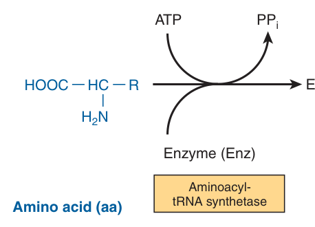
These enzymes are termed aminoacyl-tRNA synthetases. They form an activated intermediate of aminoacyl-AMP-enzyme complex.
The amino acid remains attached to its specific tRNA in an ESTER linkage.
The charging reactions have an error rate of less than 10⁻⁴ and so are extremely accurate.
Here is the fact that makes accuracy at this step non-negotiable: the ribosome inspects the codon-anticodon pairing, and nothing else. It has no way to verify that the amino acid hanging off the tRNA is the right one.
The classic demonstration is chemical. Charge a cysteine-tRNA with cysteine, then chemically reduce the cysteine to alanine while it sits on the tRNA. The ribosome inserts alanine wherever a cysteine codon appears. It read the anticodon and asked no further questions.
So the fidelity of the genetic code is established entirely by the synthetases — which is why their error rate of less than 10⁻⁴ matters, why there is one synthetase per amino acid, and why they possess proofreading sites of their own. The code is enforced at charging, not at the ribosome.
- Which enzymes charge tRNA? → Aminoacyl-tRNA synthetases
- What intermediate do they form? → An aminoacyl-AMP-enzyme complex
- What kind of linkage attaches the amino acid? → An ester linkage
- Their error rate? → Less than 10⁻⁴
- Where is the fidelity of the code enforced? → At charging — the ribosome only checks codon-anticodon pairing
Initiation ★★★
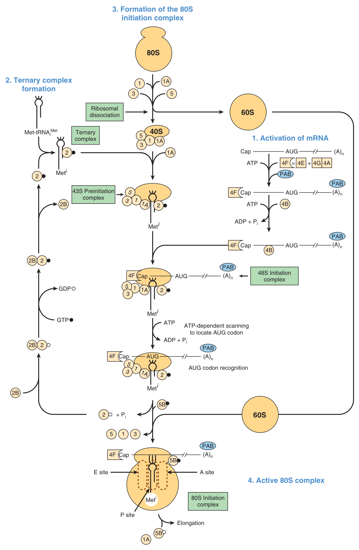
Initiation can be divided into four steps:
A · Ribosomal dissociation — dissociation of the ribosome into its 40S and 60S subunits.
B · Formation of the 43S preinitiation complex — the binding of GTP by eIF-2; this binary complex binds to met-tRNAi, a tRNA specifically involved in binding to the initiation codon AUG. eIF-2 consists of α, β and γ subunits.
C · Formation of the 43S initiation complex — binding of mRNA to the 40S preinitiation complex. The 5′ terminals of most mRNA molecules in eukaryotic cells are “capped”, and this helps direct the 40S ribosomal subunit to the 5′ end of the mRNA. Context matters: most preferred is the presence of a purine at positions −3 and +4 relative to the AUG.
D/E · Formation of the 80S initiation complex — the rapid association of the 40S and 60S subunits to form the 80S ribosome, with the met-tRNAi on the P site of the ribosome, ready for the elongation cycle to commence.
The cap and poly(A) tail structures have a synergistic effect on protein synthesis.
The 4F complex is particularly important in controlling the rate of protein translation, and the regulation of eIF-4E controls the rate of initiation — a component of the MAP kinase pathway appears to be involved in this phosphorylation reaction.
eIF-2 is a control point. PKR is particularly interesting in this regard.
Phosphorylated eIF-2α binds tightly to and inactivates the GTP-GDP recycling protein eIF-2B. This prevents formation of the 43S preinitiation complex and blocks protein synthesis.
Look carefully at the eIF-2 mechanism, because its logic is unusual and the exam rewards explaining it.
eIF-2 must be recycled from its GDP form back to GTP after every initiation event, and eIF-2B does that recycling. Crucially, eIF-2B is present in much smaller amounts than eIF-2. So phosphorylating even a modest fraction of eIF-2α produces enough inhibitor to sequester essentially all the eIF-2B — and without recycling, every eIF-2 molecule is stranded as the GDP form.
A partial modification of the abundant protein completely inactivates the scarce one. It is an amplifier working in reverse.
The physiology follows. PKR is activated by double-stranded RNA — the signature of a viral infection. An infected cell shuts down its entire translational machinery, denying the virus the ribosomes it has no choice but to borrow. The cell sacrifices itself. This is why the section on viruses (§10) belongs in the same unit: viruses co-opt the host cell's protein synthesis machinery, and the host's defence is to switch that machinery off.
- The four steps of initiation? → Ribosomal dissociation; 43S preinitiation complex; 43S initiation complex; 80S initiation complex
- What does eIF-2 bind? → GTP, then met-tRNAi
- Which subunits has eIF-2? → α, β and γ
- How does phosphorylated eIF-2α block translation? → It binds and inactivates eIF-2B, the GTP-GDP recycling protein
- Where does met-tRNAi sit in the 80S complex? → The P site
- What directs the 40S subunit to the mRNA? → The 5′ cap
Elongation ★★★
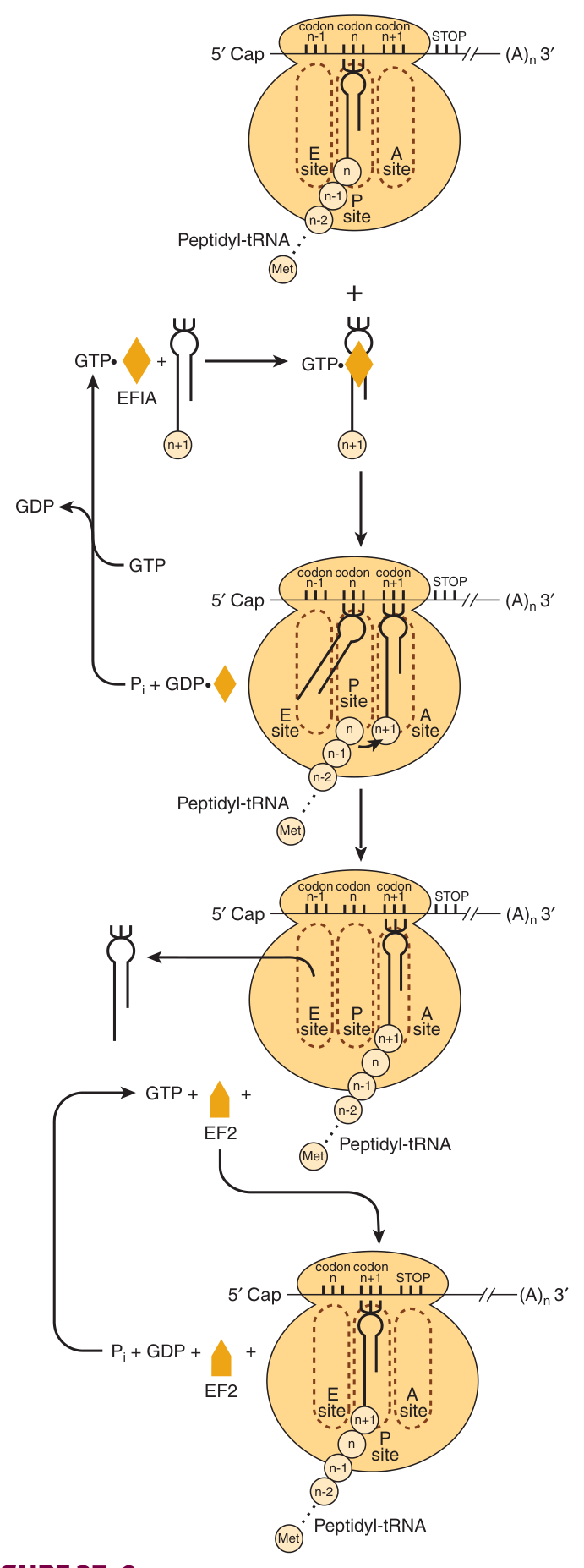
Elongation also is a multistep process.
A · Binding of aminoacyl-tRNA to the A site. At initiation, this site is occupied by aminoacyl-tRNA met-i.
B · Peptide bond formation, catalysed by peptidyl transferase — which is a ribozyme, an RNA catalyst, not a protein.
C · Translocation. The EF2-GTP complex is hydrolyzed to EF2-GDP, effectively moving the mRNA forward by one codon. Afterwards the deacylated tRNA is attached to the P site and by its open CCA tail to an exit (E) site, from which it leaves the ribosome.
A site — Aminoacyl. Where the incoming charged tRNA binds.
P site — Peptidyl. Holds the tRNA carrying the growing chain.
E site — Exit. Where the deacylated tRNA leaves.
The whole ribosome comprises a large subunit and a small subunit, with the mRNA threaded between them and the growing polypeptide emerging through an exit tunnel.
The single most important chemical reaction in the cell — the formation of the peptide bond — is catalysed by RNA, not protein. That fact was a genuine surprise, and it is worth understanding rather than merely reciting.
It resolves an obvious circularity. Proteins are made by ribosomes; if ribosomes were made of protein, then proteins would be needed to make the first proteins. RNA escapes the loop, because it can carry information AND catalyse — which is the basis of the RNA world hypothesis about the origin of life.
Note that you already met the other great example in Unit 24: the ribozymes involved in RNA splicing. The two most fundamental reactions in gene expression — cutting RNA and joining amino acids — are both done by RNA.
- The three elongation steps? → Aminoacyl-tRNA binding to the A site, peptide bond formation, translocation
- Which enzyme forms the peptide bond, and what is unusual about it? → Peptidyl transferase — a ribozyme
- Which factor drives translocation? → EF2, with GTP hydrolysis to EF2-GDP
- Name the three ribosomal sites → A (aminoacyl), P (peptidyl), E (exit)
Termination and the energy cost ★★★
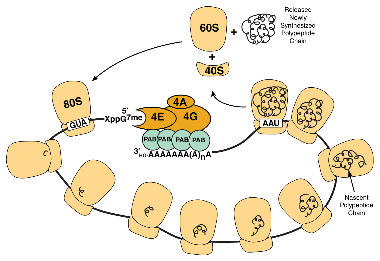
Termination occurs when a stop codon is recognized. The mRNA is read continuously from a start codon (AUG) to a termination codon (UAA, UAG, UGA).
A water molecule is added. This hydrolysis releases the protein and the tRNA from the P site. Then the 80S ribosome dissociates into its 40S and 60S subunits, which are recycled.
Hydrolysis of FOUR high-energy phosphate bonds is required per peptide bond formed.
Two at charging — the aminoacyl-tRNA synthetase converts ATP to AMP + PPi, and the pyrophosphate is hydrolysed.
One for delivery of the aminoacyl-tRNA to the A site (EF1-GTP).
One for translocation (EF2-GTP).
Four high-energy bonds per peptide bond, in a protein of 300 residues, is 1200 ATP equivalents for one molecule — before any folding, modification or transport. A growing cell may spend the majority of its energy budget here.
Why pay so much? Because the price buys accuracy. Each hydrolysis step is a point at which a wrong intermediate can be rejected — the energy is spent driving reactions irreversibly forward so that errors are discarded rather than accumulated. The same logic as the two-phosphate cost of activating a fatty acid in Unit 17: irreversibility is purchased, and here it buys fidelity.
It also explains a fact from Unit 22: in starvation, protein synthesis falls sharply. Not only is substrate scarce — the process itself is unaffordable.
Polysomes are assemblies of ribosomes — several ribosomes translating the same mRNA simultaneously, each at a different point along it.
This multiplies output without any increase in transcription: one message, many copies of the protein at once.
- The three stop codons? → UAA, UAG, UGA
- What terminates the chain chemically? → Addition of a water molecule, hydrolysing the protein from the P-site tRNA
- How many high-energy phosphate bonds per peptide bond? → Four
- Where do they go? → Two at charging, one for A-site delivery, one for translocation
- What is a polysome? → An assembly of ribosomes translating one mRNA at once
Mutations ★★★
| Type | What happens |
|---|---|
| Silent (acceptable) | The base change does not alter the amino acid, because the code is degenerate — some mutations have no apparent effect |
| Missense | Substitution of one amino acid for another. May be acceptable, partially acceptable or unacceptable depending on the residue and its position |
| Nonsense | A sense codon becomes a stop codon; the chain is truncated |
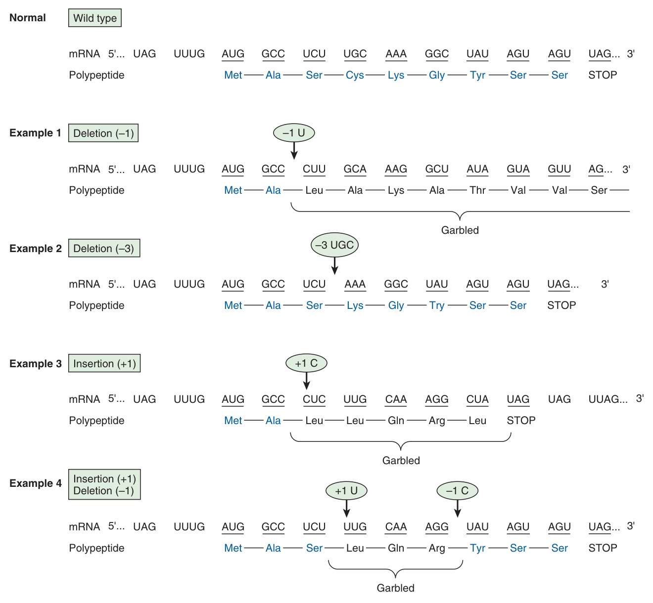
| Frameshift | Insertion or deletion of a number of bases not divisible by three; since the code is not punctuated, everything downstream is misread |
Hemoglobin illustrates the effects of single-base changes in structural genes. At position 67 of the β chain, normal haemoglobin has a valine (GUU, GUC, GUA, GUG). Single-base changes give:
Hemoglobin Milwaukee — a glutamic acid (GAA, GAG)
Hemoglobin Bristol — an aspartic acid (GAU, GAC)
Hemoglobin Sydney — an alanine (GCU, GCC, GCA, GCG)
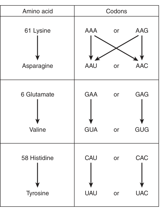
Substitution of amino acids causes MISSENSE mutations, and some mutations have no apparent effect — classified as acceptable missense mutations.
Compare the damage. A missense mutation changes one residue, and thanks to degeneracy and to the chemical clustering of similar codons, it is often tolerated — the haemoglobin variants above make the point.
A frameshift changes every residue downstream. And the reason is a feature you learned in §2: once the reading is commenced at a specific codon, there is no punctuation between codons. Nothing marks where a codon begins except the position of the start codon. Shift by one base and the ribosome reads a completely different set of triplets, until it meets a stop codon in the new frame — usually within a few dozen residues.
The absence of punctuation, which makes the code compact, is exactly what makes frameshifts catastrophic. Every feature has a cost, and this is the cost of that one.
- Why can a base change be silent? → The code is degenerate
- Define missense → Substitution of one amino acid for another
- Define nonsense → A sense codon becomes a stop codon, truncating the chain
- Why is a frameshift so damaging? → The code is unpunctuated, so everything downstream is misread
- Hb Milwaukee, Bristol and Sydney all substitute what, and where? → Valine at position 67 of the β chain — by glutamic acid, aspartic acid and alanine respectively
Post-translational processing ★★
Post-translational processing affects the activity of many proteins.
The modifications you have already met in this course:
Proteolytic cleavage — zymogen activation (Unit 7): trypsinogen → trypsin, pepsinogen → pepsin, proinsulin → insulin.
Hydroxylation — 4-hydroxyproline and 5-hydroxylysine in collagen (Unit 1), requiring vitamin C.
Glycosylation — of secreted and membrane proteins; loss of a sialic acid marks a plasma glycoprotein for degradation (Unit 20).
Phosphorylation — the reversible covalent modification of Unit 7, controlling glycogen phosphorylase, acetyl-CoA carboxylase, hormone-sensitive lipase and HMG-CoA reductase.
Ubiquitination — targeting for proteasomal degradation (Unit 20).
Formation of disulfide bonds, and proline cis-trans isomerisation during folding (Unit 3).
A cell that synthesised active trypsin would digest itself. A cell that synthesised active insulin in the wrong place would empty its own blood glucose. So a great many proteins are made deliberately inactive and switched on only at the right time and place.
Look at what that buys. Synthesis is slow — four high-energy bonds per residue and minutes per protein — whereas activation by cleavage or phosphorylation is fast, and needs no new synthesis at all. Making the protein in advance and activating it on demand separates the expensive step from the urgent one.
That is the same principle you met at the digestive zymogens, the clotting cascade, and the cAMP cascade of Unit 12. Post-translational processing is where the speed of regulation comes from.
- Name three post-translational modifications → Proteolytic cleavage, glycosylation, phosphorylation (also hydroxylation, ubiquitination, disulfide formation)
- Which vitamin is needed for collagen hydroxylation? → Vitamin C
- What does ubiquitination signal? → Degradation in the proteasome
- Why are zymogens made? → So that a destructive enzyme can be synthesised safely and activated only where needed
Antibiotics and toxins ★★★
Many antibiotics work because they selectively inhibit protein synthesis in bacteria. The selectivity comes from the difference between the 70S bacterial ribosome (50S + 30S) and the 80S eukaryotic ribosome (60S + 40S) — different enough that a drug can bind one and not the other.
| Agent | Action |
|---|---|
| Tetracycline | Prevents the binding of aminoacyl-tRNAs to the A site |
| Chloramphenicol | Inhibits peptidyl transferase of the bacterial 50S subunit |
| Erythromycin (macrolides) | Blocks translocation on the 50S subunit |
| Streptomycin (aminoglycosides) | Binds the 30S subunit, causing misreading of mRNA |
| Puromycin | An analogue of aminoacyl-tRNA; enters the A site, accepts the peptide and causes premature chain release — inhibits BOTH prokaryotic and eukaryotic synthesis, so it is a research tool, not a drug |
| Diphtheria toxin | ADP-ribosylates and inactivates EF2 in eukaryotes, blocking translocation |
| Ricin | Inactivates the eukaryotic 60S subunit by depurinating 28S rRNA |
Viruses replicate by using host cell processes, including those involved in protein synthesis; viruses co-opt the host cell's protein synthesis machinery because they possess no ribosomes of their own.
The machinery of protein synthesis can respond to environmental threats — the PKR/eIF-2α mechanism of §5 — but the protein synthesis machinery can also be modified in deleterious ways, and many viruses have evolved to disable the host's cap-dependent initiation while translating their own messages by other means.
Both block protein synthesis. The difference is entirely one of selectivity, and it is the central principle of antimicrobial chemotherapy.
Tetracycline discriminates: it prevents aminoacyl-tRNA binding at the bacterial A site and largely spares the human ribosome. That difference is the therapeutic window.
Puromycin does not discriminate, because it is an analogue of the aminoacyl end of a tRNA — a feature every ribosome shares. It enters any A site, accepts the growing peptide onto itself, and then simply falls off, releasing a truncated chain. Nothing about that mechanism could be selective, so it kills the patient as efficiently as the pathogen.
A useful antibiotic exploits a difference; a research tool exploits a similarity. Note that diphtheria toxin is the mirror image — exquisitely selective for the eukaryotic machinery, which is precisely what makes it a toxin.
- Bacterial vs eukaryotic ribosome? → 70S (50S + 30S) vs 80S (60S + 40S)
- Tetracycline blocks? → Binding of aminoacyl-tRNAs to the A site
- Chloramphenicol blocks? → Peptidyl transferase of the 50S subunit
- Why is puromycin not therapeutic? → It mimics aminoacyl-tRNA and so acts on all ribosomes
- Diphtheria toxin acts how? → ADP-ribosylates and inactivates eukaryotic EF2
Revision layer
The central dogma, one line
The flow of genetic information follows the sequence DNA → RNA → protein. The mRNA is read continuously from a start codon (AUG) to a termination codon (UAA, UAG, UGA).
Numbers to have ready
| Quantity | Value |
|---|---|
| Possible codons | 64 (4³); two-base codons would give only 16 (4²) |
| Codons specifying amino acids | 61 |
| Nonsense (stop) codons | 3 — UAA, UAG, UGA |
| Codons for serine | 6 — UCU, UCC, UCA, UCG, AGU, AGC |
| Codons for Met and Trp | 1 each |
| Cytoplasmic tRNA species | 31 |
| Mitochondrial tRNA species | 22 |
| Anticodon arm | 7 nucleotides |
| Charging error rate | < 10⁻⁴ |
| High-energy bonds per peptide bond | 4 |
| Ribosomes | Bacterial 70S = 50S + 30S; eukaryotic 80S = 60S + 40S |
The stages, in order
| Stage | Key events |
|---|---|
| Charging | Aminoacyl-tRNA synthetase forms an aminoacyl-AMP-enzyme intermediate; the amino acid is attached by an ester linkage; error rate < 10⁻⁴ |
| Initiation | Ribosomal dissociation → 43S preinitiation complex (eIF-2·GTP·met-tRNAi) → 43S initiation complex (mRNA binds, cap-directed) → 80S initiation complex, met-tRNAi in the P site |
| Elongation | Aminoacyl-tRNA to the A site → peptide bond by peptidyl transferase (a ribozyme) → translocation by EF2-GTP; deacylated tRNA leaves via the E site |
| Termination | Stop codon recognised; a water molecule is added, releasing protein and tRNA; the 80S dissociates into 40S and 60S, recycled |
Definitions the examiner has actually set
| Term | One-line answer |
|---|---|
| Degeneracy | Multiple codons decode the same amino acid |
| Anticodon | A specific tRNA sequence, complementary to a codon; the arm has 7 nucleotides and is read 3′→5′ |
| Wobble | Non-Watson-Crick pairing at the third codon base, letting one tRNA read several codons |
The five features: “Dear Uncle Ned, Never Understand” — Degenerate, Unambiguous, Nonoverlapping, Not punctuated, Universal.
The ribosomal sites in order: A-P-E — arrive, peptide, exit. The tRNA moves through them in alphabetical order.
The stop codons: U Are Away, U Are Gone, U Go Away — UAA, UAG, UGA.
- Why is the code a triplet? → 4² = 16 is too few for 20 amino acids; 4³ = 64 suffices
- Define degeneracy → Multiple codons decode the same amino acid
- Degenerate vs ambiguous? → The code is degenerate but unambiguous — one codon always means one amino acid
- Define an anticodon → A specific tRNA sequence complementary to a codon, read 3′→5′
- Where is wobble, and why does it matter? → At the third codon base; it lets 31 tRNAs read 61 codons
- Which enzymes enforce the fidelity of the code? → The aminoacyl-tRNA synthetases — the ribosome checks only codon-anticodon pairing
- Which catalyses the peptide bond? → Peptidyl transferase, a ribozyme
- How many high-energy phosphate bonds per peptide bond? → Four
- How does PKR shut down translation? → It phosphorylates eIF-2α, which sequesters and inactivates eIF-2B
- Why is puromycin not a therapeutic antibiotic? → It mimics aminoacyl-tRNA, so it inhibits eukaryotic ribosomes too