RNA Synthesis, Processing & Modification
RNA versus DNA ★★★
| DNA | RNA | |
|---|---|---|
| Sugar | 2′-deoxyribose | Ribose |
| Pyrimidines | Cytosine, thymine | Cytosine, uracil |
| Strandedness | Double-stranded | Usually single-stranded |
| Base ratios | A = T and G = C (Chargaff) | Its guanine content does not necessarily equal its cytosine content, nor does its adenine content necessarily equal its uracil content |
| Alkali | Stable | Can be hydrolyzed by alkali to 2′,3′ cyclic diesters of the mononucleotides — useful both diagnostically and analytically |
| Catalysis | None | Some RNA molecules have intrinsic catalytic activity — RIBOZYMES |
The single structural difference — a 2′-OH on the ribose — accounts for almost every entry in that table, and it is worth tracing.
Alkaline hydrolysis: the 2′-OH sits right beside the phosphodiester bond and, when deprotonated, attacks it — forming the 2′,3′ cyclic diester Harper's names. DNA, lacking that hydroxyl, cannot do this. RNA is chemically self-destructive; DNA is not.
Which is why DNA is the archive and RNA the working copy. A molecule that must survive for the lifetime of an organism cannot carry a built-in cleavage mechanism; a message that must be made, used and disposed of benefits from one.
Catalysis: the same reactive hydroxyl, plus the single-stranded chain's freedom to fold, is what lets RNA act as an enzyme at all. Two ribozymes are the peptidyl transferase that catalyzes peptide bond formation on the ribosome, and ribozymes involved in RNA splicing — the two most fundamental reactions in this module are both catalysed by RNA, not protein.
Thymine is simply 5-methyluracil. The methyl group is a tag marking the base as belonging in DNA.
It matters because cytosine spontaneously deaminates to uracil. If uracil were a normal DNA base, that damage would be invisible; because it is not, a repair glycosylase can recognise every uracil in DNA as an error and excise it. The methyl group is the difference between a repairable lesion and a silent mutation.
- Sugar difference? → 2′-deoxyribose in DNA, ribose in RNA
- Base difference? → Thymine in DNA, uracil in RNA
- Does Chargaff's rule apply to RNA? → No — G need not equal C, nor A equal U
- What does alkali do to RNA? → Hydrolyses it to 2′,3′ cyclic diesters of the mononucleotides
- What is a ribozyme? → An RNA molecule with intrinsic catalytic activity — e.g. peptidyl transferase
The classes of RNA ★★★
| Class | Function |
|---|---|
| Messenger RNA (mRNA) | Carries the coding sequence from gene to ribosome; the least stable and most heterogeneous class |
| Transfer RNA (tRNA) | The adapter molecule — recognises a codon through its anticodon and carries the corresponding amino acid |
| Ribosomal RNA (rRNA) | The structural and catalytic core of the ribosome; the most abundant class |
| Small nuclear RNAs (snRNAs) | rRNA and mRNA processing and gene regulation — the components of the spliceosome |
| Micro-RNAs (miRNAs) | Silence mRNAs by annealing to their 3′ untranslated regions |
| Small interfering RNAs (siRNAs) | RNA interference; protect the host from RNA viruses |
Harper's compares them on four axes: abundance, size, function and general stability.
rRNA is the most abundant and the most stable; mRNA is the least stable, which is exactly what a regulatory message should be — a signal that persisted indefinitely could not be switched off.
The cloverleaf, with four arms:
The acceptor arm — the 3′-CCA-OH terminus, the site of attachment of the specific amino acid.
The anticodon arm — consists of seven nucleotides and recognises the three-letter codon in mRNA.
The TψC arm — involved in binding of the aminoacyl-tRNA to the ribosomal surface at the site of protein synthesis.
The D arm — one of the sites important for the proper recognition of a given tRNA species by its proper aminoacyl-tRNA synthetase.
tRNA is rich in nontraditional nucleotides introduced by post-transcriptional modification — methylated guanosines, pseudouridine (ψ), inosine and others.
- Name the four principal RNA classes → mRNA, tRNA, rRNA and the small RNAs
- Which is most abundant? → rRNA. Least stable? → mRNA
- Where does the amino acid attach to tRNA? → The 3′-CCA-OH of the acceptor arm
- How many nucleotides in the anticodon arm? → Seven, of which three are the anticodon
- What do snRNAs do? → rRNA and mRNA processing and gene regulation — the spliceosome
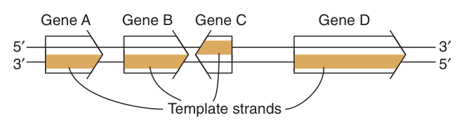
The central dogma ★★
RNA biosynthesis from a DNA template is called transcription. Its products are mRNA, tRNA and rRNA.
The synthesis of an RNA molecule from DNA is a complex process involving one of the group of RNA polymerase enzymes and a number of associated proteins. The general steps required to synthesize the primary transcript are initiation, elongation and termination.
| Prokaryotes | Eukaryotes | |
|---|---|---|
| The primary transcript | Equivalent to the mRNA molecule | A precursor (pre-mRNA) to the mRNA |
| Processing | Essentially none for mRNA | Modified at both ends, and introns are removed; occurs primarily within the nucleus |
| Coupling | Translation can begin before transcription is complete | Separated in space — after processing, the mRNA is exported to the cytoplasm for translation |
| Polymerases | One RNA polymerase | Three distinct nuclear polymerases |
In a bacterium, ribosomes attach to the 5′ end of an mRNA while its 3′ end is still being transcribed. That is fast and economical — but it makes splicing impossible. You cannot cut an intron out of a message that is already being read.
The nuclear membrane is what buys the time. By separating transcription from translation in space, it creates a compartment in which the transcript can be capped, polyadenylated, spliced and inspected before any ribosome sees it. Everything in §8 and §9 depends on that separation.
The cost is speed; the return is alternative splicing, and hence many proteins from one gene. Errors or changes in synthesis, processing, splicing, stability or function of mRNA transcripts are a cause of disease — a whole class of pathology that prokaryotes simply cannot have.
- Define transcription → RNA biosynthesis from a DNA template
- In prokaryotes, what is the primary transcript equivalent to? → The mRNA itself
- In eukaryotes? → Pre-mRNA, a precursor requiring processing
- Where does eukaryotic processing occur? → Primarily within the nucleus
The promoter and the transcription unit ⭐
A promoter is the DNA sequence to which RNA polymerase binds to initiate transcription of a gene.
DNA-dependent RNA polymerase attaches at this specific site on the template strand. This is followed by initiation of RNA synthesis at the starting point, and the process continues until a termination sequence is reached.
The promoter determines two things: where transcription is to commence along the DNA, and how frequently this event is to occur.
A transcription unit is the region of DNA that includes the signals for transcription initiation, elongation and termination.
Position +1 is the transcript initiation site.
Upstream sequences (negative numbers) — the promoter.
Downstream sequences — introns and exons.
The RNA product is the primary transcript.
Consider the search problem. E. coli has 4 × 10³ transcription initiation sites in 4.2 × 10⁶ base pairs of DNA, and humans have about 10⁵ promoters in 3 × 10⁹ base pairs. The polymerase must find a few thousand specific addresses among millions of possible ones.
It does so by a strategy worth stating in an exam: RNA polymerase can bind, with low affinity, to many regions of DNA, but it scans the DNA sequence — at a rate of ≥10³ bp per second — until it recognizes certain specific regions to which it binds with higher affinity.
So a promoter is not a lock that only one key opens; it is a region of unusually high binding affinity in a sequence the polymerase is already sliding along. And because binding affinity is a continuous quantity, a stronger promoter is transcribed more often — which is how the same sequence element sets both where and how much.
- Define a promoter → The DNA sequence to which RNA polymerase binds to initiate transcription of a gene
- Define a transcription unit → The region of DNA that includes the signals for initiation, elongation and termination
- What is position +1? → The transcript initiation site
- The two things a promoter determines? → Where transcription commences, and how frequently
- How does polymerase find a promoter? → It binds DNA weakly and scans at ≥10³ bp/s until it meets a higher-affinity region
RNA polymerase ★★★
The enzyme responsible for the polymerization of ribonucleotides into a sequence complementary to the template strand of the gene.
Four features distinguish it from DNA polymerase, and all four are examinable:
1 · It adheres to Watson-Crick base-pairing rules, using ATP, GTP, CTP and UTP — U replacing T.
2 · It synthesises with 5′→3′ polarity, reading the template strand in the 3′→5′ direction.
3 · A primer is NOT involved in RNA synthesis, as RNA polymerases have the ability to initiate synthesis de novo.
4 · Initiation requires large, multicomponent initiation complexes.
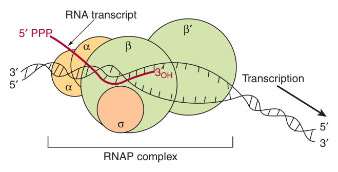
Core enzyme: α₂ββ′. Holoenzyme: α₂ββ′σ.
Functions of the subunits:
α — assembly of the tetrameric core
β — ribonucleoside triphosphate binding site
β′ — DNA template binding region
σ — helps the core enzyme recognize and bind to the promoter region
The transcription “bubble” is 20 bp of DNA, and the entire complex covers 30–75 bp.
Mammalian cells possess three distinct nuclear DNA-dependent RNA polymerases.
Pol I — most rRNA
Pol II — mRNA and most snRNAs and miRNAs
Pol III — tRNA and 5S rRNA
α-Amanitin is a specific differential inhibitor of the eukaryotic nuclear DNA-dependent RNA polymerases and as such has proved to be a powerful research tool — it blocks the translocation of RNA polymerase during phosphodiester bond formation. It distinguishes the three because they differ in sensitivity to it.
Notice the division of labour in the bacterial enzyme. The core can polymerise but cannot find a promoter; σ helps the core enzyme recognize and bind to the promoter region and is then released.
Making promoter recognition a detachable function is what makes bacterial gene regulation possible. Swap one σ factor for another and the same core polymerase transcribes an entirely different set of genes — heat-shock genes, sporulation genes, and so on. One catalytic machine, many programmes.
The eukaryotic solution to the same problem is different in form but identical in logic: instead of interchangeable σ factors, all eukaryotic RNA polymerase forms require other proteins known as general transcription factors (GTFs), and it is these — not the polymerase — that recognise the promoter.
- Does RNA synthesis need a primer? → No — RNA polymerases initiate de novo
- In which direction is the template read? → 3′→5′, while RNA is made 5′→3′
- Bacterial core enzyme vs holoenzyme? → α₂ββ′ vs α₂ββ′σ
- What does σ do? → Helps the core recognise and bind the promoter
- Size of the transcription bubble? → 20 bp; the whole complex covers 30–75 bp
- Which polymerase makes mRNA? → Pol II
- What is α-amanitin? → A differential inhibitor of the eukaryotic nuclear polymerases, blocking translocation
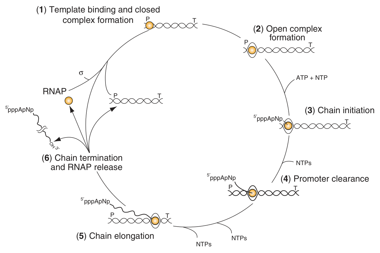
Initiation, elongation, termination ★★★
Initiation: RNA polymerase binds to the promoter of DNA, and then a transcription “bubble” is formed. The holoenzyme must bind DNA and locate a promoter; then comes localized unwinding of the two strands by RNA polymerase to provide a single-stranded template, and formation of phosphodiester bonds between the first few ribonucleotides in the nascent RNA chain. The unwound complex is the preinitiation complex (PIC).
Elongation: the polymerase catalyzes formation of 3′,5′-phosphodiester bonds in the 5′→3′ direction, using NTPs as building units. The nascent chain is attached to the polymerization site on the β subunit.
Termination: when the polymerase reaches a termination sequence on DNA, the reaction stops and the newly synthesized RNA is released.
RNA polymerase continues to incorporate nucleotides 3 to ~10, at which point the polymerase undergoes another conformational change and moves away from the promoter; this reaction is termed PROMOTER CLEARANCE.
Until it happens, the polymerase repeatedly makes and releases very short abortive transcripts. Promoter clearance is therefore the commitment step of transcription.
Intrinsic terminators. The predominant bacterial transcription termination signal contains an inverted, hyphenated repeat followed by a stretch of AT base pairs. The inverted repeat, when transcribed into RNA, generates a secondary structure — an RNA hairpin — which causes RNA polymerase to pause. The weak rU:dA hybrid that follows then lets the transcript fall off. About 50% of genes use an inverted palindrome plus poly-A.
Rho-dependent termination. Rho is an ATP-dependent, RNA-stimulated helicase that disrupts the ternary transcription elongation complex composed of RNA polymerase, nascent RNA and DNA. It interacts with the paused polymerase and induces chain termination.
It looks improbable that a fold in the product could halt the machine making it, but the geometry makes it inevitable.
The nascent RNA emerges from an exit channel in the polymerase. An inverted repeat — a sequence that reads the same on both strands — transcribes into RNA that can base-pair with itself, forming a hairpin. That hairpin is too bulky for the channel, and forming it physically wrenches RNA out of the enzyme, causing RNA polymerase to pause.
Then the stretch of AT base pairs does the rest. The RNA:DNA hybrid holding transcript to template at that point is rU:dA — the weakest hybrid there is. Stall the enzyme over the weakest possible grip and the transcript simply lets go.
A sequence, transcribed, becomes a mechanical device. That is a genuinely elegant piece of design, and it is worth being able to explain rather than merely name.
- The three stages of transcription? → Initiation, elongation, termination
- What is promoter clearance? → After ~3–10 nucleotides the polymerase changes conformation and moves away from the promoter
- The two bacterial termination mechanisms? → Intrinsic (hairpin + AT stretch) and rho-dependent
- What is rho? → An ATP-dependent, RNA-stimulated helicase that disrupts the elongation complex
- Which subunit carries the polymerisation site? → β
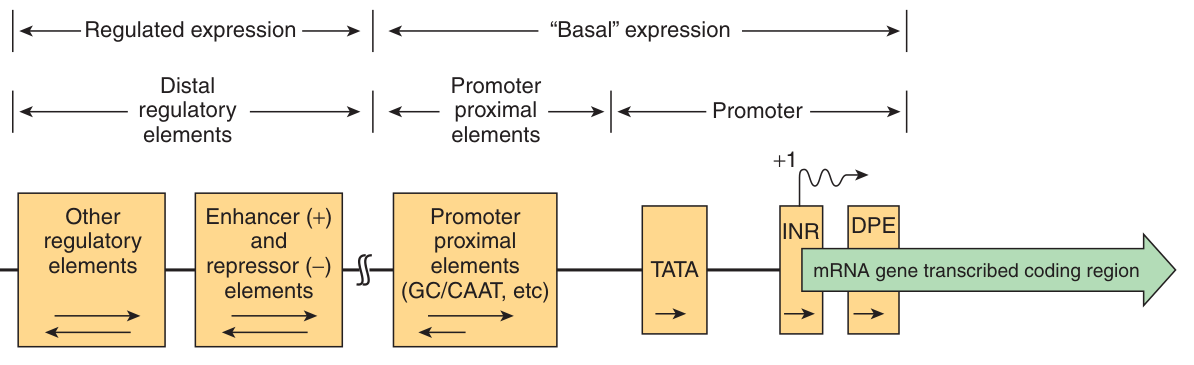
Eukaryotic promoters and transcription factors ★★★
Bacterial promoters are simple: approximately 40 nucleotides in length, with an eight-nucleotide-pair sequence about 35 bp upstream of the transcription start site and a six-nucleotide-pair A+T-rich sequence about 10 nucleotides upstream — the classical −35 and −10 (Pribnow) boxes. Eukaryotic promoters are more complex, and are built from three classes of element.
| Class | Position | Elements |
|---|---|---|
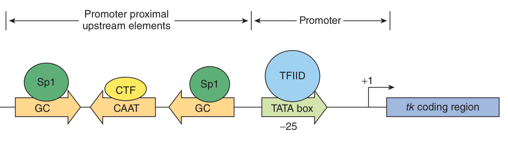
| The promoter proper | At and around +1 | TATA box, initiator sequence (Inr), downstream promoter element (DPE). The TATA box has a particularly rigid requirement for both position and orientation. |
| Promoter-proximal elements | 50–200 bp upstream | Sequence elements bound by specific transcription factors, setting the frequency of initiation |
| Distal elements | 1000–10⁵ bp away | Enhancers and repressors (silencers) — a third class that can either increase or decrease the rate of transcription initiation |
TATA box and Inr: 30% · Inr alone: 30% · Inr and DPE: 25% · all three elements: 15%.
Note what this means: the TATA box is present in only a minority of genes. The textbook picture of “every eukaryotic promoter has a TATA box” is wrong.
cis-acting elements are DNA sequences on the same molecule as the gene they control — promoters, enhancers, silencers.
trans-acting factors are diffusible proteins that bind them — the transcription factors.
Transcription factors have two functional parts: DNA-binding domains (DBDs) and activation domains (ADs).
A complex consisting of 50 unique proteins provides accurate and regulatable transcription of eukaryotic genes.
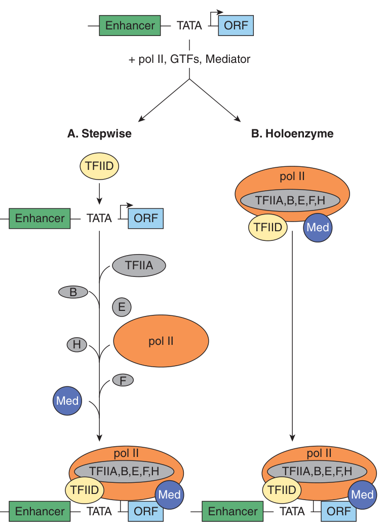
RNA polymerase II requires TFIIA, B, D (or TBP), E, F and H to both facilitate promoter-specific binding of the enzyme and formation of the preinitiation complex (PIC). RNA polymerases I and III require their own polymerase-specific GTFs.
TFIID binds to the TATA box promoter element through its TATA-binding protein (TBP) subunit; TFIID consists of 15 subunits — TBP and 14 TBP-associated factors (TAFs).
Crucially: RNA polymerase II and the GTFs can only catalyze BASAL or UNREGULATED transcription in vitro. Regulated transcription needs more.
The coactivators, or coregulators, work in conjunction with the DNA-binding transactivator proteins to communicate with Pol II and the GTFs to regulate the rate of transcription.
Promoter accessibility, and hence PIC formation, is often modulated by nucleosomes: nucleosome eviction by chromatin-active coregulators facilitates PIC formation and transcription. The machinery therefore includes Mediator, chromatin remodellers and chromatin modifying factors alongside the polymerase and GTFs.
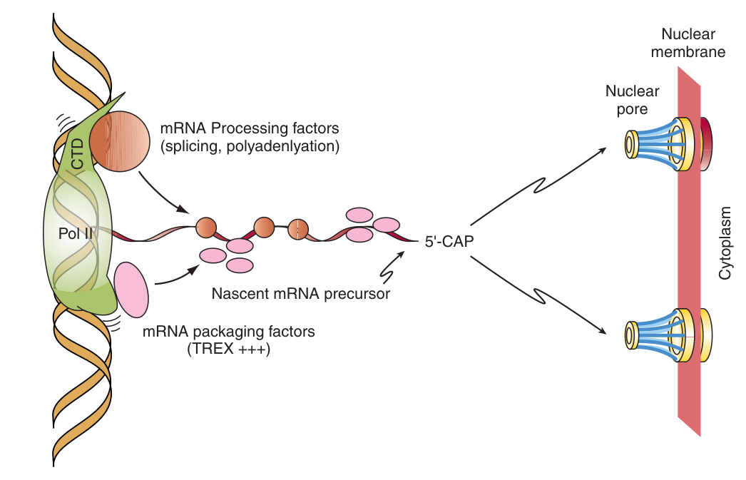
Eukaryotic pol II consists of 12 subunits. The largest carries, at its carboxyl terminus, a carboxyl terminal repeat domain (CTD) with the consensus sequence Tyr-Ser-Pro-Thr-Ser-Pro-Ser, repeated many times.
The CTD is a substrate for several enzymes, and CTD phosphorylation/dephosphorylation is critical for promoter clearance, elongation, termination, and even appropriate mRNA processing.
Ask what the cell needs. Capping must happen immediately after initiation, splicing during elongation, and polyadenylation at termination. Each processing enzyme must arrive at exactly the right moment.
The CTD solves this by acting as a moving scaffold whose phosphorylation state encodes the stage of transcription. Different patterns of phosphorylation on the repeated heptapeptide recruit different sets of enzymes, and the pattern changes as the polymerase progresses.
That is why Harper's can say the CTD is critical for promoter clearance, elongation, termination AND mRNA processing — four apparently separate jobs. It is also why eukaryotic transcription and processing are described as cotranscriptionally coupled: the processing machinery rides on the polymerase. One tail, carrying a clock.
- The three classes of eukaryotic promoter element? → The promoter proper, promoter-proximal elements (50–200 bp), distal elements (1000–10⁵ bp)
- Which element has a rigid position and orientation requirement? → The TATA box
- What fraction of genes have all three core elements? → 15%
- cis vs trans? → cis = DNA sequences; trans = diffusible protein factors
- Which GTF binds the TATA box, and through what? → TFIID, through its TBP subunit
- How many subunits has TFIID? → 15 — TBP plus 14 TAFs
- What can Pol II + GTFs achieve alone? → Only basal, unregulated transcription
- What is the CTD consensus sequence? → Tyr-Ser-Pro-Thr-Ser-Pro-Ser
Capping and polyadenylation ★★★
Mammalian mRNA molecules contain a 7-methylguanosine cap structure at their 5′ terminal.
The 5′ cap of the RNA transcript is required both for efficient translation initiation and protection of the 5′ end of mRNA from attack by 5′→3′ exonucleases.
Most mRNAs have a poly(A) tail at the 3′ terminal.
The mRNA is first cleaved about 20 nucleotides downstream from an AAUAAA sequence; poly(A) polymerase then adds a poly(A) tail, which is subsequently extended to about 200 A residues.
The poly(A) tail both protects the 3′ end of mRNA from 3′→5′ exonuclease attack and facilitates translation.
Note the exception worth knowing: histone mRNA lacks a poly(A) tail.
Read the two definitions side by side and the symmetry is exact. The cap protects against 5′→3′ exonucleases and promotes translation; the tail protects against 3′→5′ exonucleases and promotes translation. Both ends are capped against attack from the direction they are exposed to.
And the two cooperate: the cap and poly(A) tail structures have a synergistic effect on protein synthesis, because initiation factors bridge them and effectively circularise the message. A ribosome finishing at the 3′ end is handed straight back to the 5′ end.
That circularisation is also a quality check. Only a message with both a cap and a tail can be circularised — that is, only a transcript that was completed and processed properly. A truncated or damaged mRNA fails the test and is not translated.
Processing — cleavage of the 45S rRNA precursor, and base modifications of tRNAs and rRNAs
Capping — mRNAs and snRNAs
Polyadenylation — mRNAs
Splicing — mRNAs, some tRNAs
Both ribosomal RNAs and most transfer RNAs are processed from larger precursors; the 45S transcript is cleaved to yield the 18S, 5.8S and 28S rRNAs.
- What is the 5′ cap? → A 7-methylguanosine structure
- Its two functions? → Efficient translation initiation, and protection from 5′→3′ exonucleases
- Where is the mRNA cleaved before polyadenylation? → About 20 nucleotides downstream of AAUAAA
- Length of the poly(A) tail? → Extended to about 200 A residues
- Which mRNA lacks a poly(A) tail? → Histone mRNA
- What is the rRNA precursor? → The 45S transcript
Introns, exons and splicing ★★★
The RNA sequences that appear in mature RNAs are termed EXONS.
In mRNA-encoding genes, exons are often interrupted by long sequences of DNA that neither appear in mature mRNA, nor contribute to the genetic information ultimately translated into the amino acid sequence of a protein molecule. These intervening sequences are termed INTRONS.
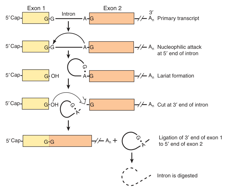
The intron RNA sequences are cleaved out of the transcript, and the exons are appropriately spliced together in the nucleus before the resulting mRNA molecule appears in the cytoplasm for translation.
Splicing depends on consensus sequences at the splice junctions and on an internal branch site.
The spliceosome is assembled from snRNAs (small nuclear RNAs, the U series) and snRNPs (small nuclear ribonucleoprotein particles).
1 · Pre-mRNA combines with the snRNPs and other proteins to form a spliceosome.
2 · Within the spliceosome, snRNA base-pairs with nucleotides at the ends of the intron.
3 · The RNA transcript is cut to release the intron, and the exons are spliced together; the spliceosome then comes apart, releasing mRNA, which now contains only exons.
Introns look wasteful. A human gene may be tens of kilobases long and encode a protein from a few kilobases of exon; the rest is transcribed at full metabolic cost and then thrown away. Why tolerate that?
Alternative splicing provides for different mRNAs. By joining the same exons in different combinations, one gene yields several proteins. That is how roughly 20,000 human genes specify a far larger proteome — and it is a capability a prokaryote, with no nucleus and no time to splice, simply cannot have.
A related device operates at the other end. Alternative promoter utilization provides a form of regulation: in the glucokinase gene, the β-cell promoter and exon 1B are located about 30 kbp upstream from the liver promoter and exon 1L; each promoter has a unique structure and is regulated differently, while exons 2–10 are identical and the proteins have identical kinetic properties.
So the same enzyme you met in Unit 22 — glucokinase, sensing glucose in the β cell and trapping it in the liver — is one protein transcribed from two independently regulated promoters. Two jobs, two control systems, one coding sequence.
- Define exon and intron → Exons appear in the mature RNA; introns are intervening sequences removed from it
- What removes introns? → The spliceosome, built from snRNAs and snRNPs
- Where does splicing occur? → In the nucleus, before export
- What does alternative splicing achieve? → Different mRNAs, and hence different proteins, from one gene
- Give an example of alternative promoter use → The glucokinase gene — separate liver and β-cell promoters ~30 kbp apart

Small RNAs and RNA editing ★★
The majority of miRNAs are transcribed by RNA pol II into primary transcripts termed pri-miRNAs. These are cut by Drosha into hairpins, transported into the cytoplasm and cut by Dicer, and the product anneals to the 3′ untranslated region of the target mRNA and interferes with protein translation.
Three mechanisms: (a) promoting mRNA degradation directly; (b) stimulating CCR4/NOT complex-mediated poly(A) tail degradation; (c) inhibition of translation by targeting the 5′-methyl cap binding translation factor eIF4.
RNA interference is triggered by the Dicer ribonuclease, which generates short interfering RNAs (siRNAs) of 21–28 bp. These are used to degrade target RNA by the RNA-induced silencing complex (RISC), and protect the host from RNA viruses.
RNA editing refers to the reactions that can change the nucleotide sequence of an mRNA molecule by non-splicing mechanisms. The change may include nucleotide change, deletion or insertion.
The classic example: the mRNA for apolipoprotein B in the liver is translated to apolipoprotein B100, while in the small intestine the mRNA is changed to yield a new termination codon (UAA), resulting in a much shorter protein, apolipoprotein B48.
In Unit 18 you learned that apo B-48 is 48% of the length of apo B-100, from the same gene, and that this matters because B-48 lacks the LDL-receptor-binding domain — which is why chylomicron remnants must be cleared through apo E instead.
Here is the mechanism behind that sentence. A single C→U change in the intestinal transcript converts a glutamine codon (CAA) into a stop codon (UAA). The ribosome stops halfway, and the receptor-binding domain — encoded downstream — is never made.
One base, edited in one tissue, redirects an entire lipoprotein pathway. It is worth carrying as an example because it demonstrates the whole point of post-transcriptional control: the gene is identical in liver and intestine, and the difference is imposed entirely after transcription.
- Which polymerase transcribes miRNAs? → Pol II, as pri-miRNAs
- Which two enzymes process them? → Drosha in the nucleus, Dicer in the cytoplasm
- Where does an miRNA bind its target? → The 3′ untranslated region
- Size of siRNAs, and what degrades the target? → 21–28 bp; the RNA-induced silencing complex (RISC)
- Define RNA editing → Change of an mRNA's nucleotide sequence by non-splicing mechanisms
- The classic example? → apo B-100 in liver vs apo B-48 in intestine, via a new UAA stop codon
Revision layer
Transcription versus replication — the contrasts examiners set
| Replication (Unit 23) | Transcription (Unit 24) | |
|---|---|---|
| Product | DNA | RNA |
| Substrates | dNTPs | ATP, GTP, CTP, UTP |
| Primer | Required — RNA, made by primase | NOT required — initiation is de novo |
| Extent | The whole genome | Selected genes only |
| Strands copied | Both | One — the template strand, and not necessarily the same strand for every gene |
| Direction | 5′→3′ | 5′→3′ (template read 3′→5′) |
| Proofreading | Yes — 3′→5′ exonuclease | Much less accurate; errors matter less, since transcripts are disposable |
Numbers to have ready
| Quantity | Value |
|---|---|
| Transcription bubble | 20 bp; whole complex 30–75 bp |
| Bacterial promoter length | ~40 nucleotides; boxes at −35 (8 bp) and −10 (6 bp, A+T-rich) |
| Promoter clearance | After nucleotides 3 to ~10 |
| Polymerase scanning rate | ≥10³ bp/s |
| Transcription initiation sites | 4 × 10³ in E. coli; ~10⁵ in humans |
| Eukaryotic promoter element combinations | TATA+Inr 30% · Inr 30% · Inr+DPE 25% · all three 15% |
| Promoter-proximal / distal elements | 50–200 bp / 1000–10⁵ bp |
| TFIID | 15 subunits — TBP + 14 TAFs |
| Pol II subunits | 12; CTD consensus Tyr-Ser-Pro-Thr-Ser-Pro-Ser |
| Poly(A) tail | ~200 A residues, cleaved ~20 nt downstream of AAUAAA |
| siRNAs | 21–28 bp |
| rRNA precursor | 45S |
Prokaryotic versus eukaryotic transcription
| Prokaryote | Eukaryote | |
|---|---|---|
| Polymerases | One, core α₂ββ′ + σ | Three — Pol I, II, III |
| Promoter recognition | σ factor | General transcription factors (GTFs) |
| Promoter | ~40 nt; −35 and −10 boxes | TATA / Inr / DPE + proximal + distal elements |
| Primary transcript | Is the mRNA | Pre-mRNA, requiring processing |
| Processing | None for mRNA | Cap, poly(A), splicing |
| Coupling | Translation before transcription ends | Separated by the nuclear membrane |
| Inhibitor | Rifampicin (β subunit) | α-Amanitin |
“Promoter = where the polymerase parks.” The full mark-earning sentence is “the DNA sequence to which RNA polymerase binds to initiate transcription of a gene” — write that verbatim, since it was set in both papers.
“Cap guards the 5′, tail guards the 3′” — each end is protected against the exonuclease that attacks from its own direction, and both help translation.
- Define a promoter → The DNA sequence to which RNA polymerase binds to initiate transcription of a gene
- Define a transcription unit → The region of DNA including the signals for initiation, elongation and termination
- Does transcription need a primer? → No — RNA polymerase initiates de novo
- Bacterial holoenzyme composition, and σ's job? → α₂ββ′σ; σ helps the core recognise the promoter
- Which eukaryotic polymerase makes mRNA, and what inhibits it? → Pol II; α-amanitin
- Which GTF binds the TATA box? → TFIID, via its TBP subunit
- What does the CTD do? → Its phosphorylation state controls promoter clearance, elongation, termination and mRNA processing
- The two bacterial termination mechanisms? → Intrinsic hairpin plus AT stretch, and rho-dependent
- The three eukaryotic mRNA processing events? → 5′ capping, 3′ polyadenylation, and splicing
- What does alternative splicing achieve? → Several proteins from one gene