DNA: Structure, Organisation & Replication
The double helix ★★★
Everything in Module E follows from one structure, so learn its dimensions precisely. The numbers are examinable in their own right, and they also explain why the molecule behaves as it does.
The monomeric units — deoxyadenylate, deoxyguanylate, deoxycytidylate and thymidylate — are held in polymeric form by 3′,5′-phosphodiester bonds, with the bases attached to the 2-deoxyribose by an N-glycosidic bond.
The informational content of DNA (the genetic code) resides in the sequence in which these monomers are ordered.
The polymer possesses a polarity: one end has a 5′-hydroxyl or phosphate terminal, the other a 3′-phosphate or hydroxyl terminal. Convention dictates that a single-stranded DNA sequence is written in the 5′ to 3′ direction. Note also that the phosphodiester backbone is negatively charged.
The B form is usually found under physiologic conditions (low salt, high degree of hydration).
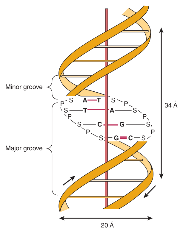
One turn of B-DNA about the long axis contains 10 bp.
The distance spanned by one turn is 3.4 nm (34 Å) — so the rise is 3.4 Å per base pair.
The width (helical diameter) is 2 nm (20 Å).
The helix is right-handed, because as one looks down the double helix the base residues form a spiral in a clockwise direction. It has a major groove and a minor groove, and in the test tube double-stranded DNA can exist in at least six forms (A–E and Z).
The two strands are held in register by both hydrogen bonds between the purine and pyrimidine bases of the respective linear molecules AND by van der Waals and hydrophobic interactions between the stacked adjacent base pairs.
Note that hydrogen bonding is only half the answer — base stacking contributes at least as much, and it is the half candidates usually forget.
A protein that must switch a gene on has a problem: it has to recognise a specific DNA sequence without unwinding the helix. The bases are on the inside, and the sugar-phosphate backbone is the same everywhere.
The grooves are the solution. The edges of the base pairs are exposed at the floor of the major groove, and each of the four possible pairs presents a distinguishable pattern of hydrogen-bond donors and acceptors there. A protein can therefore read the sequence from the outside.
That is exactly what the helix-turn-helix, zinc finger and leucine zipper motifs of Unit 26 do — each inserts a recognition α-helix into the major groove. When you meet them, remember they are solving this problem.
- Base pairs per turn of B-DNA? → 10
- Length of one turn? → 3.4 nm (34 Å); rise 3.4 Å per bp
- Helical diameter? → 2 nm (20 Å)
- Which bond joins the nucleotides? → The 3′,5′-phosphodiester bond
- Which two forces hold the strands together? → Hydrogen bonds AND base stacking (van der Waals and hydrophobic)
- Conditions favouring B-DNA? → Low salt, high degree of hydration
Base pairing and the two strands ★★★
Chargaff observed that in DNA molecules the concentration of deoxyadenosine (A) nucleotides equals that of thymidine (T) nucleotides (A = T), while the concentration of deoxyguanosine (G) equals that of deoxycytidine (C) (G = C).
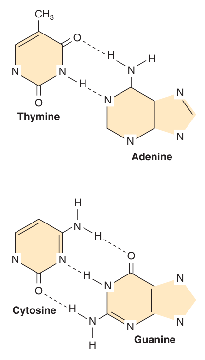
Three hydrogen bonds hold the deoxyguanosine nucleotide to the deoxycytidine nucleotide, whereas the A-T pair is held together by two hydrogen bonds.
Thus the G-C bonds are more resistant to denaturation, or strand separation — termed “melting” — than A-T-rich regions of DNA.
Chargaff's observation came before the structure, and it is worth seeing why it practically forced the answer.
If A always equals T and G always equals C, then A must be paired with T and G with C. And the geometry explains why: a purine (two rings) paired with a pyrimidine (one ring) always spans the same distance. Purine-purine would be too wide, pyrimidine-pyrimidine too narrow — and the helix would bulge and pinch instead of holding a constant 20 Å diameter.
So the pairing rule is not arbitrary chemistry; it is what a uniform-diameter helix requires. Harper's adds the further constraints: restrictions imposed by rotation about the phosphodiester bond, the favored anti-configuration of the glycosidic bond, and the predominant tautomers of the four bases allow A to pair only with T, and G only with C.
The two strands, each of which possesses a polarity, are ANTIPARALLEL: one strand runs in the 5′ to 3′ direction and the other in the 3′ to 5′ direction.
Within a particular gene:
The TEMPLATE strand — the strand whose sequence carries the genetic information, and the strand of DNA that is copied during RNA synthesis. It is sometimes called the noncoding strand.
The CODING strand — the opposite strand, so called because it matches the sequence of the RNA transcript, but containing thymine in place of uracil.
Note the point students most often miss: the template strand for each gene will not necessarily be the same strand of the DNA double helix.
- Chargaff's rule? → A = T and G = C
- How many hydrogen bonds in G-C? → Three. A-T? → Two
- Which region melts at a higher temperature? → G-C-rich, because of the third hydrogen bond
- Why must a purine pair with a pyrimidine? → Only that combination keeps the helix diameter constant at 20 Å
- Which strand is copied into RNA? → The template (noncoding) strand
- Which strand matches the RNA sequence? → The coding strand, but with T for U
Denaturation and Tm ★★★
The double-stranded structure of DNA can be separated into two component strands in solution by increasing the temperature or decreasing the salt concentration. Not only do the two stacks of bases pull apart, but the bases themselves unstack while still connected in the polymer by the phosphodiester backbone.
Concomitant with denaturation is an increase in the optical absorbance of the purine and pyrimidine bases — a phenomenon referred to as HYPERCHROMICITY OF DENATURATION.
Because of the stacking of the bases and the hydrogen bonding between the stacks, the double-stranded DNA molecule exhibits properties of a rigid rod and in solution is a viscous material that loses its viscosity upon denaturation.
The strands of a given molecule of DNA separate over a temperature range; the midpoint is called the melting temperature, or Tm.
Three things influence it:
1 · Base composition. DNA rich in G-C pairs, which have three hydrogen bonds, melts at a higher temperature than that rich in A-T pairs, which have two.
2 · Salt concentration. A 10-fold increase of monovalent cation concentration increases the Tm by 16.6 °C, by neutralizing the intrinsic interchain repulsion between the highly negatively charged phosphates of the phosphodiester backbone.
3 · Solvent. Formamide destabilizes hydrogen bonding between bases, thereby lowering the Tm — used in recombinant DNA work to separate strands at much lower temperatures, minimising the phosphodiester bond breakage that can occur on extended incubation at higher temperatures.
Hyperchromicity looks like an odd laboratory curiosity until you see the reason, and then it becomes obvious.
In the intact double helix the bases are stacked face to face, and their electron clouds interact — which suppresses their absorption of ultraviolet light at 260 nm. Pull the strands apart and the bases unstack, the suppression is lost, and the same amount of DNA now absorbs more light.
So the absorbance at 260 nm is a direct read-out of how much of the DNA is double-stranded. That single fact turns a spectrophotometer into a melting-curve machine, and it is the basis of every hybridisation experiment in the next section.
Separated strands of DNA will renature or reassociate when appropriate physiologic temperature and salt conditions are achieved; this reannealing process is often referred to as HYBRIDIZATION.
The rate of reassociation depends upon the concentration of the complementary strands. Renaturation requires base pair matching — which is precisely what makes it useful, because a labelled probe will bind only to a sequence complementary to it.
Southern blotting — DNA/DNA. Northern blotting — RNA/DNA. (Western blotting, by the same joke, detects protein with antibody.)
- What is hyperchromicity of denaturation? → The rise in optical absorbance when DNA strands separate and the bases unstack
- Define T_m → The midpoint of the temperature range over which the strands separate
- Which DNA has the higher T_m? → G-C-rich
- Effect of a 10-fold rise in monovalent cation? → T_m rises by 16.6 °C
- Effect of formamide? → Lowers T_m by destabilising base hydrogen bonding
- Southern vs Northern blot? → DNA/DNA vs RNA/DNA
Supercoiling and topoisomerases ★★
DNA exists in relaxed and supercoiled forms. Supercoiling arises in a covalently closed circle — or in a linear molecule anchored at both ends, as chromosomal DNA is — because the two strands cannot rotate freely about each other.
Negative supercoils are underwound and favour strand separation; positive supercoils are overwound and oppose it.
The nicking-resealing enzymes that relieve torsional strain.
The nicks are quickly resealed without requiring energy input, because of the formation of a high-energy covalent bond between the nicked phosphodiester backbone and the nicking-sealing enzyme. Contrast this with DNA ligase, whose resealing is ATP-dependent.
Among the classes of proteins involved in replication, topoisomerases relieve torsional strain that results from helicase-induced unwinding.
Take a length of twisted rope, hold both ends fixed, and try to separate the two strands in the middle. You can do it — but the twist you remove there has to go somewhere, and it piles up ahead of your hands as ever-tighter overwinding.
That is exactly the replication fork's problem. The helicase separates the strands, and positive supercoils accumulate ahead of it. Without relief, the DNA would simply seize up. Topoisomerases cut, let the strain unwind, and reseal.
This also explains their pharmacology: topoisomerases are targets of fluoroquinolone antibiotics (bacterial DNA gyrase) and of several anticancer drugs. Trap the enzyme mid-cut and you leave a permanent break in the DNA of a dividing cell.
- What do topoisomerases do? → Nick and reseal DNA to relieve torsional strain
- Why do they need no ATP? → A high-energy covalent enzyme-DNA bond is formed at the nick
- Which enzyme's resealing IS ATP-dependent? → DNA ligase
- What generates the strain at a replication fork? → Helicase-induced unwinding
Chromatin and the nucleosome ⭐
Start with the problem, because the definition is the answer to it. Human genomic DNA, if extended end-to-end, would be metres in length, yet must fit within a nucleus only microns in diameter — Harper's puts the total length at thousands of times the diameter of the cell nucleus.
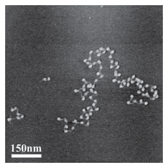
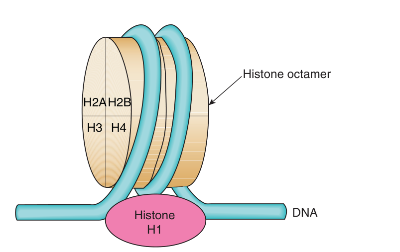
Dense spherical particles, approximately 10 nm in diameter, connected by DNA filaments, composed of DNA wound around an octameric complex of histone molecules.
Structure: the histone octamer is two each of H2A, H2B, H3 and H4. In the nucleosome, the DNA is supercoiled in a LEFT-HANDED helix over the surface of the disk-shaped histone octamer. 1.75 superhelical turns of DNA are wrapped around the surface of the histone octamer, protecting 145 to 150 bp of DNA and forming the nucleosome core particle.
Assembly: the (H3-H4)₂ tetramer itself can confer nucleosome-like properties on DNA and thus has a central role in the formation of the nucleosome; the addition of two H2A-H2B dimers stabilizes the primary particle.
H1 is NOT part of the core. H1 histones are the ones least tightly bound to chromatin and are therefore easily removed with a salt solution; neither histone H1 nor the nonhistone proteins are necessary for the reconstitution of the nucleosome core. H1 binds the linker DNA between core particles.
Strings of nucleosomes form along the linear sequence of genomic DNA to form chromatin, which itself can be more tightly packaged and condensed, ultimately leading to the formation of the chromosomes.
Histones are a small family of closely related basic proteins — that is, highly positively charged, which is what allows them to bind the negatively charged phosphodiester backbone.
The human haploid genome consists of about 3 × 10⁹ bp and about 1.7 × 10⁷ nucleosomes. The length of each DNA molecule must be compressed about 8000-fold to generate the structure of a condensed metaphase chromosome.
It would be easy to read the nucleosome as mere storage — a spool for a very long thread. Harper's warns explicitly against that reading: the histones also integrally participate in gene regulation; indeed histones contribute importantly to all DNA-directed molecular transactions.
The mechanism is electrostatic and beautifully simple. Histones are basic — positively charged — and DNA is negatively charged. Acetylation of lysine residues in the amino-terminal tails reduces the positive charge, decreases the binding affinity of histone for DNA, and disrupts nucleosomal structure, giving transcription factors access. Deacetylation has the opposite effect.
So a gene wrapped tightly in nucleosomes is off, and one whose nucleosomes have been loosened or evicted is available. Packing and permission are the same thing. This is where Unit 26 begins.
- Define a nucleosome → DNA wound around an octameric complex of histones, ~10 nm in diameter
- Which histones form the octamer? → Two each of H2A, H2B, H3 and H4
- How much DNA is protected? → 145–150 bp, in 1.75 superhelical turns
- Is the DNA supercoiled left- or right-handed on the octamer? → LEFT-handed
- Where is H1? → Not in the core — it binds linker DNA and is least tightly bound
- Nucleosomes in the human haploid genome? → About 1.7 × 10⁷, from 3 × 10⁹ bp
- Compression to a metaphase chromosome? → About 8000-fold
Chromosomes, centromeres and telomeres ⭐
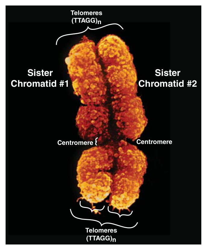
At metaphase, mammalian chromosomes possess a twofold symmetry, with the identical duplicated sister chromatids connected at a centromere, the relative position of which is characteristic for a given chromosome.
Each sister chromatid contains one dsDNA molecule. During interphase the packing of the DNA molecule is less dense than in the condensed chromosome at metaphase; metaphase chromosomes are nearly completely transcriptionally inactive.
In metaphase chromosomes, the 30-nm chromatin fibers are also folded into a series of looped domains. Humans have 23 pairs of chromosomes: 22 autosomes and two sex chromosomes.
An adenine-thymine (A-T)-rich region containing repeated DNA sequences that range in size from 10² (brewers' yeast) to 10⁶ (mammals) base pairs.
Metazoan centromeres are bound by nucleosomes containing the histone H3 variant protein CENP-A and other specific centromere-binding proteins. This complex, called the KINETOCHORE, provides the anchor for the mitotic spindle. It thus is an essential structure for chromosomal segregation during mitosis.
The structures at the ends of each chromosome.
Telomeres consist of short TG-rich repeats. Human telomeres have a variable number of repeats of the sequence 5′-TTAGGG-3′, which can extend for several kilobases.
Telomerase — a multisubunit RNA template-containing complex related to viral RNA-dependent DNA polymerases (reverse transcriptases) — is the enzyme responsible for telomere synthesis and thus for maintaining the length of the telomere.
Since telomere shortening has been associated with both malignant transformation and aging, this enzyme has become an attractive target for cancer chemotherapy and drug development.
The reason telomeres exist is a direct mechanical consequence of §8, so it is worth anticipating here.
DNA polymerases synthesise only in the 5′ to 3′ direction and cannot start a chain — they must extend an RNA primer. On the lagging strand, the final primer at the very end of a linear chromosome is removed and there is nothing upstream to fill the gap from. Every round of replication therefore shortens the chromosome. This is the end-replication problem.
Telomeres solve it by being expendable: a long stretch of repeated, non-coding TTAGGG that can be eaten away without losing genes. And telomerase restores it — carrying its own RNA template, which is why it is a reverse transcriptase rather than an ordinary polymerase.
The clinical consequence follows immediately. Most somatic cells lack telomerase, so their telomeres shorten and they eventually stop dividing — a limit on lifespan. Most cancers reactivate it, which is precisely why it is a drug target. One mechanical limitation of a polymerase, and it reaches all the way to ageing and cancer.
- Define telomeres → Structures at chromosome ends, consisting of short TG-rich repeats
- Human telomere repeat sequence? → 5′-TTAGGG-3′, extending for several kilobases
- Which enzyme maintains them? → Telomerase, an RNA template-containing reverse transcriptase
- Why is telomerase a drug target? → Telomere shortening is associated with malignant transformation and ageing
- What is the centromere? → An A-T-rich repeated region; with CENP-A nucleosomes it forms the kinetochore
- What does the kinetochore do? → Anchors the mitotic spindle for chromosomal segregation
Semiconservative replication ★★★
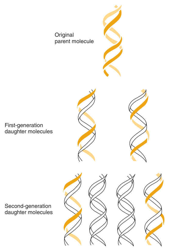
During a round of replication, each of the two strands of DNA is used as a template for synthesis of a new, complementary strand.
Each daughter duplex therefore consists of one strand derived from the original parental DNA duplex and one strand that was newly synthesized.
This is what makes faithful transmission possible: since the genetic information resides in the order of the monomeric units within the polymers, there must exist a mechanism of reproducing or replicating this specific information with a high degree of fidelity.
The famously understated line at the end of Watson and Crick's 1953 paper — that the pairing they had proposed immediately suggests a copying mechanism — points at exactly this.
Because A pairs only with T and G only with C, each strand already contains the full information needed to rebuild the other. Separate them, and each is a complete template. No additional information store is required, and no interpretation step: the structure is the copying mechanism.
Everything difficult about replication — primers, Okazaki fragments, topoisomerases, telomeres — is engineering, not information. The information problem was solved by the base pairing rule.
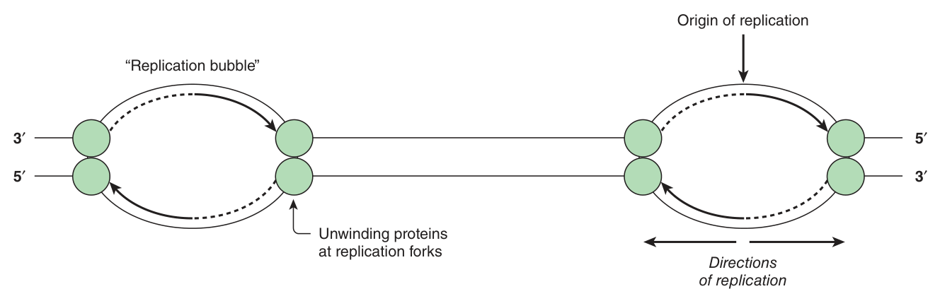
At the origin of replication (ori) there is an association of sequence-specific dsDNA-binding proteins with a series of direct repeat DNA sequences.
In E. coli the oriC is bound by the protein dnaA; in eukaryotes the group of proteins is the origin recognition complex (ORC), and ORC homologs have been found in all eukaryotes examined. The ORE lies adjacent to an approximately 80-bp A+T-rich sequence that is easy to unwind, called the DNA unwinding element (DUE).
Replication then proceeds bidirectionally, generating “replication bubbles” with a fork at each end.
- Define semiconservative replication → Each daughter duplex has one parental and one newly synthesised strand
- What is bound at oriC in E. coli? → The protein dnaA
- The eukaryotic equivalent? → The origin recognition complex (ORC)
- What is the DUE? → An ~80-bp A+T-rich DNA unwinding element, easy to melt
- Why A+T-rich? → Only two hydrogen bonds per pair, so it separates most readily
The replication fork ★★★
| Protein | Function |
|---|---|
| DNA polymerases | Deoxynucleotide polymerization |
| Helicases | ATP-driven processive unwinding of DNA |
| Topoisomerases | Relieve torsional strain that results from helicase-induced unwinding |
| DNA primase | Initiates synthesis of RNA primers |
| Single-strand binding proteins (SSBs) | Prevent premature reannealing of dsDNA |
| DNA ligase | Seals the single-strand nick between the nascent chain and Okazaki fragments on the lagging strand |
DNA polymerases only synthesize DNA in the 5′ to 3′ direction, and only one of the several different types of polymerases is involved at the replication fork. Because the DNA strands are antiparallel, the polymerase functions ASYMMETRICALLY.
On the leading (forward) strand, the DNA is synthesized continuously.
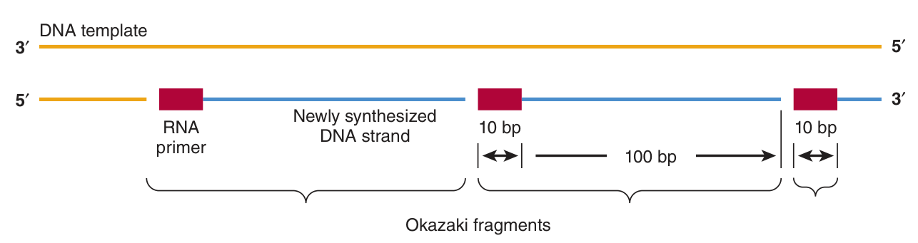
On the lagging (retrograde) strand, the DNA is synthesized in short (1–5 kb) fragments, the so-called OKAZAKI fragments — several Okazaki fragments (up to a thousand) must be sequentially synthesized for each replication fork.
The helicase acts on the lagging strand to unwind dsDNA in a 5′ to 3′ direction, and associates with the primase to afford the latter proper access to the template. This allows the RNA primer to be made and, in turn, the polymerase to begin.
The segments of DNA attached to an RNA initiator component are the Okazaki fragments. In mammals, after many Okazaki fragments are generated, the replication complex begins to remove the RNA primers, to fill in the gaps left by their removal with the proper base-paired deoxynucleotide, and then to seal the fragments of newly synthesized DNA by enzymes referred to as DNA ligases.
The chemistry of chain growth: the template dictates which deoxyribonucleoside triphosphate is complementary and by hydrogen bonding holds it in place while the 3′-hydroxyl group of the growing strand attacks and incorporates the new nucleotide into the polymer.
The whole of this section is deducible from two facts about DNA polymerase, so learn the facts rather than the list.
Constraint 1: synthesis is 5′→3′ only. The strands are antiparallel, so as the fork opens, one template runs the right way and the other runs backwards. The right way gives the continuous leading strand; the backwards one must be built in short pieces, each started afresh as new template appears — the Okazaki fragments. Every fragment then needs its own primer, its own primer removal, its own gap fill, and its own ligase seal.
Constraint 2: polymerase cannot start a chain, only extend one. Hence primase, and hence the curious fact that DNA synthesis begins with RNA. Why RNA? Because a primer made of RNA is recognisably foreign and can be excised and replaced with high-fidelity DNA — a deliberate error-avoidance design, since the very first nucleotides of a chain are laid down without the benefit of proofreading.
And constraint 2, applied to the end of a linear chromosome, is the end-replication problem that telomeres exist to solve (§6).
- In which direction do DNA polymerases synthesise? → 5′ to 3′ only
- Why is the lagging strand discontinuous? → The strands are antiparallel, so the polymerase must work backwards in pieces
- Size of an Okazaki fragment? → 1–5 kb; up to a thousand per fork
- What does primase make? → RNA primers, because polymerase cannot initiate a chain
- What do SSBs do? → Prevent premature reannealing of dsDNA
- What does ligase do? → Seals the nick between the nascent chain and the Okazaki fragments
DNA repair ★★
Replication is the most accurate copying process in biology, and it still is not accurate enough on its own. Fidelity comes from three successive layers: correct base pairing, polymerase proofreading, and post-replication repair.
| Mechanism | What it corrects |
|---|---|
| Base-pair selection | Ensures the incoming nucleotide is complementary before the bond is made |
| Proofreading | The polymerase's own 3′→5′ exonuclease removes a mismatched nucleotide immediately after it is added |
| Mismatch repair | Corrects mismatches that survive proofreading; its failure causes hereditary nonpolyposis colon cancer |
| Base excision repair | Single damaged or inappropriate bases — a glycosylase removes the base, leaving an AP site |
| Nucleotide excision repair | Bulky lesions distorting the helix, notably UV-induced pyrimidine dimers; its failure causes xeroderma pigmentosum |
| Double-strand break repair | Breaks in both strands, by homologous recombination or non-homologous end joining |
Fidelity is multiplicative, and that is why the layered design wins.
Base pairing alone gives roughly one error in 10⁴ or 10⁵. Add proofreading — the polymerase pausing to excise a wrong nucleotide before moving on — and you gain about two more orders of magnitude. Add mismatch repair, which sweeps up afterwards, and you reach roughly one error per 10⁹ or 10¹⁰ base pairs.
No single mechanism could plausibly achieve that. But three independent checks, each catching most of what the previous one missed, multiply together — the same reason three independent safety systems beat one very reliable one.
And note what happens when a layer fails: not immediate death, but cancer. Mismatch repair failure gives hereditary nonpolyposis colon cancer; nucleotide excision repair failure gives xeroderma pigmentosum, with extreme sensitivity to sunlight and early skin cancers. A repair defect is a mutation-rate defect, and a mutation-rate defect is a cancer predisposition.
- Which activity performs proofreading? → The polymerase's 3′→5′ exonuclease
- Which repair pathway handles UV pyrimidine dimers? → Nucleotide excision repair
- Failure of that pathway causes? → Xeroderma pigmentosum
- Failure of mismatch repair causes? → Hereditary nonpolyposis colon cancer
- What starts base excision repair? → A glycosylase removing the damaged base
Revision layer
Numbers to have ready
| Quantity | Value |
|---|---|
| Base pairs per turn of B-DNA | 10 |
| Length of one turn | 3.4 nm (34 Å); rise 3.4 Å per bp |
| Helical diameter | 2 nm (20 Å) |
| Hydrogen bonds, G-C and A-T | 3 and 2 |
| Effect of 10-fold monovalent cation increase on Tm | +16.6 °C |
| Nucleosome core particle | 145–150 bp, 1.75 superhelical turns, ~10 nm diameter |
| Histone octamer | 2 each of H2A, H2B, H3, H4 |
| Human haploid genome | 3 × 10⁹ bp, 1.7 × 10⁷ nucleosomes |
| Compression to a metaphase chromosome | ~8000-fold |
| Human telomere repeat | 5′-TTAGGG-3′ |
| Okazaki fragment | 1–5 kb; up to 1000 per fork |
| Human chromosomes | 23 pairs — 22 autosomes and two sex chromosomes |
Pairs that are easy to confuse
| This one | Not this one | |
|---|---|---|
| Template vs coding strand | Template — copied into RNA; also called noncoding | Coding — matches the RNA sequence, with T for U |
| Topoisomerase vs ligase | Topoisomerase — relieves torsion, no ATP needed | Ligase — seals nicks, ATP-dependent |
| Leading vs lagging | Leading — continuous | Lagging — Okazaki fragments, many primers |
| Centromere vs telomere | Centromere — A-T-rich, mid-chromosome, kinetochore, spindle attachment | Telomere — TG-rich TTAGGG, chromosome END, telomerase |
| Core histones vs H1 | H2A, H2B, H3, H4 — the octamer | H1 — linker DNA, least tightly bound, not needed for reconstitution |
“G-C = 3 letters, 3 hydrogen bonds. A-T = 2 letters in the pair name you say first, 2 bonds.” From there, G-C-rich = higher Tm follows automatically.
For the telomere repeat: TTAGGG — “Two Ts, A, three Gs.”
- Dimensions of B-DNA? → 10 bp per turn, 34 Å per turn, 20 Å wide
- Why must a purine pair with a pyrimidine? → To keep the helix diameter constant
- What is hyperchromicity? → The rise in UV absorbance on denaturation, because the bases unstack
- Define a nucleosome → DNA supercoiled left-handed around an octamer of H2A, H2B, H3, H4 — 145–150 bp, 1.75 turns
- Which histone is NOT in the core? → H1 — it binds linker DNA
- Define telomeres → Short TG-rich repeats at chromosome ends; in humans 5′-TTAGGG-3′
- Which enzyme maintains them, and what is unusual about it? → Telomerase — it carries its own RNA template and is a reverse transcriptase
- Define semiconservative replication → Each daughter duplex retains one parental strand
- Why is the lagging strand discontinuous? → Polymerase works only 5′→3′ and the strands are antiparallel
- Why does DNA synthesis begin with an RNA primer? → Polymerase cannot initiate a chain; RNA is recognisably foreign and can be excised and replaced accurately