Nucleic Acids & Nucleotide Metabolism
The vocabulary ★★
Three words are used loosely in conversation and precisely in examinations, so fix them now. A base is the ring alone. A nucleoside is base + sugar. A nucleotide is base + sugar + phosphate — nucleotides are phosphorylated nucleosides.
| Purines | Pyrimidines | |
|---|---|---|
| Ring | Two fused rings | One ring |
| Bases | Adenine, guanine (and hypoxanthine, xanthine, uric acid) | Cytosine, uracil, thymine |
| Where the ring is built | On the ribose — PRPP is the scaffold | Free, then attached — PRPP joins only after the ring is complete |
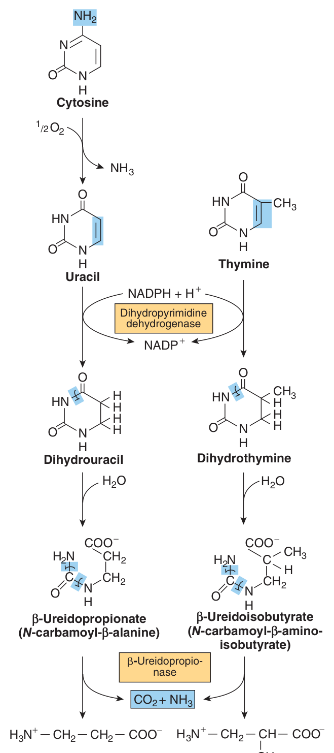
| End product of catabolism in humans | Uric acid — poorly soluble | CO₂, NH₃, β-alanine, β-aminoisobutyrate — highly water-soluble |
| Clinical consequence | Gout and the stones | Essentially none — see §10 |
Look at the last two rows and notice that they are the same fact stated twice.
A purine is a fused bicyclic system — flat, aromatic, and stubbornly insoluble once oxidised to uric acid. A pyrimidine is a single small ring that can be opened and dismantled into small, freely soluble fragments.
So purine overproduction precipitates — literally — as crystals in joints and kidney; pyrimidine overproduction simply washes out in the urine. Harper's says it plainly: since the end products of pyrimidine catabolism are highly water-soluble, pyrimidine overproduction results in few clinical signs or symptoms. When you are asked why gout has no pyrimidine equivalent, that is the answer — and it is a structural answer, not a physiological one.
Purine and pyrimidine nucleotides are synthesised in the CYTOSOL.
Ingested nucleic acids are degraded to purines and pyrimidines. Purines and pyrimidines are formed from amphibolic intermediates and are thus dietarily nonessential — the body makes its own, and does not depend on dietary nucleic acid at all.
- Nucleoside vs nucleotide? → Nucleoside = base + sugar; nucleotide = base + sugar + phosphate
- Where are nucleotides synthesised? → In the cytosol
- Are purines and pyrimidines dietary essentials? → No — they are made from amphibolic intermediates
- End product of purine catabolism in humans? → Uric acid
- End products of pyrimidine catabolism? → CO₂, NH₃, β-alanine and β-aminoisobutyrate
PRPP — the common precursor ★★★
An important precursor of both purine and pyrimidine nucleotides (and of some amino acids), formed from ribose 5-phosphate + ATP by PRPP synthase.
The overall determinant of the rate of de novo purine nucleotide biosynthesis is the concentration of PRPP. Its rate of synthesis depends on the availability of ribose 5-phosphate and on the activity of PRPP synthase, which is feedback inhibited by AMP, ADP, GMP and GDP.
Trace it back one step and you land in Unit 14: ribose 5-phosphate is the product of the pentose phosphate pathway. That connection is not decorative — it explains two diseases later in this unit.
Von Gierke disease (glucose-6-phosphatase deficiency) causes hyperuricemia because glucose 6-phosphate cannot be dephosphorylated and is shunted into the pentose phosphate pathway, generating extra ribose 5-phosphate, hence extra PRPP, hence purine overproduction. And Lesch-Nyhan syndrome overproduces purines for a mirror-image reason: salvage fails, so PRPP is not consumed and accumulates.
Whenever PRPP rises, purine synthesis rises and urate follows. Hold that one sentence and half of §9 and §10 becomes deducible rather than memorised.
- What is PRPP made from? → Ribose 5-phosphate + ATP, by PRPP synthase
- What determines the rate of de novo purine synthesis? → The concentration of PRPP
- PRPP synthase is feedback inhibited by? → AMP, ADP, GMP and GDP
- Where does ribose 5-phosphate come from? → The pentose phosphate pathway
De novo purine synthesis ★★★
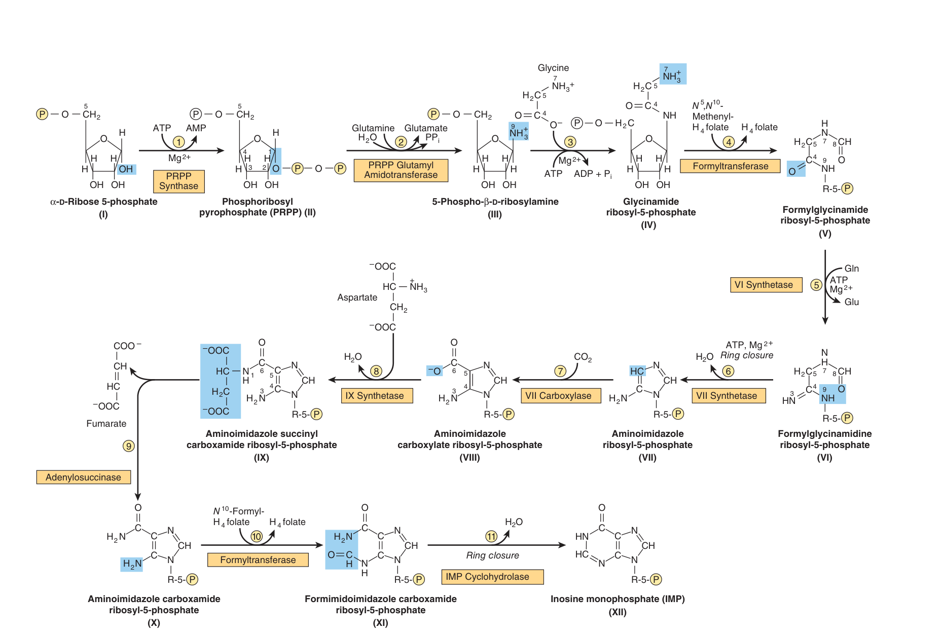
The defining feature, and the one examiners test: the purine ring is assembled directly on the ribose. PRPP is not a reagent added at the end; it is the scaffold on which the ring is built. Compare §6, where the pyrimidine ring is finished first and attached to PRPP afterwards.
The ring is assembled from small pieces contributed by several pathways:
Glycine — contributes C4, C5 and N7 (an entire fragment)
Glutamine — contributes N3 and N9
Aspartate — contributes N1
CO₂ — contributes C6
N¹⁰-formyl-tetrahydrofolate — contributes C2 and C8
Hence Harper's summary: several reactions of IMP biosynthesis require folate derivatives and glutamine — consequently, antifolate drugs and glutamine analogs inhibit purine biosynthesis.
PRPP glutamyl amidotransferase converts PRPP to 5-phosphoribosylamine (5-phospho-β-D-ribosylamine) using the amide nitrogen of glutamine. This is reaction ② of the pathway and the committed step, feedback inhibited by AMP and GMP.
Eleven reactions later the pathway arrives at inosine monophosphate (IMP), the parent purine nucleotide. Multifunctional catalysts carry out reactions 3/4/6, 7/8, and 10/11 — several enzyme activities on a single polypeptide, exactly the channelling principle of fatty acid synthase in Unit 16.
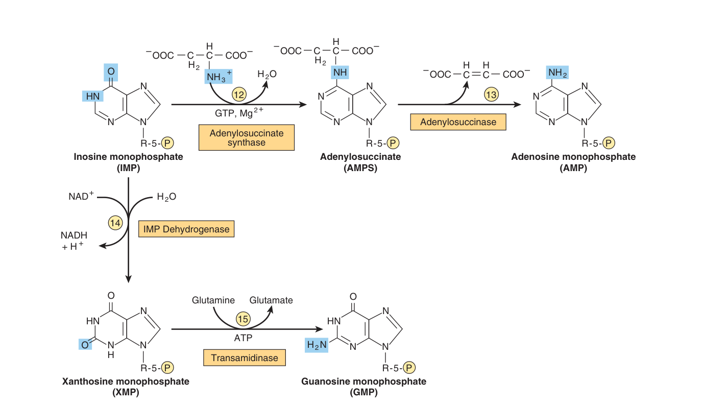
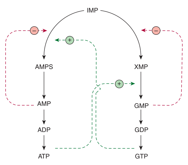
IMP is a precursor both of AMP and of GMP. The two branches are elegantly cross-wired:
To AMP: IMP + aspartate + GTP → adenylosuccinate (adenylosuccinate synthase) → AMP + fumarate (adenylosuccinase). Aspartate provides the 6-amino group of AMP.
To GMP: IMP + NAD⁺ → XMP (xanthosine monophosphate) (IMP dehydrogenase) → GMP, using glutamine and ATP. Glutamine provides the 2-amino group of GMP.
Read the two branches again and notice which nucleotide each one spends.
Making AMP requires GTP. Making GMP requires ATP. Each branch is paid for in the currency of the other.
Now think about what happens if guanine nucleotides run low. GTP falls, so the AMP branch slows — and IMP is pushed towards GMP instead. The shortage corrects itself. Harper's states the principle exactly: this cross-regulation between the pathways of IMP metabolism serves to balance the biosynthesis of purine nucleoside triphosphates by decreasing the synthesis of one purine nucleotide when there is a deficiency of the other.
This is not feedback inhibition in the ordinary sense — it is reciprocal funding, and it keeps A and G in the proportions that DNA and RNA actually need.
- On what is the purine ring assembled? → Directly on the ribose, with PRPP as the scaffold
- Which amino acids donate atoms to the purine ring? → Glycine, glutamine and aspartate
- Which one-carbon donor is required? → N¹⁰-formyl-tetrahydrofolate (C2 and C8)
- Committed step? → PRPP glutamyl amidotransferase, forming 5-phosphoribosylamine
- What is the parent purine nucleotide? → IMP
- Aspartate provides which group? → The 6-amino group of AMP. Glutamine? → The 2-amino group of GMP
- AMP synthesis requires which nucleotide? → GTP — and GMP synthesis requires ATP
The salvage pathway ★★★
Reactions that convert purines, their ribonucleosides and their deoxyribonucleosides to mononucleotides, and which require far less energy than de novo synthesis.
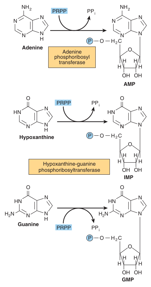
The more important mechanism is phosphoribosylation by PRPP of a free purine:
Pu + PRPP → Pu-RP + PPi
catalysed by adenine phosphoribosyltransferase (APRT) and hypoxanthine-guanine phosphoribosyltransferase (HGPRT), which convert adenine → AMP, hypoxanthine → IMP and guanine → GMP.
A second mechanism is phosphoryl transfer from ATP to a purine ribonucleoside:
Pu-R + ATP → PuR-P + ADP
catalysed by adenosine kinase (adenosine and deoxyadenosine → AMP and dAMP) and deoxycytidine kinase (deoxycytidine and 2′-deoxyguanosine → dCMP and dGMP).
De novo synthesis is expensive: in addition to ATP, glycine, glutamine, aspartate and reduced tetrahydrofolate derivatives all are consumed. Not every tissue can pay.
Liver is the major site of purine nucleotide biosynthesis, and provides purines and purine nucleosides for salvage and for utilization by tissues incapable of their biosynthesis. Two groups depend on that supply:
Brain — has a low level of PRPP glutamyl amidotransferase and hence depends in part on exogenous purines.
Erythrocytes and polymorphonuclear leukocytes — cannot synthesise 5-phosphoribosylamine at all, and therefore also use exogenous purines.
Now hold that fact next to Lesch-Nyhan syndrome in §10. If the brain depends on salvage, and salvage is what HGPRT does, then a child born without HGPRT has a brain starved of purines. That is why a disease of urate chemistry produces a neurological syndrome — the tissue distribution of the enzyme predicts the phenotype.
- The two salvage mechanisms? → Phosphoribosylation by PRPP, and phosphoryl transfer from ATP
- HGPRT converts? → Hypoxanthine to IMP and guanine to GMP
- APRT converts? → Adenine to AMP
- Which tissue is the major site of de novo purine synthesis? → The liver
- Which cells cannot synthesise 5-phosphoribosylamine? → Erythrocytes and polymorphonuclear leukocytes
Regulation ★★★
Harper's gives the reason for tight control in one sentence: biosynthesis of IMP is energetically expensive… it thus is of survival advantage to closely regulate purine biosynthesis in response to varying physiologic need. Control operates at three levels.
| Level | Mechanism |
|---|---|
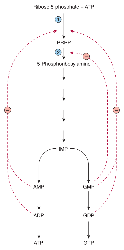
| 1 · PRPP supply | PRPP synthase is feedback inhibited by AMP, ADP, GMP and GDP — “elevated levels of these nucleoside phosphates signal a physiologically appropriate overall decrease in their biosynthesis” |
| 2 · The committed step | AMP and GMP feedback inhibit PRPP glutamyl amidotransferase |
| 3 · The branch point | AMP inhibits adenylosuccinate synthase; GMP inhibits IMP dehydrogenase; and each branch requires the nucleotide of the other (AMP needs GTP, GMP needs ATP). AMP and GMP also inhibit HGPRT, and GMP feedback inhibits PRPP glutamyl amidotransferase |
Purine and pyrimidine biosynthesis parallel one another quantitatively — that is, mole for mole — suggesting coordinated control of their biosynthesis.
The pivot is PRPP synthase, which forms a precursor essential for both processes and is feedback inhibited by both purine and pyrimidine nucleotides.
The purpose, in Harper's words: coordinated regulation ensures their presence in proportions appropriate for nucleic acid biosynthesis and other metabolic needs.
Ask what the nucleotides are for. DNA and RNA are built from purines and pyrimidines in a 1:1 ratio — every A pairs with a T, every G with a C. A cell that made twice as many purines as pyrimidines would waste half of them.
So the regulation is not an accident of enzyme chemistry; it is dictated by the base pairing rule of Unit 23. The product determines the proportions of its own precursors. This is the same reasoning that makes ribonucleotide reductase in §7 so elaborately regulated — balanced production of dNTPs is the requirement, and every control loop exists to enforce it.
- The overall determinant of de novo purine synthesis rate? → The concentration of PRPP
- Which enzyme is the pivot of coordinated purine/pyrimidine control? → PRPP synthase
- AMP inhibits which branch enzyme? → Adenylosuccinate synthase. GMP? → IMP dehydrogenase
- In what ratio do purine and pyrimidine synthesis run? → Mole for mole
Pyrimidine synthesis ★★★
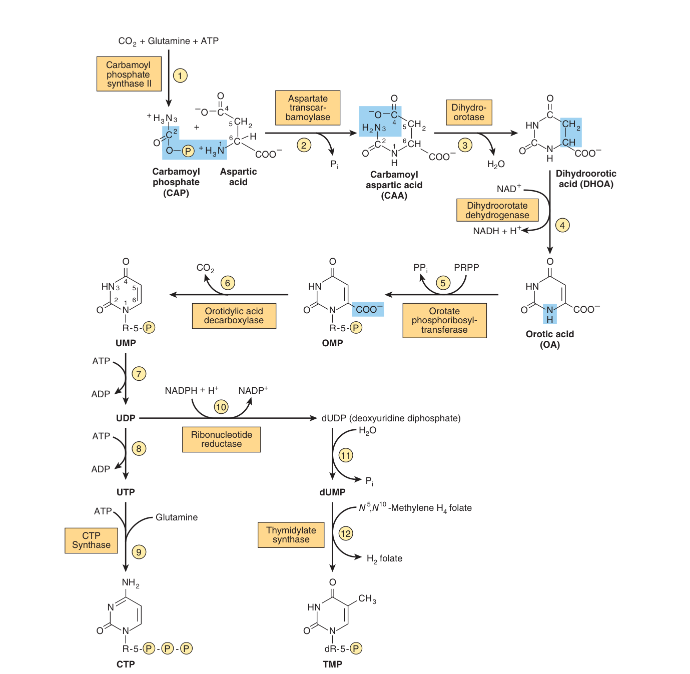
Unlike in purine biosynthesis, where PRPP serves as a scaffold for assembly of the purine ring, PRPP participates in pyrimidine biosynthesis only SUBSEQUENT to assembly of the pyrimidine ring.
The ring is built free in the cytosol from carbamoyl phosphate and aspartate, and only then attached to ribose 5-phosphate.
| # | Step | Enzyme |
|---|---|---|
| 1 | CO₂ + glutamine + ATP → carbamoyl phosphate | Carbamoyl phosphate synthase II — cytosolic |
| 2 | + aspartate → carbamoyl aspartate | Aspartate transcarbamoylase |
| 3 | → dihydroorotate (ring closure) | Dihydroorotase |
| 4 | → orotic acid | Dihydroorotate dehydrogenase |
| 5 | + PRPP → OMP | Orotate phosphoribosyltransferase |
| 6 | → UMP (−CO₂) | Orotidylic acid decarboxylase |
| 7–9 | UMP → UDP → UTP → CTP (using glutamine) | CTP synthase |
| 10–12 | UDP → dUDP → dUMP → TMP | Ribonucleotide reductase, then thymidylate synthase |
The catalyst for the initial reaction is cytosolic carbamoyl phosphate synthase II, a DIFFERENT enzyme from the mitochondrial carbamoyl phosphate synthase I of urea synthesis. Compartmentation thus provides an independent pool of carbamoyl phosphate for each process.
CPS-I — mitochondrial · nitrogen from ammonia · urea cycle · requires N-acetylglutamate
CPS-II — cytosolic · nitrogen from glutamine · pyrimidines · inhibited by UTP and purine nucleotides, activated by PRPP
Five of the first six enzyme activities of pyrimidine biosynthesis reside on multifunctional polypeptides — one catalyses the first three reactions, a second bifunctional enzyme catalyses reactions 5 and 6. The close proximity of multiple active sites facilitates efficient channeling of the intermediates.
Regulation: carbamoyl phosphate synthase II is inhibited by UTP and purine nucleotides but activated by PRPP; aspartate transcarbamoylase is inhibited by CTP but activated by ATP. In addition, the first three and the last two enzymes of the pathway are regulated by coordinate repression and derepression.
- When does PRPP enter pyrimidine synthesis? → Only after the ring is complete — at OMP formation
- First enzyme of pyrimidine synthesis? → Cytosolic carbamoyl phosphate synthase II, using glutamine
- Which intermediate gives its name to a disease? → Orotic acid
- CPS-II is inhibited by? → UTP and purine nucleotides; activated by PRPP
- Aspartate transcarbamoylase is inhibited by? → CTP; activated by ATP
Deoxyribonucleotides ★★
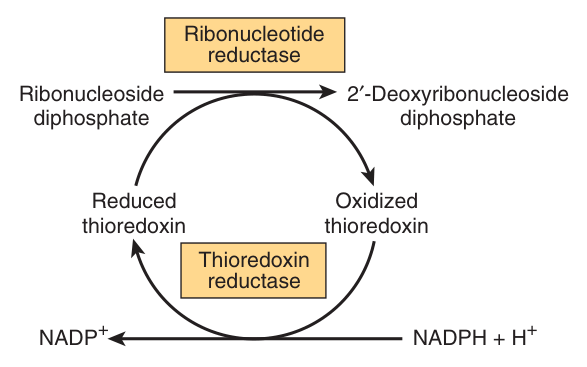
The enzyme complex that reduces the 2′-hydroxyl of purine and pyrimidine ribonucleotides, providing the deoxyribonucleoside diphosphates (dNDPs) needed for both the synthesis and repair of DNA.
The enzyme complex is functional only when cells are actively synthesizing DNA.
Reduction requires reduced thioredoxin, thioredoxin reductase and NADPH — the immediate reductant is reduced thioredoxin, regenerated by NADPH-dependent reduction of the oxidised form.
The reaction is subject to complex regulatory controls that achieve balanced production of dNTPs for synthesis of DNA.
Two design points hide in that definition.
First, the enzyme acts at the diphosphate level — NDP → dNDP, not on the triphosphate. So there is a single gateway between the RNA world and the DNA world, and it can be shut. The enzyme is functional only when cells are actively synthesizing DNA — a non-dividing cell simply does not make deoxyribonucleotides.
Second, the four dNTPs must be produced in balanced amounts. An excess of one dNTP relative to the others is mutagenic, because DNA polymerase misincorporates when the pool is skewed. That is why the enzyme carries an unusually elaborate set of allosteric sites — and, remarkably, it is also the mechanism of a disease: in adenosine deaminase and purine nucleoside phosphorylase deficiency, the immune dysfunctions appear to result from accumulation of dGTP and dATP, which inhibit ribonucleotide reductase and thereby deplete cells of DNA precursors (§10).
- What does ribonucleotide reductase do? → Reduces the 2′-OH of ribonucleoside DIphosphates to deoxyribonucleoside diphosphates
- What does it require? → Reduced thioredoxin, thioredoxin reductase and NADPH
- When is it functional? → Only when cells are actively synthesising DNA
The antimetabolite drugs ★★★
Nucleotide synthesis is the classic target of cancer chemotherapy, for a simple reason: a dividing cell must replicate its DNA, and cannot do so without dNTPs. Block the supply and the fastest-dividing cells are hit hardest. Every drug below is a molecule that looks enough like a natural substrate to be mistaken for it.
| Drug | Target | Class |
|---|---|---|
| Methotrexate | Dihydrofolate reductase | Antifolate |
| 5-Fluorouracil | Thymidylate synthase; also an alternate substrate for orotate phosphoribosyltransferase | Pyrimidine analog |
| 6-Mercaptopurine | Converted to a nucleotide which inhibits purine biosynthesis | Purine analog |
| Mycophenolic acid | IMP dehydrogenase — the GMP branch | Purine analog |
| Azaserine | Reaction 5 of purine synthesis | Glutamine analog |
| Diazanorleucine | Reaction 2 of purine synthesis | Glutamine analog |
| Allopurinol | Xanthine oxidase (§9); also an alternate substrate for orotate phosphoribosyltransferase | Hypoxanthine analog |
| 6-Azauridine | Orotidylate decarboxylase | Pyrimidine analog |
The reaction catalysed by thymidylate synthase is the only reaction of pyrimidine nucleotide biosynthesis that requires a tetrahydrofolate derivative. During it, the methylene group of N⁵,N¹⁰-methylene-tetrahydrofolate is reduced to the methyl group transferred to the 5-position of the ring, and tetrahydrofolate is oxidised to dihydrofolate.
For further pyrimidine synthesis to occur, dihydrofolate must be reduced back to tetrahydrofolate — and this reduction, catalysed by dihydrofolate reductase, is inhibited by methotrexate.
Dividing cells, which must generate TMP and dihydrofolate, are thus especially sensitive to inhibitors of dihydrofolate reductase such as the anticancer drug methotrexate.
Follow the folate. Thymidylate synthase does not merely use tetrahydrofolate as a coenzyme — it consumes it, leaving dihydrofolate behind. It is the only reaction in the pathway that does so.
That makes the cell's supply of TMP absolutely dependent on a recycling step. Block dihydrofolate reductase and the folate pool is trapped in the dihydro form after a single round. The cell can no longer make thymine — and thymine is the one base for which there is no alternative source, since it appears in DNA only.
This is why the effect is called “thymineless death”, and why methotrexate's toxicity falls on exactly the tissues that divide fastest: bone marrow, gut mucosa, hair follicles. The side effects are the mechanism. The same logic explains why disorders of folate and vitamin B₁₂ metabolism result in deficiencies of TMP — and hence in megaloblastic anaemia, cells that grow but cannot divide.
- Methotrexate inhibits? → Dihydrofolate reductase
- Which is the only pyrimidine synthesis reaction requiring a folate? → Thymidylate synthase
- 5-Fluorouracil targets? → Thymidylate synthase
- Azaserine and diazanorleucine are analogs of? → Glutamine
- Mycophenolic acid inhibits? → IMP dehydrogenase
Purine catabolism, uric acid and gout ★★★
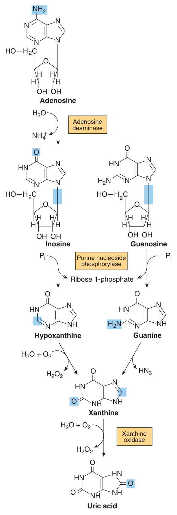
Humans convert adenosine and guanosine to uric acid.
Adenosine → (adenosine deaminase) → inosine → (purine nucleoside phosphorylase) → hypoxanthine → (xanthine oxidase) → xanthine → (xanthine oxidase) → URIC ACID
Guanosine → (purine nucleoside phosphorylase) → guanine → xanthine → uric acid
All the enzymes exist in the mucosa of the mammalian gastrointestinal tract, and purine deoxyribonucleosides are degraded by the same pathway.
In mammals other than higher primates, uricase converts uric acid to the water-soluble product allantoin. However, since humans lack uricase, the end product of purine catabolism in humans is uric acid.
Uric acid has a pKa of 5.8 — present as the relatively insoluble acid at acidic pH, or as its more soluble sodium urate salt at a pH near neutrality.
That single number explains where gout deposits and why.
At plasma pH 7.4, uric acid is largely ionised as the more soluble sodium urate. But drop the pH — or the temperature — and the balance shifts towards the relatively insoluble free acid.
Two consequences follow, and both are examinable. First, the urine is acidic, so uric acid precipitates there as stones — uric acid lithiasis. Second, peripheral joints are cooler than core body temperature, and solubility falls with temperature — which is why classical gout strikes the first metatarsophalangeal joint of the big toe, the coolest large joint in the body.
Alkalinising the urine is therefore rational therapy for the stones, and it works directly from the pKa.
A metabolic disorder of purine catabolism.
Various genetic defects in PRPP synthase present clinically as gout — an elevated Vmax, increased affinity for ribose 5-phosphate, or resistance to feedback inhibition — each resulting in overproduction and overexcretion of purine catabolites.
When serum urate levels exceed the solubility limit, sodium urate crystalizes in soft tissues and joints and causes an inflammatory reaction, gouty arthritis.
But note the qualification Harper's adds, which candidates routinely omit: most cases of gout reflect abnormalities in RENAL HANDLING of uric acid, not overproduction. Urate crystals are diagnostic of gout.
A hypoxanthine analog that inhibits xanthine oxidase, blocking the last two steps of purine catabolism.
The result is that hypoxanthine and xanthine accumulate instead of uric acid — and both are considerably more soluble than urate, so they are excreted without crystallising.
Allopurinol is also an alternate substrate for orotate phosphoribosyltransferase, and the nucleotide formed inhibits orotidylate decarboxylase, resulting in orotic aciduria and orotidinuria — a drug-induced version of the disease in §10.
- Adenosine deaminase converts? → Adenosine to inosine
- Which enzyme acts twice in purine catabolism? → Xanthine oxidase — hypoxanthine→xanthine→uric acid
- Why is uric acid the human end product? → Humans lack uricase, which would convert it to allantoin
- pKa of uric acid? → 5.8
- Most cases of gout are due to? → Abnormalities in renal handling of uric acid
- Allopurinol inhibits? → Xanthine oxidase
Other disorders ★★
An overproduction hyperuricemia characterized by frequent episodes of uric acid lithiasis and a bizarre syndrome of self-mutilation, reflecting a defect in hypoxanthine-guanine phosphoribosyl transferase (HGPRT), an enzyme of purine salvage.
The accompanying rise in intracellular PRPP results in purine overproduction. Salvage normally consumes PRPP; when it fails, PRPP accumulates and drives the de novo pathway harder — so the patient both fails to recycle purines and makes too many new ones.
Causative mutations include deletions, frameshift mutations, base substitutions and aberrant mRNA splicing.
| Disorder | Defective enzyme | Key features |
|---|---|---|
| Gout | PRPP synthase (in the genetic form) | Gouty arthritis; most cases are actually renal in origin |
| Lesch-Nyhan syndrome | HGPRT | Uricemia, uric acid lithiasis, self-mutilation |
| Von Gierke disease | Glucose-6-phosphatase | Purine overproduction secondary to enhanced generation of ribose 5-phosphate; an associated lactic acidosis elevates the renal threshold for urate |
| Hypouricemia | Xanthine oxidase | Increased excretion of hypoxanthine and xanthine; severe cases show xanthinuria and xanthine lithiasis. May also follow severe liver damage |
| Adenosine deaminase deficiency | Adenosine deaminase | Both T cells and B cells sparse and dysfunctional — severe immunodeficiency; without enzyme replacement or bone marrow transplantation, infants often succumb to fatal infections |
| Purine nucleoside phosphorylase deficiency | Purine nucleoside phosphorylase | Severe deficiency of T cells, but apparently normal B-cell function |
| Orotic aciduria type I | Orotate phosphoribosyltransferase AND orotidylate decarboxylase | Orotic aciduria with megaloblastic anaemia |
| Orotic aciduria type II | Orotidylate decarboxylase only | Rarer |
| β-Hydroxybutyric aciduria | Dihydropyrimidine dehydrogenase | Also called combined uraciluria-thyminuria; when inborn, serious neurological complications. A nongenetic form is triggered by 5-fluorouracil in patients with low enzyme levels |
Adenosine deaminase deficiency is worth understanding rather than memorising, because the mechanism links three separate sections of this unit.
Without ADA, adenosine and deoxyadenosine cannot be degraded, so they are phosphorylated instead, and dATP accumulates. Harper's gives the consequence: the immune dysfunctions appear to result from accumulation of dGTP and dATP, which inhibit ribonucleotide reductase and thereby deplete cells of DNA precursors (§7).
Now, why lymphocytes and not every cell? Because lymphocytes have the highest ADA activity and must undergo enormous clonal proliferation — they are the tissue most dependent on a rapid dNTP supply. A general block on DNA precursors falls hardest on the cells that divide most.
The result is severe combined immunodeficiency, and it is historically important: ADA-SCID was the first human disease treated by gene therapy.
Unlike the low-solubility products of purine catabolism, catabolism of the pyrimidines forms highly water-soluble products — CO₂, NH₃, β-alanine, and β-aminoisobutyrate.
Humans transaminate β-aminoisobutyrate to methylmalonate semialdehyde, which then forms succinyl-CoA. Excretion of β-aminoisobutyrate increases in leukemia and severe x-ray radiation exposure, due to increased destruction of DNA — though many persons of Chinese or Japanese ancestry routinely excrete β-aminoisobutyrate.
Pseudouridine is a curiosity worth knowing: no human enzyme catalyses its hydrolysis or phosphorolysis, so it is excreted unchanged in the urine of normal subjects — it was indeed first isolated from human urine.
- Lesch-Nyhan — enzyme and triad? → HGPRT; uricemia, uric acid lithiasis, self-mutilation
- Why does HGPRT deficiency cause overproduction? → Unconsumed PRPP accumulates and drives de novo synthesis
- Why does von Gierke disease cause hyperuricemia? → Excess ribose 5-phosphate, hence PRPP; plus lactic acidosis raising the renal urate threshold
- ADA deficiency affects which cells? → Both T and B cells — severe immunodeficiency
- PNP deficiency affects? → T cells severely, B cells apparently normally
- Orotic aciduria type I vs II? → I = both orotate phosphoribosyltransferase and orotidylate decarboxylase; II = decarboxylase only
- Which nucleoside is excreted unchanged? → Pseudouridine
Heme catabolism and jaundice ★★★
Lecture 20 closes with the other great ring-degradation pathway of the body. The logic is the same as §9's: a hydrophobic ring system that the body cannot burn must be converted into something excretable — and the disease appears at the point where the conversion fails.
Catabolism of the heme ring, initiated by the mitochondrial enzyme heme oxygenase, produces the linear tetrapyrrole biliverdin. Subsequent reduction of biliverdin in the cytosol forms bilirubin.
This occurs in the reticuloendothelial system. The iron of heme is released and reutilized, and the globin is degraded to free amino acids.
Since 1 g of hemoglobin yields about 35 mg of bilirubin, human adults form 250 to 350 mg of bilirubin per day, principally from haemoglobin but also from ineffective erythropoiesis and other heme proteins.
The conversion can be observed visually as the purple colour of the heme in a hematoma slowly converts to the yellow pigment of bilirubin.
Bilirubin is only sparingly water-soluble, but bilirubin bound to serum albumin is readily transported to the liver. Albumin's high-affinity site can bind approximately 25 mg of bilirubin per 100 mL of plasma; more loosely bound bilirubin can detach and diffuse into tissues, and antibiotics and certain other drugs can compete with and displace bilirubin from the high-affinity site.
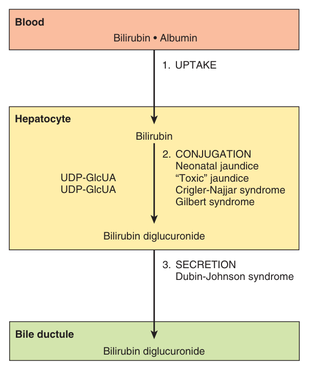
Hepatic catabolism of bilirubin takes place in three stages: uptake by the liver, conjugation with glucuronic acid, and secretion in the bile.
1 · Uptake — at the sinusoidal surface, by a large capacity, saturable facilitated transport system; once inside, bilirubin binds cytosolic proteins such as glutathione S-transferase (ligandin), preventing its return to the blood.
2 · Conjugation — bilirubin UDP-glucuronosyl transferase of the endoplasmic reticulum transfers two glucuronosyl moieties from UDP-glucuronate, giving bilirubin diglucuronide.
3 · Secretion — active transport via the multispecific organic anion transporter (MOAT) of the bile canaliculus, probably rate-limiting for the entire process.
Adding glucuronate does one thing: it makes a hydrophobic molecule water-soluble so it can be excreted. That is exactly the uronic acid pathway of Unit 14, which supplies the glucuronate used to excrete drugs and metabolites as glucuronides.
And notice how the properties of the two forms determine the entire clinical picture:
Unconjugated bilirubin is hydrophobic. It therefore travels bound to albumin, cannot appear in the urine, and can cross the blood-brain barrier.
Conjugated bilirubin is water-soluble. It therefore can appear in the urine, and cannot cross into the brain.
Harper's states both consequences directly: encephalopathy due to hyperbilirubinemia (kernicterus) thus occurs only with unconjugated bilirubin, and choluric jaundice occurs only in regurgitation hyperbilirubinemia. Two solubilities, two diseases. If you remember only that, you can reconstruct the whole of the table below.
When conjugated bilirubin reaches the terminal ileum and the large intestine, the glucuronosyl moieties are removed by specific bacterial β-glucuronidases. Subsequent reduction by the fecal flora forms a group of colorless tetrapyrroles called urobilinogens.
Small portions are reabsorbed in the terminal ileum and large intestine and reexcreted via the enterohepatic urobilinogen cycle; under abnormal conditions urobilinogen also appears in the urine. Most of the colorless urobilinogens formed in the colon are oxidized there to colored urobilins and excreted in the feces — and fecal darkening on standing in air results from the oxidation of residual urobilinogens to urobilins.
Quantitation uses the reddish-purple color formed when bilirubin reacts with diazotized sulfanilic acid.
“Direct bilirubin” — assayed without added methanol — is bilirubin glucuronide, i.e. CONJUGATED.
Total bilirubin — assayed with added methanol.
“Indirect bilirubin” — total minus direct — is UNCONJUGATED.
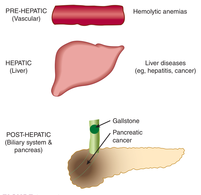
Hyperbilirubinemia is a blood level exceeding 1 mg of bilirubin per dL (17 μmol/L). It may result from production of more bilirubin than the normal liver can excrete, from failure of a damaged liver to excrete normal amounts, or from obstruction of the excretory ducts.
When the blood concentration reaches 2 to 2.5 mg/dL it diffuses into the tissues, which turn yellow — a condition termed jaundice or icterus.
Retention hyperbilirubinemia — due to overproduction, unconjugated.
Regurgitation hyperbilirubinemia — due to reflux into the bloodstream because of biliary obstruction, conjugated.
| Prehepatic — haemolytic anaemia | Hepatic — hepatitis | Posthepatic — obstruction | |
|---|---|---|---|
| Serum bilirubin | ↑ Indirect | ↑ Direct and indirect | ↑ Direct |
| Urine urobilinogen | Increased | Decreased if micro-obstruction is present | Absent |
| Urine bilirubin | Absent (acholuric) | Present if micro-obstruction occurs | Present (choluric) |
| Fecal urobilinogen | Increased | Decreased | Trace to absent — pale stools |
| Serum enzymes | — | ALT and AST markedly elevated | Alkaline phosphatase elevated |
The unconjugated hyperbilirubinemia of neonatal “physiologic jaundice” results from accelerated hemolysis and an immature hepatic system for the uptake, conjugation and secretion of bilirubin — bilirubin-glucosyltransferase activity, and probably also synthesis of UDP-glucuronate, are reduced.
When the plasma concentration of unconjugated bilirubin exceeds that which can be tightly bound by albumin (20–25 mg/dL), bilirubin can penetrate the blood-brain barrier. If left untreated, the resulting hyperbilirubinemic toxic encephalopathy, or KERNICTERUS, can result in mental retardation.
Treatment: exposure to blue light (phototherapy) promotes hepatic excretion of unconjugated bilirubin by converting some to derivatives that are excreted in the bile; phenobarbital, a promoter of bilirubin metabolism, may be administered.
| Disorder | Nature | Note |
|---|---|---|
| Gilbert syndrome | Unconjugated | About 30% of transferase activity is retained — the condition is harmless |
| Crigler-Najjar type I | Unconjugated | Complete absence of hepatic UDP-glucuronosyl transferase; over 20 mg/dL, brain damage, often fatal within the first 15 months. Phototherapy helps a little; phenobarbital has no beneficial effect |
| Crigler-Najjar type II | Unconjugated | Some activity retained; more benign, responds to large doses of phenobarbital |
| Dubin-Johnson syndrome | Conjugated | Benign autosomal recessive; mutation in the protein that secretes conjugated bilirubin into bile |
| Rotor syndrome | Conjugated | Benign |
| Toxic hyperbilirubinemia | Unconjugated | Chloroform, carbon tetrachloride, acetaminophen, hepatitis virus, cirrhosis, Amanita mushroom poisoning |
- Which enzyme initiates heme catabolism, and where? → Heme oxygenase, mitochondrial; it gives biliverdin
- How much bilirubin is formed daily? → 250–350 mg; 1 g haemoglobin yields ~35 mg
- How is bilirubin transported to the liver? → Bound to serum albumin
- The three hepatic stages? → Uptake, conjugation with glucuronic acid, secretion in bile
- Which step is rate-limiting? → Secretion into the bile
- Direct bilirubin = ? → Conjugated. Indirect = total − direct = unconjugated
- Which form crosses the blood-brain barrier? → Unconjugated only — hence kernicterus
- Which form appears in urine? → Conjugated only — hence choluric jaundice
- Jaundice becomes visible at what level? → 2–2.5 mg/dL
- Crigler-Najjar type I vs II? → I = complete absence, fatal, no response to phenobarbital; II = partial, responds
Revision layer
The contrast that organises the whole unit
| Purines | Pyrimidines | |
|---|---|---|
| Ring assembly | Built ON PRPP (the ribose is the scaffold) | Ring built FIRST, PRPP added at OMP |
| First committed enzyme | PRPP glutamyl amidotransferase | Carbamoyl phosphate synthase II (cytosolic, glutamine) |
| Parent nucleotide | IMP | UMP |
| Nitrogen donors | Glycine, glutamine, aspartate | Glutamine, aspartate |
| Folate needed? | Yes — C2 and C8 from N¹⁰-formyl-THF | Only at thymidylate synthase |
| Catabolic end product | Uric acid — insoluble | CO₂, NH₃, β-alanine, β-aminoisobutyrate — soluble |
| Clinical burden | Gout, stones, Lesch-Nyhan, SCID | Orotic aciduria only, and it is rare |
Numbers to have ready
| Quantity | Value |
|---|---|
| Bilirubin from 1 g haemoglobin | ~35 mg |
| Bilirubin formed daily | 250–350 mg |
| Albumin high-affinity binding capacity | ~25 mg/100 mL plasma |
| Definition of hyperbilirubinemia | >1 mg/dL (17 μmol/L) |
| Bilirubin level at which jaundice is visible | 2–2.5 mg/dL |
| Level at which kernicterus threatens | >20–25 mg/dL unconjugated |
| pKa of uric acid | 5.8 |
| Transferase activity retained in Gilbert syndrome | ~30% |
| Crigler-Najjar type I survival | often fatal within the first 15 months |
Enzyme → disease, at a glance
| Enzyme | Disorder |
|---|---|
| PRPP synthase (defects) | Gout |
| HGPRT | Lesch-Nyhan syndrome |
| Adenosine deaminase | Severe combined immunodeficiency (T and B) |
| Purine nucleoside phosphorylase | T-cell deficiency |
| Xanthine oxidase | Hypouricemia, xanthinuria |
| Glucose-6-phosphatase | Von Gierke disease with hyperuricemia |
| Orotate PRTase + orotidylate decarboxylase | Orotic aciduria type I |
| Orotidylate decarboxylase | Orotic aciduria type II |
| Dihydropyrimidine dehydrogenase | β-Hydroxybutyric aciduria; 5-FU toxicity |
| Ornithine transcarbamoylase | Hyperammonemia type 2 and orotic aciduria |
| Bilirubin UDP-glucuronosyl transferase | Gilbert, Crigler-Najjar I and II |
“Purines are built on the sugar; pyrimidines are built then sugared.”
For the bilirubin lab table: “Direct = conjugated = in the urine.” All three words describe the water-soluble form, and the unconjugated form is the opposite of all three.
- What determines the rate of de novo purine synthesis? → The concentration of PRPP
- Which is the committed step of purine synthesis? → PRPP glutamyl amidotransferase → 5-phosphoribosylamine
- Parent nucleotides of the two pathways? → IMP for purines, UMP for pyrimidines
- CPS-I vs CPS-II? → I mitochondrial/ammonia/urea; II cytosolic/glutamine/pyrimidines
- Which enzyme provides all dNTPs? → Ribonucleotide reductase, using thioredoxin and NADPH
- Methotrexate's target and consequence? → Dihydrofolate reductase; thymineless death of dividing cells
- Why do humans make uric acid? → They lack uricase
- Two reasons a Lesch-Nyhan patient overproduces purines? → Salvage fails, and unconsumed PRPP drives de novo synthesis
- Why is there no pyrimidine equivalent of gout? → Pyrimidine catabolites are highly water-soluble
- Which bilirubin crosses into the brain, and which into the urine? → Unconjugated into the brain, conjugated into the urine