Metabolism of Proteins & Amino Acids
Nitrogen balance and protein turnover ★★
Every other fuel in this course has a store. Glucose has glycogen, fat has the adipocyte — and if you eat more than you need, the surplus is put away for later. Amino acids have no such arrangement. Harper's states it flatly: excess free amino acids are not stored for future use. Whatever is not incorporated into new protein within a short time is rapidly degraded.
That single fact organises the whole of this unit. Because there is no amino acid store, the body must dispose of surplus nitrogen continuously — and the nitrogen it must dispose of is ammonia, which is highly toxic to the central nervous system. So the design problem is not storage but safe disposal: collect the nitrogen, carry it to the liver without poisoning anything on the way, and convert it into something harmless that the kidney can excrete. Sections 4 to 7 are that journey, one step at a time.
Nitrogen balance = nitrogen ingested (primarily as protein) − nitrogen excreted (primarily as urea).
Nitrogen equilibrium (balance = 0): protein synthesis = protein degradation. The normal state of a healthy adult.
Positive nitrogen balance: an excess of ingested over excreted nitrogen; synthesis exceeds degradation. Accompanies growth and pregnancy.
Negative nitrogen balance: output exceeds intake; degradation exceeds synthesis. May follow surgery, advanced cancer, and the nutritional disorders kwashiorkor and marasmus.
The continuous degradation and synthesis of cellular proteins, which occurs in all forms of life.
Each day humans turn over 1% to 2% of their total body protein, principally muscle protein. High rates of degradation occur in tissues undergoing structural rearrangement — uterine tissue during pregnancy, skeletal muscle in starvation, tadpole tail tissue during metamorphosis.
About 75% of the amino acids liberated by protein degradation are reutilized; the remainder are degraded, the carbon skeletons to amphibolic intermediates and the nitrogen to urea.
The time required to lower its concentration to half of its initial value — the measure of a protein's susceptibility to degradation.
Half-lives of liver proteins range from under 30 minutes to over 150 hours. Typical “housekeeping” enzymes, such as those of glycolysis, have t½ values of over 100 hours; key regulatory enzymes may have t½ values as low as 0.5 to 2 hours.
This is not an accident of chemistry — it is a design requirement, and you have already met its consequences half a dozen times in this course.
A regulatory enzyme exists in order to change. If the cell wants to double the amount of HMG-CoA reductase when sterols run low, or halve it when they are plentiful, then transcription alone is not enough: a long-lived protein would sit there for days after the signal had passed. A short half-life is what makes an enzyme's concentration responsive. A housekeeping enzyme, by contrast, is wanted at a steady level all the time, and replacing it constantly would be pure waste.
So the rule to carry away is: the more tightly regulated an enzyme, the shorter its half-life. PEST sequences — regions rich in proline (P), glutamate (E), serine (S) and threonine (T) — target some proteins for rapid degradation, and it is the regulatory proteins that carry them.
- Nitrogen balance = ? → Nitrogen ingested − nitrogen excreted
- Positive nitrogen balance occurs in? → Growth and pregnancy
- Negative nitrogen balance occurs in? → Surgery, advanced cancer, kwashiorkor and marasmus
- How much body protein is turned over daily? → 1–2%, principally muscle protein
- What proportion of liberated amino acids is reutilized? → About 75%
- t½ of a housekeeping enzyme vs a regulatory enzyme? → Over 100 hours vs 0.5–2 hours
- What are PEST sequences? → Regions rich in Pro, Glu, Ser and Thr that target proteins for rapid degradation
How proteins are degraded ★★
There are two routes, and the distinction between them is worth getting right because it is exactly the kind of paired contrast an examiner can set in one line: one costs ATP and one does not.
| ATP-independent — lysosomal | ATP- and ubiquitin-dependent — cytosolic | |
|---|---|---|
| Substrates | Extracellular, membrane-associated and long-lived intracellular proteins | Regulatory proteins with short half-lives, and abnormal or misfolded proteins |
| Where | The lysosome | The cytosol, then the proteasome |
| Energy | None required | Requires ATP and ubiquitin |
| Tagging | Loss of a sialic acid moiety marks blood glycoproteins; the resulting asialoglycoproteins are internalized by liver-cell asialoglycoprotein receptors | Polyubiquitination — four or more ubiquitin molecules |
A small (8.5 kDa, 76 residue) polypeptide, named for its presence in all eukaryotic cells, that targets intracellular proteins for degradation.
Its primary structure is highly conserved — only three of the 76 residues differ between yeast and human ubiquitin. It is attached by a non-α-peptide bond between the carboxyl terminal of ubiquitin and the ε-amino group of a lysyl residue in the target protein.
Three enzymes are involved: E1 an activating enzyme, E2 a ligase, and E3 a transferase. The residue at the target's amino terminus matters — Met or Ser retard, whereas Asp or Arg accelerate ubiquitination.
A macromolecular, cylindrical complex of proteins, ubiquitous in eukaryotic cells, whose stacked rings form a central pore that harbors the active sites of proteolytic enzymes.
For degradation a protein must first enter the central pore, and entry is gated by two outer regulatory rings that recognize polyubiquitinated proteins and reject everything else.
Think about what a free protease loose in the cytosol would do. It would digest the cell. The proteasome solves that by putting the blades inside a tube and guarding the door — proteolysis is not restrained by regulating the enzyme's activity but by controlling access to it.
You have seen this architectural idea before, in a different guise: the fatty acid synthase complex of Unit 16 keeps its intermediates on the protein so they cannot go astray. Same principle — geometry doing the work that regulation would otherwise have to do.
Genetic defects in the genes encoding ubiquitin, ubiquitin ligases, or deubiquitinating enzymes include Angelman syndrome, autosomal recessive juvenile Parkinson's disease, von Hippel-Lindau syndrome, and congenital polycythemia.
Ciechanover, Hershko and Rose received the 2004 Nobel Prize in Chemistry for the discovery of ubiquitin-mediated protein degradation.
- Which pathway degrades short-lived regulatory and abnormal proteins? → The ATP- and ubiquitin-dependent cytosolic pathway
- What size is ubiquitin? → 8.5 kDa, 76 residues
- Ubiquitin attaches to which group? → The ε-amino group of a lysyl residue
- Name the three enzymes of ubiquitination → E1 activating, E2 ligase, E3 transferase
- What gates entry into the proteasome? → Two outer rings that recognise polyubiquitinated proteins only
Interorgan amino acid exchange ★★
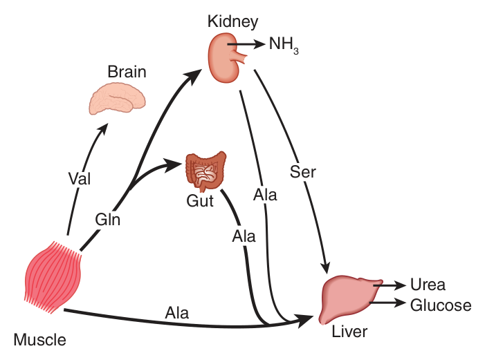
Two organs dominate. Muscle generates over half of the total body pool of free amino acids, and the liver is the site of the urea cycle enzymes necessary for disposal of excess nitrogen. Everything else is traffic between them.
| State | What moves where |
|---|---|
| Postabsorptive (between meals) | Free amino acids — particularly alanine and glutamine — are released from muscle. Alanine is extracted primarily by the liver; glutamine is extracted by the gut and the kidney, both of which convert a significant portion to alanine. The kidney provides a major source of serine. Branched-chain amino acids, particularly valine, are released by muscle and taken up predominantly by the brain. |
| After a protein-rich meal | The splanchnic tissues release amino acids while the peripheral muscles extract them — in both cases predominantly branched-chain amino acids, which have been spared by the liver. |
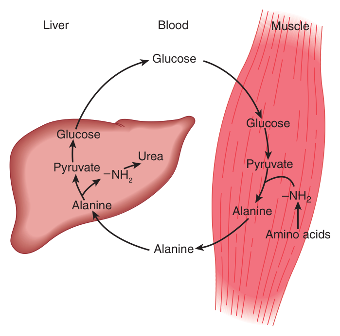
Alanine is synthesized in muscle by transamination of glucose-derived pyruvate, released into the bloodstream, and taken up by the liver. In the liver the carbon skeleton of alanine is reconverted to glucose and released into the bloodstream, where it is available for uptake by muscle and resynthesis of alanine.
Alanine is a key gluconeogenic amino acid — the rate of hepatic gluconeogenesis from alanine is far higher than from all other amino acids, and the liver's capacity does not saturate until the alanine concentration reaches 20 to 30 times its normal physiologic level.
Look at what the muscle needs to get rid of and what the liver needs to receive, and the glucose-alanine cycle almost designs itself.
Working muscle has surplus nitrogen from protein breakdown and surplus pyruvate from glycolysis. It cannot make urea — it has no urea cycle — and it cannot release free ammonia into the blood without poisoning the brain. So it does the obvious thing: it staples the nitrogen onto the pyruvate, and alanine, a perfectly harmless molecule, carries both to the liver.
The liver then takes the two apart and uses each: the nitrogen enters the urea cycle, the carbon skeleton enters gluconeogenesis, and the glucose goes back to the muscle. Compare the Cori cycle of Unit 13, which does the same trick with lactate — but lactate carries carbon only. The glucose-alanine cycle carries carbon AND nitrogen in one vehicle.
- Which tissue generates over half the total body pool of free amino acids? → Muscle
- Which two amino acids are released from muscle postabsorptively? → Alanine and glutamine
- Which organs extract glutamine? → The gut and the kidney
- Which amino acids are taken up predominantly by the brain? → Branched-chain, particularly valine
- What does the glucose-alanine cycle carry that the Cori cycle does not? → Nitrogen as well as carbon
Transamination ⭐
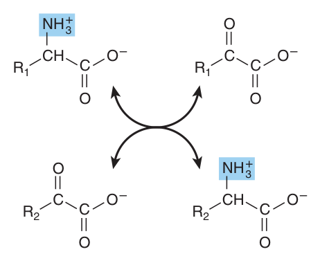
The reversible transfer of an α-amino group from an α-amino acid to an α-keto acid, interconverting pairs of α-amino acids and α-keto acids, catalysed by aminotransferases (transaminases) with pyridoxal phosphate (PLP), a derivative of vitamin B₆, at the catalytic site.
The reaction is freely reversible, with an equilibrium constant close to unity.
All of the common amino acids except lysine, threonine, proline and hydroxyproline participate in transamination. Transamination is not restricted to α-amino groups — the δ-amino group of ornithine (but not the ε-amino group of lysine) readily undergoes transamination.
Two aminotransferases matter most. Alanine aminotransferase transfers amino groups to pyruvate, forming alanine; glutamate aminotransferase transfers them to α-ketoglutarate, forming glutamate. Each aminotransferase is specific for one pair of substrates but nonspecific for the other pair.
Here is the piece of reasoning that makes this section make sense, and it is worth following slowly because the examiner is really testing whether you have followed it.
L-glutamate is the only amino acid that undergoes oxidative deamination at an appreciable rate in mammalian tissues. That is the constraint. There is essentially one door out of the amino acid pool, and it is glutamate's door.
So the nitrogen of every other amino acid has to be moved into glutamate before it can leave. How? Since alanine is also a substrate for glutamate aminotransferase, the α-amino nitrogen from all amino acids that undergo transamination can be concentrated in glutamate. Whatever the nitrogen started on, a chain of freely reversible transaminations delivers it to α-ketoglutarate.
The formation of ammonia from α-amino groups thus occurs mainly via the α-amino nitrogen of L-glutamate. Transamination is the collection system; §5 is the release step. Say those two sentences in that order and you have the mechanism.
Transamination occurs via a “ping-pong” mechanism, characterized by the alternate addition of a substrate and release of a product.
PLP serves as a “carrier” of amino groups. An enzyme-bound Schiff base forms between the oxo group of enzyme-bound PLP and the α-amino group of the amino acid; rearrangement then yields an α-keto acid and enzyme-bound pyridoxamine phosphate, which passes the amino group to the second keto acid.
The enzyme therefore never holds both substrates at once — the first product leaves before the second substrate arrives. That is what “ping-pong” means, and it is why the enzyme itself alternates between the PLP and pyridoxamine phosphate forms.
This is Unit 7's diagnostic-enzyme principle applied: an enzyme normally confined to a cell appears in the plasma when that cell is damaged.
Aspartate aminotransferase (AST, or SGOT) — myocardial infarction.
Alanine aminotransferase (ALT, or SGPT) — viral hepatitis.
Both are widely used liver function tests, and both derive their value from the fact that transamination is a cytosolic and mitochondrial intracellular process.
- Define transamination → The reversible transfer of an α-amino group from an α-amino acid to an α-keto acid
- Which coenzyme is required? → Pyridoxal phosphate, from vitamin B₆
- Which four amino acids do NOT undergo transamination? → Lysine, threonine, proline and hydroxyproline
- Why does all amino nitrogen collect in glutamate? → Because glutamate is the only amino acid that undergoes oxidative deamination at an appreciable rate
- What is the equilibrium constant? → Close to unity — the reaction is freely reversible
- AST is raised in? → Myocardial infarction. ALT? → Viral hepatitis

Glutamate dehydrogenase — the central position ★★★
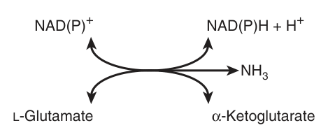
The hepatic enzyme that releases the nitrogen of glutamate as ammonia, converting L-glutamate + NAD(P)⁺ + H₂O → α-ketoglutarate + NH₃ + NAD(P)H + H⁺. It can use either NAD⁺ or NADP⁺.
The reaction is reversible, but strongly favours glutamate formation. It is allosterically inhibited by ATP, GTP and NADH, and activated by ADP.
Because it is reversible, it functions in both nitrogen catabolism and amino acid biosynthesis — which is precisely why Harper's says it occupies a central position in nitrogen metabolism.
The conversion of α-amino nitrogen to ammonia by the concerted action of glutamate aminotransferase and glutamate dehydrogenase.
Transamination collects the nitrogen onto α-ketoglutarate to form glutamate; oxidative deamination then strips it off as ammonia and regenerates the α-ketoglutarate. The two reactions coupled together are transdeamination.
Inhibited by ATP, GTP and NADH; activated by ADP. Translate that: GDH runs when the cell is energy-poor and stops when it is energy-rich.
Which makes sense of what the reaction actually does. Deaminating glutamate produces α-ketoglutarate — a citric acid cycle intermediate, a fuel. A cell with plenty of ATP has no reason to dismantle amino acids for energy; a cell running short has every reason. So the same signal that switches on isocitrate dehydrogenase in Unit 11 switches on GDH here. It is the same logic, applied to a different substrate.
And note the direction of the equilibrium: it strongly favours glutamate formation. The reaction that releases ammonia is running uphill against its own equilibrium. What pulls it forward is the fact that the ammonia is removed immediately — straight into carbamoyl phosphate (§7). The urea cycle is what makes deamination possible.
An enzyme of liver and kidney which converts an amino acid to an α-imino acid, an unstable intermediate that decomposes to an α-keto acid with release of ammonium ion.
The reduced flavin is reoxidized by molecular oxygen, forming hydrogen peroxide (H₂O₂), which is then split to O₂ and H₂O by catalase.
Note that this is a minor route in humans — the main path to ammonia is via glutamate dehydrogenase.
- What does glutamate dehydrogenase do? → Releases the nitrogen of glutamate as ammonia, forming α-ketoglutarate
- Which coenzymes can it use? → Either NAD⁺ or NADP⁺
- Inhibited by? → ATP, GTP and NADH. Activated by? → ADP
- Define transdeamination → The concerted action of glutamate aminotransferase and glutamate dehydrogenase converting α-amino nitrogen to ammonia
- What disposes of the H₂O₂ made by L-amino acid oxidase? → Catalase
Ammonia — transport and toxicity ★★★
Ammonia has now been released. It must reach the liver — and it must do so without ever reaching a toxic concentration in the blood. Only traces (10–20 μg/dL) are normally present in peripheral blood.
The mitochondrial enzyme that fixes ammonia as glutamine: glutamate + NH₄⁺ + ATP → glutamine + ADP + Pi.
Because amide bond synthesis is coupled to the hydrolysis of ATP to ADP and Pi, the reaction strongly favours glutamine synthesis.
Besides providing glutamine as a carrier of nitrogen, carbon and energy between organs, glutamine synthase plays a major role in ammonia detoxification and acid-base homeostasis. A rare neonatal deficiency causes severe brain damage, multiorgan failure and death.
The mitochondrial enzyme that hydrolytically releases the amide nitrogen of glutamine as ammonia: glutamine + H₂O → glutamate + NH₄⁺. The reaction proceeds essentially irreversibly in the direction of glutamate and ammonium formation.
Note carefully: it is the amide nitrogen, not the α-amino nitrogen, that is removed.
There are two human isoforms, liver-type and renal-type, products of different genes and differing in structure, kinetics and regulation. Hepatic glutaminase levels rise in response to high protein intake; renal kidney-type glutaminase increases in metabolic acidosis.
The concerted action of glutamine synthase and glutaminase catalyses the interconversion of free ammonium ion and glutamine. Read that as a transport system.
In the peripheral tissue, glutamine synthase wraps the toxic ammonia into a harmless amide — at the cost of one ATP, which is the price of safe carriage. Glutamine travels in the blood, where it is one of the most abundant amino acids precisely because it is doing this job. In the liver, glutaminase unwraps it, releasing the ammonia exactly where the urea cycle is waiting for it.
Two enzymes catalysing opposite reactions in the same cell would be a futile cycle. Two enzymes catalysing opposite reactions in different organs is a delivery service. Compare the liver-versus-periphery split of the ketone bodies in Unit 17 — the same architecture, a different cargo.
Excretion into urine of ammonia produced by renal tubular cells facilitates cation conservation and regulation of acid-base balance.
Ammonia production from intracellular renal amino acids, especially glutamine, increases in metabolic acidosis and decreases in metabolic alkalosis. NH₃ buffers a secreted proton as NH₄⁺, allowing H⁺ to be excreted without the loss of Na⁺ or K⁺.
Ammonia produced by enteric bacteria and absorbed into portal venous blood, and ammonia produced by tissues, are rapidly removed from the circulation by the liver and converted to urea. Should portal blood bypass the liver, systemic blood ammonia may reach toxic levels — as in severely impaired hepatic function, or the development of collateral links between the portal and systemic veins in cirrhosis.
Symptoms: tremor, slurred speech, blurred vision, coma, and ultimately death.
Mechanism: ammonia may be toxic to the brain in part because it reacts with α-ketoglutarate to form glutamate. The resulting depletion of α-ketoglutarate then impairs function of the tricarboxylic acid cycle in neurons.
Look at what is actually happening: a metabolite is being drained out of the citric acid cycle and not replaced. That is the anaplerosis problem of Unit 11 — the cycle is catalytic, so removing an intermediate stops it turning.
And the neuron is uniquely exposed. It depends almost entirely on oxidative phosphorylation, it cannot use fatty acids, and it has no reserve. Stall its citric acid cycle and you stall its entire ATP supply within minutes — hence tremor, slurred speech, coma, in that order, as function fails from the most demanding tissue downwards.
This is also why hyperammonemia and hypoglycemia produce such similar neurological pictures: both are, in the end, failures of neuronal ATP supply.
| Animal | End product | Term | Why |
|---|---|---|---|
| Fish (teleostean) | Ammonia | Ammonotelic | Their aqueous environment permits them to excrete water continuously, diluting the highly toxic ammonia |
| Birds | Uric acid | Uricotelic | Birds must conserve water and maintain low weight; nitrogen-rich uric acid is excreted as semisolid guano |
| Humans and many land animals | Urea | Ureotelic | Urea is nontoxic and highly water-soluble |
- Normal peripheral blood ammonia? → Only traces, 10–20 μg/dL
- Which enzyme fixes ammonia as glutamine? → Glutamine synthase, mitochondrial, ATP-dependent
- Which nitrogen does glutaminase remove? → The amide nitrogen, not the α-amino nitrogen
- Renal glutaminase increases in? → Metabolic acidosis
- Why is ammonia toxic to the brain? → It reacts with α-ketoglutarate to form glutamate, depleting the TCA cycle in neurons
- Fish, birds and humans are? → Ammonotelic, uricotelic and ureotelic respectively
The urea cycle ★★★
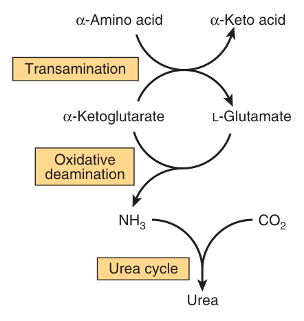
Urea biosynthesis occurs in four stages: (1) transamination, (2) oxidative deamination of glutamate, (3) ammonia transport, and (4) the reactions of the urea cycle.
Sections 4, 5 and 6 were stages 1, 2 and 3. This section is stage 4.
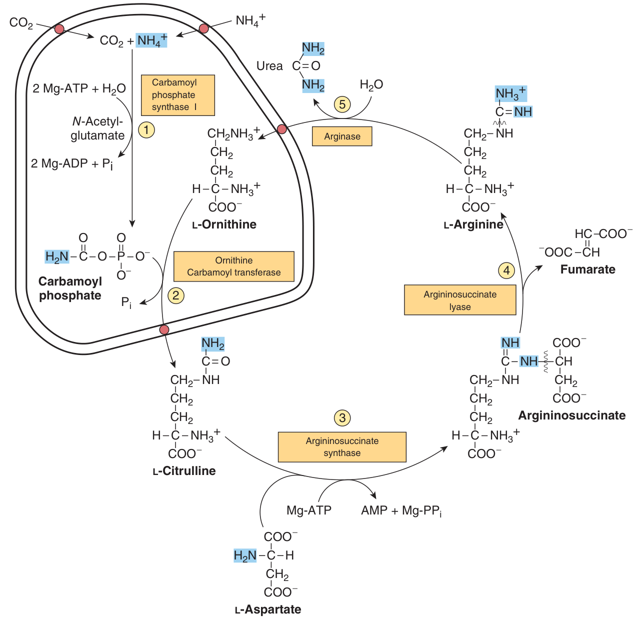
Synthesis of 1 mol of urea requires 3 mol of ATP, 1 mol each of ammonium ion and of aspartate, and employs five enzymes.
NH₃, CO₂, and the amide nitrogen of aspartate provide the atoms of urea.
Of the six participating amino acids, N-acetylglutamate functions solely as an enzyme activator; the others carry the atoms that become urea. The major metabolic role of ornithine, citrulline and argininosuccinate in mammals is urea synthesis.
| # | Reaction | Enzyme | Where |
|---|---|---|---|
| 1 | CO₂ + NH₄⁺ + 2 ATP → carbamoyl phosphate | Carbamoyl phosphate synthase I — requires N-acetylglutamate | Mitochondrial matrix |
| 2 | Carbamoyl phosphate + ornithine → citrulline + Pi | Ornithine transcarbamoylase (ornithine carbamoyl transferase) | Mitochondrial matrix |
| 3 | Citrulline + aspartate + ATP → argininosuccinate + AMP + PPi | Argininosuccinate synthase | Cytosol |
| 4 | Argininosuccinate → arginine + fumarate | Argininosuccinate lyase | Cytosol |
| 5 | Arginine + H₂O → urea + ornithine | Arginase | Cytosol |
Reactions 1 and 2 occur in the matrix of liver mitochondria; reactions 3, 4 and 5 in liver cytosol. Because of that split, CO₂ (as bicarbonate), ammonium ion, ornithine and citrulline enter or leave the matrix via specific carriers in the inner membrane — entry of ornithine and exodus of citrulline involve mitochondrial inner membrane permeases.
This is the point on which most urea cycle answers are won or lost, so trace it once deliberately.
The first nitrogen enters as free ammonium ion, at reaction 1, built into carbamoyl phosphate. That is the nitrogen that came off glutamate in §5.
The second nitrogen enters at reaction 3, as the amino group of aspartate — argininosuccinate synthase links aspartate and citrulline via the amino group of aspartate and provides the second nitrogen of urea. It never appears as free ammonia at all.
Now watch what happens to the aspartate skeleton. Reaction 4 proceeds with retention of all three nitrogens in arginine and release of the aspartate skeleton as fumarate. The fumarate is hydrated to L-malate and oxidised to oxaloacetate by cytosolic fumarase and malate dehydrogenase — reactions analogous to the citric acid cycle but in the cytosol — and transamination of oxaloacetate by glutamate aminotransferase then re-forms aspartate.
So the carbon skeleton of aspartate-fumarate acts as a carrier of the nitrogen of glutamate into a precursor of urea. Aspartate is not consumed; it is a shuttle. And that fumarate is the link between the urea cycle and the citric acid cycle — the two are sometimes drawn together as the “Krebs bicycle” for exactly this reason.
The rate-limiting enzyme of the urea cycle. It condenses CO₂, ammonia and ATP, and is active only in the presence of N-acetylglutamate, an allosteric activator that enhances the affinity of the synthase for ATP.
Synthesis of 1 mol of carbamoyl phosphate requires 2 mol of ATP — one as the phosphoryl donor for the mixed acid anhydride bond, one to drive synthesis of the amide bond; the products are 2 ADP and 1 Pi.
Do not confuse with carbamoyl phosphate synthase II, which is cytosolic, uses glutamine rather than ammonia as the nitrogen donor, and functions in pyrimidine biosynthesis (Unit 21).
Changes in enzyme levels and allosteric regulation of carbamoyl phosphate synthase I by N-acetylglutamate regulate urea biosynthesis.
The steady-state level of N-acetylglutamate is set by the balance between N-acetylglutamate synthase (NAGS), which makes it from acetyl-CoA and glutamate, and N-acetylglutamate deacylase, which hydrolyses it back.
Major changes in diet can increase the concentrations of individual urea cycle enzymes 10- to 20-fold. Starvation elevates enzyme levels, presumably to cope with the ammonia produced by starvation-induced protein degradation; expression of the RNAs for all the urea cycle enzymes increases several-fold in starvation.
It is made from acetyl-CoA and glutamate — and think about what each of those reports.
Glutamate is the collection point for all amino acid nitrogen (§4). A high glutamate concentration means nitrogen is arriving fast. Acetyl-CoA means fuel is available to pay the 3 ATP the cycle costs. N-acetylglutamate therefore rises exactly when there is both a nitrogen load to dispose of and the energy to dispose of it, and it switches on the rate-limiting enzyme.
This is the same design you met at pyruvate carboxylase in Unit 13, where acetyl-CoA signals “there is fuel, start gluconeogenesis.” A metabolite that is a by-product of the situation becomes the signal for the response to it. Once you see that pattern you will find it everywhere in this course.
- The four stages of urea biosynthesis? → Transamination, oxidative deamination of glutamate, ammonia transport, and the urea cycle
- How much ATP per mole of urea? → 3 mol ATP, and 5 enzymes
- Where do the atoms of urea come from? → NH₃, CO₂ and the amide nitrogen of aspartate
- Which reactions are mitochondrial? → 1 and 2; reactions 3, 4 and 5 are cytosolic
- Rate-limiting enzyme and its activator? → Carbamoyl phosphate synthase I, activated by N-acetylglutamate
- What links the urea cycle to the citric acid cycle? → Fumarate, released at reaction 4
- Which amino acid is regenerated in reaction 5? → Ornithine
- CPS-II differs how? → Cytosolic, uses glutamine, and serves pyrimidine biosynthesis
Urea cycle disorders ★★
Urea cycle disorders are characterized by hyperammonemia, encephalopathy and respiratory alkalosis.
Clinical symptoms common to all: vomiting, avoidance of high-protein foods, intermittent ataxia, irritability, lethargy, and severe mental retardation. The most dramatic presentation is in full-term infants who initially appear normal, then exhibit progressive lethargy, hypothermia and apnea due to high plasma ammonia.
The clinical features and treatment of all five disorders are similar.
| Enzyme | Disorder | Note |
|---|---|---|
| Carbamoyl phosphate synthase I | Hyperammonemia type 1 | Estimated frequency 1:62,000 |
| N-acetylglutamate synthase (NAGS) | Clinically indistinguishable from CPS-I deficiency | May respond to administered N-acetylglutamate |
| Ornithine permease (ORNT1) | HHH syndrome — hyperornithinemia, hyperammonemia, homocitrullinuria | Ornithine cannot enter the matrix; in the absence of its normal acceptor, carbamoyl phosphate carbamoylates lysine to homocitrulline |
| Ornithine transcarbamoylase | Hyperammonemia type 2 — X-chromosome linked | The mothers also exhibit hyperammonemia and an aversion to high-protein foods. Glutamine is elevated in blood, CSF and urine |
| Argininosuccinate synthase | Citrullinemia | Plasma and CSF citrulline elevated; 1 to 2 g of citrulline excreted daily. A 25-fold elevated Km for citrulline has also been reported |
| Argininosuccinate lyase | Argininosuccinic aciduria | Associated with friable, tufted hair (trichorrhexis nodosa). Diagnosis by erythrocyte enzyme assay on cord blood or amniotic fluid cells |
| Arginase | Hyperargininemia — autosomal recessive | Unlike other urea cycle disorders, the first symptoms typically do not appear until age 2 to 4 years |
The obvious treatment for a child who cannot dispose of amino nitrogen is to stop giving them protein. It does not work, and the reason is a fact from Unit 1: mammals are incapable of synthesizing all 20 amino acids. The nutritionally essential ones must come from the diet.
So treatment is a balance rather than an elimination. Harper's puts it precisely: the goal of dietary therapy is to provide sufficient protein, arginine and energy to promote growth and development while simultaneously minimizing the metabolic perturbations. In practice that means a low-protein diet ingested as frequent small meals, to avoid sudden increases in blood ammonia levels — and doing so brings significant improvement and minimization of brain damage.
Note why arginine is supplied: in a cycle that is not turning properly, arginine becomes conditionally essential, and it also drives what remains of the cycle forward.
Tandem mass spectrometry can in a few minutes detect over 40 analytes of significance in the detection of metabolic disorders, and is the technique of choice for screening neonates for organic acidemias, aminoacidemias, disorders of fatty acid oxidation, and defects in the enzymes of the urea cycle.
This is Unit 2's mass spectrometry put to clinical work — and the justification is simple: early dietary intervention can in many instances ameliorate the otherwise inevitable dire effects.
- The triad of urea cycle disorders? → Hyperammonemia, encephalopathy and respiratory alkalosis
- Which urea cycle disorder is X-linked? → Ornithine transcarbamoylase deficiency (hyperammonemia type 2)
- Which is associated with friable tufted hair? → Argininosuccinate lyase deficiency
- Which presents late, at age 2–4 years? → Hyperargininemia (arginase deficiency)
- Why is a block at reaction 1 or 2 worse? → Because if citrulline can be made, some ammonia is already covalently trapped
- Why can a protein-free diet not be used? → Mammals cannot synthesise all 20 amino acids
The carbon skeletons ★★★
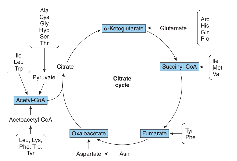
The nitrogen has gone. What remains is the carbon “skeleton” — an α-keto acid — and its fate is the second half of amino acid catabolism. Excess amino acids are catabolized to amphibolic intermediates used as sources of energy or for carbohydrate and lipid biosynthesis.
Glucogenic (glycogenic): the carbon skeleton is degraded to pyruvate or a citric acid cycle intermediate — α-ketoglutarate, succinyl-CoA, fumarate or oxaloacetate — and can therefore give rise to glucose by gluconeogenesis.
Ketogenic: the carbon skeleton is degraded to acetyl-CoA, acetoacetyl-CoA or acetoacetate, and can therefore give rise to ketone bodies and fat, but never to glucose.
The dividing line is not arbitrary; it is a single irreversible reaction, and you have met it twice already.
Pyruvate dehydrogenase is essentially irreversible. Carbon that enters as acetyl-CoA cannot come back out as pyruvate. And the two carbons of acetyl-CoA that enter the citric acid cycle are balanced by two carbons leaving as CO₂ in the same turn — so acetyl-CoA produces no net oxaloacetate, and therefore no net glucose.
That is why fatty acids are not glucogenic (Unit 17) and why an amino acid degraded to acetyl-CoA is not glucogenic either. One reaction explains both facts. If you can state the reason rather than reciting the list, you will never get this wrong.
| Classification | Amino acids |
|---|---|
| Exclusively ketogenic | Leucine and lysine — only these two |
| Both glucogenic and ketogenic | Isoleucine, phenylalanine, tryptophan, tyrosine |
| Glucogenic | All the rest — Ala, Arg, Asn, Asp, Cys, Gln, Glu, Gly, His, Hyp, Met, Pro, Ser, Thr, Val |
| Entry point | Amino acids |
|---|---|
| Pyruvate | Alanine, tryptophan, cysteine, serine, glycine, threonine — the six degraded to pyruvate |
| Oxaloacetate | Asparagine, aspartate — all four carbons, via asparaginase and transamination |
| α-Ketoglutarate | Arginine, histidine, glutamine, proline, glutamate |
| Succinyl-CoA | Isoleucine, methionine, valine |
| Fumarate | Phenylalanine, tyrosine |
| Acetyl-CoA / acetoacetyl-CoA | Leucine, lysine, phenylalanine, tryptophan, tyrosine |
1 · To pyruvate. Serine hydroxymethyltransferase converts glycine to serine, which is then converted to pyruvate.
2 · Oxidative cleavage. The glycine cleavage enzyme (glycine synthase) splits glycine to CO₂, NH₄⁺ and a methylene group. This pathway is critical in mammals; defects cause elevated serum glycine and mental retardation (nonketotic hyperglycinemia).
3 · To glyoxylate, by D-amino acid oxidase, and thence to oxalate — crystals of calcium oxalate account for 75% of all kidney stones (primary hyperoxaluria).
Isoleucine, leucine and valine are degraded only in extrahepatic tissues — they are oxidised as fuels in muscle, adipose tissue and kidney — because the branched-chain α-ketoacid dehydrogenase complex is absent from the liver.
That complex is closely analogous to pyruvate dehydrogenase, using the same five coenzymes; and the subsequent reactions of branched-chain catabolism are analogous to the reactions of fatty acid β-oxidation.
This is why branched-chain amino acids are spared by the liver after a meal and taken up by muscle (§3), and why valine is a fuel for the brain in fasting.
- Which amino acids are exclusively ketogenic? → Leucine and lysine only
- Which are both glucogenic and ketogenic? → Isoleucine, phenylalanine, tryptophan, tyrosine
- Why can a ketogenic amino acid never form glucose? → Pyruvate dehydrogenase is irreversible, so acetyl-CoA gives no net oxaloacetate
- Name the six amino acids degraded to pyruvate → Alanine, tryptophan, cysteine, serine, glycine, threonine
- Which amino acids enter at succinyl-CoA? → Isoleucine, methionine, valine
- Why are branched-chain amino acids degraded outside the liver? → The branched-chain α-ketoacid dehydrogenase complex is absent from liver
- What accounts for 75% of kidney stones? → Calcium oxalate crystals, from the glyoxylate route of glycine degradation
Disorders of amino acid catabolism ★★
Type I (classic) phenylketonuria is a defect in phenylalanine hydroxylase, with a frequency of 1 in 10,000 births. The enzyme normally converts phenylalanine to tyrosine.
Consequences: phenylalanine accumulates and undergoes transamination to phenylpyruvate, which accumulates in blood and tissues and is excreted in the urine. Some phenylpyruvate is reduced to phenylacetate, which gives the characteristic odor to the urine — traditionally used by nurses to detect PKU in infants.
Mechanism of the brain damage: accumulation of phenylalanine in early life impairs normal development of the brain, in part because excess phenylalanine competes with other amino acids for transport across the blood-brain barrier, creating a deficit of required metabolites.
Treatment: mental retardation can be prevented by a rigid diet providing only enough phenylalanine for growth — a dietary requirement, not an elimination, since phenylalanine is nutritionally essential.
| Type | Defect |
|---|---|
| Type I — classic PKU | Phenylalanine hydroxylase |
| Types II and III | Dihydrobiopterin reductase |
| Types IV and V | Dihydrobiopterin biosynthesis |
Phenylalanine hydroxylase does not work alone. It needs the cofactor tetrahydrobiopterin, which must be made and then regenerated after each reaction.
So there are three ways to break the same step: destroy the enzyme (type I), fail to regenerate the cofactor (types II and III, dihydrobiopterin reductase), or fail to make the cofactor at all (types IV and V). All five raise plasma phenylalanine — hence hyperphenylalaninemias as the collective name.
The clinical difference matters: the cofactor types also impair other biopterin-dependent hydroxylases, including those making dopamine and serotonin, so they are not corrected by dietary phenylalanine restriction alone. Two patients with the same blood test can need entirely different treatment — which is exactly why newborn screening distinguishes them.
A defect in the branched-chain α-ketoacid dehydrogenase complex, leading to accumulation of leucine, isoleucine and valine and their keto acids in the blood, excreted in the urine, which smells like maple syrup.
Untreated it leads to abnormal development of the brain, mental retardation and death in early infancy. Treatment includes limiting the intake of valine, isoleucine and leucine.
A defect in homogentisate oxidase, in the catabolism of tyrosine. Homogentisate is excreted, and the urine darkens on exposure to air due to its oxidation.
Late in the disease there is arthritis and connective tissue pigmentation (ochronosis), due to oxidation of homogentisate to benzoquinone acetate, which polymerizes and binds to connective tissue.
First described in the sixteenth century on the observation that the urine darkened in air, alkaptonuria provided the basis for Sir Archibald Garrod's early twentieth-century classic ideas concerning heritable metabolic disorders — the original “inborn error of metabolism”. The earliest known case is its 1977 detection in an Egyptian mummy dating from 1500 BC.
| Disorder | Defective enzyme |
|---|---|
| Classic PKU | Phenylalanine hydroxylase |
| Alkaptonuria | Homogentisate oxidase |
| Type II tyrosinemia (Richner-Hanhart) | Tyrosine aminotransferase |
| Neonatal tyrosinemia | p-Hydroxyphenylpyruvate hydroxylase |
| Type I tyrosinemia (tyrosinosis) | Fumarylacetoacetate hydrolase |
| Maple syrup urine disease | Branched-chain α-ketoacid dehydrogenase complex |
| Isovaleric acidemia | Isovaleryl-CoA dehydrogenase |
| Homocystinuria | Cystathionine-β-synthase |
| Histidinemia | Histidine ammonia lyase (histidase) |
- PKU — enzyme and frequency? → Phenylalanine hydroxylase; 1 in 10,000 births
- What gives the urine its odour in PKU? → Phenylacetate, reduced from phenylpyruvate
- How does phenylalanine damage the brain? → It competes with other amino acids for transport across the blood-brain barrier
- Maple syrup urine disease — enzyme? → The branched-chain α-ketoacid dehydrogenase complex
- Alkaptonuria — enzyme and sign? → Homogentisate oxidase; urine darkens on standing, later ochronosis and arthritis
- Which disorder founded the concept of inborn errors of metabolism? → Alkaptonuria, through Garrod
Revision layer
The whole unit in one line
Transamination collects all amino nitrogen into glutamate → glutamate dehydrogenase releases it as ammonia → glutamine carries the ammonia safely to the liver → the urea cycle converts NH₃ + CO₂ + the amide N of aspartate into urea at a cost of 3 ATP → and the carbon skeletons left behind enter the citric acid cycle as glucogenic or ketogenic intermediates.
Numbers to have ready
| Quantity | Value |
|---|---|
| Body protein turned over daily | 1–2%, principally muscle |
| Amino acids from degradation that are reutilized | ~75% |
| t½, housekeeping enzyme | over 100 hours |
| t½, key regulatory enzyme | 0.5 to 2 hours |
| Ubiquitin | 8.5 kDa, 76 residues; only 3 differ between yeast and human |
| Normal peripheral blood ammonia | 10–20 μg/dL |
| ATP per mole of urea | 3 mol, and 5 enzymes |
| ATP per mole of carbamoyl phosphate | 2 mol |
| CPS-I deficiency frequency | 1:62,000 |
| Classic PKU frequency | 1 in 10,000 births |
| Citrulline excreted in citrullinemia | 1–2 g daily |
| Calcium oxalate share of kidney stones | 75% |
Pairs that are easy to confuse
| This one | Not this one | |
|---|---|---|
| CPS-I vs CPS-II | I — mitochondrial, uses ammonia, urea cycle, needs N-acetylglutamate | II — cytosolic, uses glutamine, pyrimidine synthesis |
| Glutamine synthase vs glutaminase | Synthase — makes glutamine, costs ATP, in peripheral tissue | Glutaminase — releases NH₃, irreversible, in liver and kidney |
| Transamination vs deamination | Transamination — transfers the group, freely reversible, no ammonia released | Oxidative deamination — removes it as free ammonia, glutamate only |
| Alanine vs glutamine as carriers | Alanine — muscle to liver, glucose-alanine cycle | Glutamine — all tissues to gut and kidney; the ammonia carrier |
| Glucogenic vs ketogenic | Glucogenic — to pyruvate or a cycle intermediate | Ketogenic — to acetyl-CoA or acetoacetate; Leu and Lys only |
Questions the lecturer set at the end of Lecture 19
Ordinarily, Careless Crappers Are Also Frivolous About Urination — Ornithine → Carbamoyl phosphate → Citrulline → Aspartate → Argininosuccinate → Fumarate → Arginine → Urea.
For the ketogenic pair, note that Leucine and Lysine are the only two amino acids beginning with L — “the two L's are the two ketogenics.”
- Define transamination → The reversible transfer of an α-amino group from an α-amino acid to an α-keto acid, requiring pyridoxal phosphate
- Why does nitrogen collect in glutamate? → It is the only amino acid oxidatively deaminated at an appreciable rate
- Define transdeamination → The concerted action of glutamate aminotransferase and glutamate dehydrogenase
- Where do the two nitrogens of urea come from? → Free NH₄⁺ at reaction 1, and the amino group of aspartate at reaction 3
- Rate-limiting enzyme of the urea cycle and its activator → Carbamoyl phosphate synthase I; N-acetylglutamate
- Which reactions are mitochondrial? → 1 and 2 only
- What connects the urea cycle to the citric acid cycle? → Fumarate from reaction 4
- Why is ammonia neurotoxic? → It consumes α-ketoglutarate, impairing the TCA cycle in neurons
- Triad of urea cycle disorders → Hyperammonemia, encephalopathy, respiratory alkalosis
- Exclusively ketogenic amino acids → Leucine and lysine