Lipid Transport & Storage
Digestion and absorption ★★
An adult ingests about 60–150 g of lipid per day, of which 90% is triacylglycerol, the rest cholesterol, cholesteryl esters, phospholipids and free fatty acids. Around 40% of human energy comes from dietary triacylglycerol, and more than 50% of the energy of liver, heart and resting skeletal muscle.
| Site | What happens |
|---|---|
| Stomach | Triacylglycerols from milk, containing short- to medium-chain fatty acids, are degraded by the acid lipases — lingual lipase and gastric lipase |
| Small intestine — emulsification | Two mechanisms: the detergent properties of the bile salts, and mechanical mixing due to peristalsis |
| Small intestine — digestion | Pancreatic lipase, phospholipase and cholesterol esterase act on TAG containing long-chain fatty acids, phospholipids and cholesteryl esters |
| Products | Monoacylglycerol, unesterified cholesterol, free fatty acids and fragments from phospholipid digestion — which, with the fat-soluble vitamins, form micelles |
Absorbed lipid is then reassembled and packaged: TAG + cholesteryl ester + phospholipid + apolipoprotein B-48 → chylomicrons → lymph → blood → peripheral tissues.
Pancreatic lipase is a water-soluble enzyme; its substrate is an oil droplet. Enzymes can only work at the surface, so a single large droplet presents almost no substrate.
Bile salts are amphipathic (Unit 15 §9) and act as detergents, breaking the droplet into countless small ones and multiplying the surface area enormously. Then the products are carried to the enterocyte in micelles — the same amphipathic structure again.
This is why fat malabsorption follows biliary obstruction, and why it takes the fat-soluble vitamins with it.
- How much lipid is ingested daily, and what proportion is TAG? → 60–150 g, of which 90% is triacylglycerol
- Which lipases act in the stomach? → Lingual and gastric (acid) lipases, on short- and medium-chain TAG from milk
- What emulsifies fat in the small intestine? → The detergent properties of bile salts, plus mechanical mixing by peristalsis
- Name the three pancreatic enzymes → Pancreatic lipase, phospholipase, cholesterol esterase
- Which apolipoprotein makes chylomicrons? → apoB-48
What a lipoprotein is ⭐
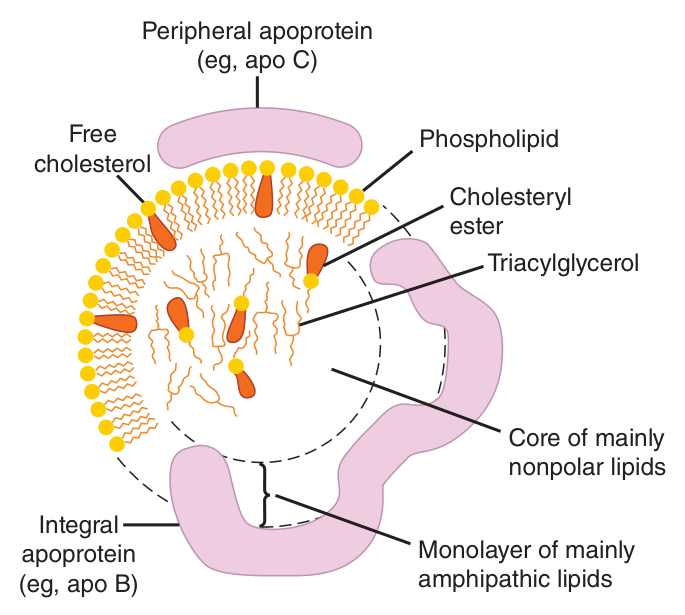
Specific combinations of non-covalently associated lipids and protein.
They are the form of transportation of lipids in plasma, and they help maintain the lipids in emulsified form.
⭐ “Lipoproteins” was a Section I term in the 2020/21 paper. Open with that definition.
1 · A lipid CORE of non-polar triacylglycerol and cholesteryl ester.
2 · A single SURFACE LAYER of phospholipid and free cholesterol.
3 · Polar groups facing OUTWARD.
4 · A protein moiety known as an APOLIPOPROTEIN.
An amphipathic molecule in water arranges itself heads-out, tails-in. Put a droplet of neutral fat in the middle and wrap it in a single layer of amphipathic phospholipid, and you have solved the transport problem: a water-soluble particle with an oily interior.
That is why the core carries the non-polar lipids — TAG and cholesteryl ester — while the free cholesterol and phospholipid, which have a polar group, sit at the surface. The chemistry is not arbitrary; you could have deduced it.
Some scale from your slides: about 500 mg of total lipid per 100 mL of human blood in the postabsorptive state — TAG 120 mg, cholesterol 220 mg (two-thirds as cholesteryl ester, one-third free), and phospholipid 160 mg.
- Define a lipoprotein → Specific combinations of non-covalently associated lipids and protein; the transport form of lipid in plasma, maintaining lipids in emulsified form
- Give the four structural features → A core of non-polar TAG and cholesteryl ester; a single surface layer; polar groups outward; a protein moiety, the apolipoprotein
- Which lipids sit in the core and which at the surface? → Non-polar TAG and cholesteryl ester in the core; phospholipid and free cholesterol at the surface
The four classes ★★★
| Class | Electrophoretic name | Origin | Dominant cargo / role |
|---|---|---|---|
| Chylomicron (CM) | remains at the origin | Intestine | Absorption of triacylglycerol — intestine to tissue. Transports all dietary lipid into the circulation |
| VLDL | pre-β-lipoprotein | Liver | Export of triacylglycerol from liver to extrahepatic tissues |
| LDL | β-lipoprotein | Final stage in the catabolism of VLDL | Cholesterol is dominant |
| HDL | α-lipoprotein | Liver and intestine | Phospholipids are dominant. Involved in VLDL and chylomicron metabolism, and in cholesterol transport |
Protein is denser than fat. So the more protein a particle carries relative to lipid, the higher its density and the smaller it is.
Chylomicrons are almost all triacylglycerol — enormous and least dense. HDL is about half protein — tiny and most dense. VLDL and LDL lie in between, in that order.
So a single spectrum — CM → VLDL → LDL → HDL — simultaneously runs decreasing size, increasing density, decreasing triacylglycerol and increasing protein. Learn the order once and you have four facts.
Lipoproteins may be separated according to their electrophoretic properties into α-, β- and pre-β-lipoproteins — the older nomenclature, still used clinically, which is why the second column is worth knowing.
- Name the four classes and their origins → Chylomicrons (intestine), VLDL (liver), LDL (from VLDL catabolism), HDL (liver and intestine)
- Give the electrophoretic names → VLDL = pre-β, LDL = β, HDL = α
- Which carries dietary TAG, and which carries hepatic TAG? → Chylomicrons and VLDL respectively
- Which is dominated by cholesterol, and which by phospholipid? → LDL and HDL
Apolipoproteins ⭐
The protein component of lipoproteins.
⭐ That is the 2019 answer sheet's wording exactly — “the protein component of lipoproteins”. Open with it, then give the three functions below.
1 · Form part of the structure of the lipoprotein.
2 · Act as enzyme cofactors — apoC-II for lipoprotein lipase, apoA-I for LCAT.
3 · Act as ligands for interaction with lipoprotein receptors in tissues — apoB-100 and apoE for the LDL receptor, apoA-I for the HDL receptor.
| Apolipoprotein | Found in | Note |
|---|---|---|
| apoA | HDL | apoA-I activates LCAT |
| apoB | LDL, VLDL and CM | B-48 synthesised in the INTESTINE · B-100 synthesised in the LIVER |
| apoC-I, C-II, C-III | Several lipoproteins | Smaller polypeptides, transferable between different lipoproteins. apoC-II activates lipoprotein lipase |
| apoE | VLDL and HDL | Ligand for remnant uptake by the liver |
apoB-48 is 48% of the length of apoB-100 — the same gene, edited differently in the two tissues. The intestine makes the short version, the liver the full-length one.
And that difference has a direct consequence, which §6 will use: the LDL receptor recognises apoB-100 but NOT apoB-48. The missing 52% contains the receptor-binding region. So chylomicrons cannot be cleared by the LDL receptor — they need apoE instead.
One number explains an entire difference in metabolic fate.
apoB is essential for chylomicron and VLDL formation and functions as a triacylglycerol transfer protein. A defect of apoB leads to lipid droplets accumulating in the intestine and liver — the particles cannot be assembled, so the fat cannot leave.
Three common alleles exist: APOE3 (about 78%), APOE4 (15%) and APOE2 (7%).
The APOE4 allele is particularly common in humans with Alzheimer's disease, and the link is highly predictive. Individuals homozygous for APOE4 have a 16-fold increased risk of late-onset Alzheimer's, with a mean age of onset just under 70 years; for people inheriting two copies of APOE3, the mean age of onset exceeds 90 years.
The molecular basis is not yet known. Speculation focuses on a role for apoE in stabilising the cytoskeletal structure of neurons: apoE2 and apoE3 bind a number of proteins associated with neuronal microtubules, whereas apoE4 does not, which may accelerate neuronal death. Note the honest hedge — this is a hypothesis, not a mechanism.
- Define apolipoproteins → The protein component of lipoproteins
- Give their three functions → Form part of the lipoprotein; act as enzyme cofactors; act as ligands for lipoprotein receptors
- Which apolipoprotein activates LPL, and which LCAT? → apoC-II and apoA-I
- Where are apoB-48 and apoB-100 made? → Intestine and liver respectively
- What is the risk in APOE4 homozygotes? → A 16-fold increased risk of late-onset Alzheimer's disease, with mean onset just under 70
Chylomicrons and VLDL ★★★
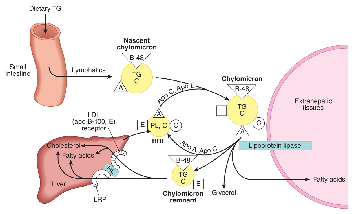
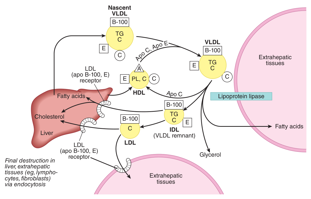
Both carry triacylglycerol, and both are emptied by the same enzyme. Chylomicrons are formed in the intestine and are responsible for transport of all dietary lipids into the circulation; VLDL is of hepatic origin and transports TAG from liver to extrahepatic tissues.
The enzyme that hydrolyses the triacylglycerol of chylomicrons and VLDL.
Location: the walls of blood capillaries.
Activators: phospholipids and apoC-II.
By Unit 6's rule, a low Km means high affinity. So cardiac LPL is saturated and extracting fatty acids even when plasma triacylglycerol is low — the heart gets fed first, always. Adipose LPL, with its high Km, only works hard when TAG is abundant — after a meal.
Your slide states the consequence directly: this causes uptake to shift from adipose tissue toward the heart as circulating TAG falls. Fuel goes to the organ that cannot stop working; storage happens only when there is surplus.
This is precisely the hexokinase/glucokinase logic of Unit 10, applied to fat. And note the regulation: in adipose tissue, insulin enhances lipoprotein lipase synthesis — the fed-state hormone increases the storage tissue's capacity to take up fat.
Remnant uptake
After LPL has stripped the triacylglycerol, what is left is a remnant. Chylomicron remnants are taken up by the liver by receptor-mediated endocytosis, via a receptor specific for apoE.
What drives hepatic VLDL secretion
Hepatic triacylglycerols are the immediate precursors of the TAG in plasma VLDL, and their formation offers the immediate stimulus for the formation and secretion of VLDL. The fatty acids used come from acetyl-CoA derived from carbohydrate (predominant in the well-fed state) or from uptake of free fatty acids from the circulation during fasting and on high-fat diets.
| Factors enhancing TAG synthesis and VLDL secretion by the liver |
|---|
| 1 · The fed state |
| 2 · Diets high in carbohydrate → high rate of lipogenesis and esterification of fatty acids |
| 3 · High levels of circulating FFA |
| 4 · Ingestion of ethanol |
| 5 · High insulin, low glucagon |
- Which enzyme empties CM and VLDL, and where is it? → Lipoprotein lipase, in the walls of blood capillaries
- What activates LPL? → Phospholipids and apoC-II
- Contrast LPL in heart and adipose tissue → Low Km in heart, high in adipose — so uptake shifts toward the heart as TAG falls
- How are chylomicron remnants cleared? → Receptor-mediated endocytosis by the liver, via a receptor specific for apoE
- Name five factors increasing VLDL secretion → The fed state, high-carbohydrate diet, high circulating FFA, ethanol, high insulin with low glucagon
LDL and the LDL receptor ★★★
| Fact | Detail |
|---|---|
| Origin | Formed from VLDL — the final stage in its catabolism |
| Receptor specificity | The liver has a specific receptor for apoB-100 and apoE. The receptor is NOT specific for apoB-48 |
| Distribution of catabolism | Approximately 30% of LDL is degraded in extrahepatic tissues, 70% in the liver |
| Consequence of receptor loss | Loss of the receptor can cause hypercholesterolaemia |
If 70% of LDL is cleared by hepatic LDL receptors, then losing those receptors leaves LDL circulating. That is familial hypercholesterolaemia: plasma cholesterol is high not because too much is made but because too little is removed.
Note the mechanistic link back to §4: the receptor recognises apoB-100 and apoE but not apoB-48. So it clears LDL and remnants, but chylomicrons — which carry apoB-48 — must be cleared by the separate apoE route.
Unit 19 will show how statins exploit this: inhibiting cholesterol synthesis makes the liver upregulate its LDL receptors, and that receptor upregulation, not the synthesis block itself, is what lowers plasma LDL.
- Where does LDL come from? → It is formed from VLDL, the final stage of its catabolism
- Which apolipoproteins does the LDL receptor recognise? → apoB-100 and apoE — NOT apoB-48
- What proportion of LDL is cleared by the liver? → About 70%; the remaining 30% in extrahepatic tissues
- What happens if the receptor is lost? → Hypercholesterolaemia
HDL and reverse cholesterol transport ★★★
| Fact | Detail |
|---|---|
| Origin | Synthesised and secreted from both liver and intestine |
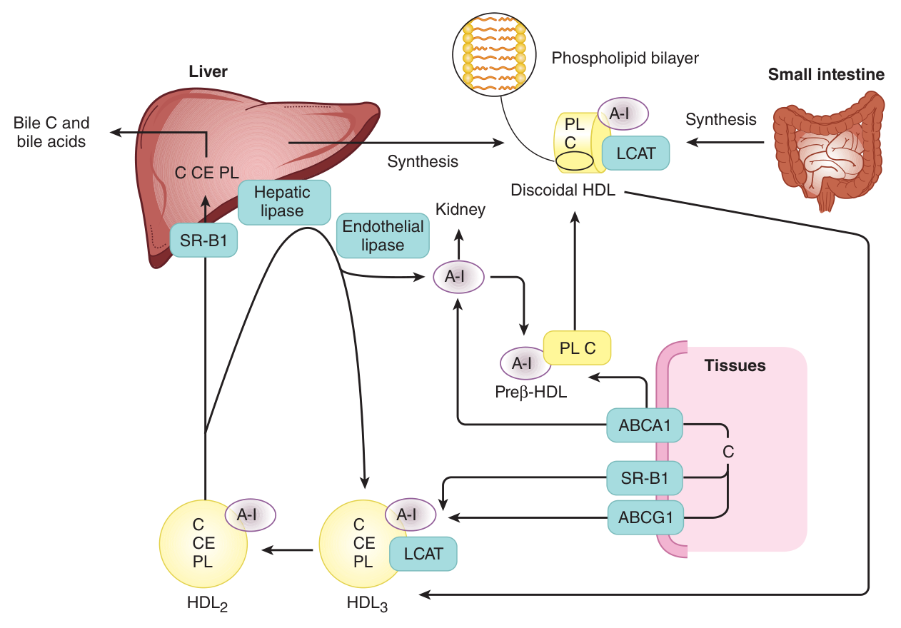
| Nascent HDL | That from the intestine does not contain apoC or apoE, only apoA. apoC and apoE are synthesised in the liver and transferred from hepatic HDL to intestinal HDL |
| Major function | To act as a repository for apoC and apoE for chylomicrons and VLDL |
| apoA-I | Activator of LCAT — lecithin:cholesterol acyltransferase, which carries out plasma esterification of cholesterol |
| LCAT | Involved in the removal of unesterified cholesterol |
| Final site of degradation | The liver, for HDL cholesteryl ester |
Uptake and esterification of cholesterol by HDL₃ → forming HDL₂ → hepatic lipase hydrolyses phospholipid and triacylglycerol → allowing release of cholesteryl ester → re-forming HDL₃ → and again.
HDL's job is to collect cholesterol from peripheral tissues. But free cholesterol is amphipathic — it sits at the surface of a particle (Unit 15 §9), and a surface fills up.
LCAT esterifies it, and a cholesteryl ester is entirely non-polar. So it sinks into the core, freeing the surface to accept more. The particle can go on loading, growing from small dense HDL₃ into larger HDL₂.
Esterification is what makes HDL a container rather than a coating. That is why LCAT — and therefore its activator apoA-I — is central to reverse cholesterol transport, and why HDL cholesterol is the “good” cholesterol: it measures how much is being carried back to the liver.
- Where is HDL made? → Liver and intestine
- What is HDL's major function? → To act as a repository for apoC and apoE for chylomicrons and VLDL
- What does apoA-I activate, and what does that enzyme do? → LCAT, which esterifies cholesterol in plasma and removes unesterified cholesterol
- Describe reverse cholesterol transport → HDL₃ takes up and esterifies cholesterol → HDL₂ → hepatic lipase releases the cholesteryl ester → HDL₃ re-forms
- Where is HDL cholesteryl ester finally degraded? → The liver
Free fatty acid transport ★★
| Stage | How FFA is carried |
|---|---|
| In plasma | Bound to albumin |
| Crossing the membrane | Bound to a membrane fatty acid-transport protein, with Na⁺ |
| In the cytosol | Bound to fatty acid-binding protein |
The rate of removal of FFA is rapid. In starvation, 25–50% of uptake is used for energy requirement and the remainder is esterified; in heart and skeletal muscle during starvation, more esterified fatty acids are oxidised. Crucially, the rate of FFA production determines the FFA uptake by tissues.
Notice that at every single stage the fatty acid is bound to a protein — albumin, then a transport protein, then a binding protein. It is never free in an aqueous compartment.
That is not fussiness. Free fatty acids are detergents: at any concentration they would disrupt membranes. Keeping them protein-bound is a safety mechanism as much as a transport one — and it is why plasma FFA is measured as an albumin-bound fraction.
- How is FFA carried in plasma, across the membrane, and in cytosol? → Bound to albumin; to a membrane fatty acid-transport protein with Na⁺; to fatty acid-binding protein
- What proportion of FFA uptake is used for energy in starvation? → 25–50%; the remainder is esterified
- What determines FFA uptake by tissues? → The rate of FFA production
Adipose tissue and the control of lipolysis ★★★
In adipose tissue, triacylglycerol is synthesised from acyl-CoA and glycerol-3-phosphate. But there is a catch worth knowing:
Instead, adipose glycerol-3-phosphate is obtained from glycolysis, via dihydroxyacetone phosphate, which requires a supply of glucose by the GLUT1 and GLUT4 transporters.
The consequence is large: adipose tissue can only store fat if glucose is available. No glucose → no glycerol-3-phosphate → no esterification → fatty acids are released instead of stored. This is a major reason why insulin, which drives glucose into adipocytes, is the great fat-storing hormone, and why uncontrolled diabetes causes lipolysis despite abundant circulating fuel.
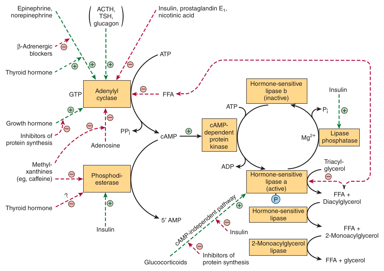
The enzyme controlling lipolysis in adipose tissue, forming FFA + glycerol — distinct from lipoprotein lipase.
| Hormone-sensitive lipase | Lipoprotein lipase | |
|---|---|---|
| Location | Inside the adipocyte | Walls of blood capillaries |
| Substrate | Stored triacylglycerol | Circulating TAG in CM and VLDL |
| Direction | Releases fat from the cell | Delivers fat into the cell |
| Activated by | cAMP / PKA — glucagon, epinephrine | apoC-II; insulin increases its synthesis |
There is a continuous cycle of lipolysis and re-esterification: the released fatty acid can be reactivated by acyl-CoA synthetase and re-esterified with glycerol-3-phosphate. Whether fat leaves the cell depends on the balance of the two.
The hormonal control
| Hormone | Effect | Mechanism |
|---|---|---|
| Insulin | REDUCES the output of FFA | Inhibits the release of FFA from adipose tissue, lowering plasma FFA; enhances lipogenesis and acylglycerol synthesis; increases oxidation of glucose via the pentose phosphate pathway; and raises pyruvate dehydrogenase, acetyl-CoA carboxylase and glycerol phosphate acyltransferase activity |
| Epinephrine / norepinephrine | PROMOTE lipolysis | Hormone → adenylyl cyclase → cAMP → cAMP-dependent protein kinase → converts the lipase to its active form. Norepinephrine has a central role in mobilisation of FFA |
| Thyroid hormones | Promote lipolysis | Possibly by increasing cAMP (receptor → adenylyl cyclase) and by inhibiting phosphodiesterase |
| Glucocorticoids | Promote lipolysis | Via synthesis of new lipase protein — a slow, long-term mechanism |
| Growth hormone | Promotes lipolysis | Slow action |
Note the permissive requirement: most of these lipolytic processes require the presence of glucocorticoids and thyroid hormones.
Epinephrine works through cAMP and phosphorylation — seconds. Glucocorticoids work by synthesising new lipase protein — hours.
That is exactly Unit 7's distinction between short-term regulation by covalent modification and long-term regulation by changing enzyme quantity. The same tissue is controlled on both timescales, by different hormones, for different physiological purposes — a fright versus a famine.
- Why can adipose tissue not re-use its glycerol? → It does not express glycerol kinase; glycerol-3-phosphate comes from glycolysis via DHAP, requiring glucose uptake by GLUT1 and GLUT4
- Contrast hormone-sensitive lipase with lipoprotein lipase → HSL releases stored fat from inside the adipocyte and is activated by cAMP/PKA; LPL delivers circulating TAG into the cell from the capillary wall and is activated by apoC-II
- How does insulin reduce FFA output? → Inhibits FFA release, enhances lipogenesis and acylglycerol synthesis, increases the pentose phosphate pathway, and raises PDH, acetyl-CoA carboxylase and glycerol phosphate acyltransferase
- By what mechanism do glucocorticoids promote lipolysis? → Synthesis of new lipase protein — a slow mechanism
Fatty liver ★★
Lipid (triacylglycerol) accumulation in liver, leading to fatty liver, cirrhosis and impaired liver function.
The mechanism is a simple imbalance: the rate of triacylglycerol formation is faster than its export by VLDL.
Ethanol is oxidised by dehydrogenase and produces NADH, which inhibits the oxidation of fatty acids and causes increased esterification of fatty acids.
Think about what a raised NADH/NAD⁺ ratio does across the units you have already built. It inhibits β-hydroxyacyl-CoA dehydrogenase, step 3 of β-oxidation (Unit 17 §6). It inhibits the citric acid cycle, whose three dehydrogenases all need NAD⁺ (Unit 11 §9). So fatty acids can neither be burned nor fed into the cycle — and the only remaining route is esterification to triacylglycerol, which accumulates faster than VLDL can carry it away.
Alcoholic fatty liver is a redox problem, not a fat problem. And note that ethanol also appears in §5's list of factors increasing VLDL secretion — the liver tries to export the surplus and cannot keep up.
The other route to fatty liver is a defect of apoB (§4), which prevents the particles being assembled at all — lipid droplets accumulate in intestine and liver.
- Define fatty liver and give the basic mechanism → TAG accumulation because the rate of formation exceeds export by VLDL; it can progress to cirrhosis and impaired function
- How does ethanol cause it? → Its oxidation produces NADH, which inhibits fatty acid oxidation and increases esterification
- What else can cause it? → A defect of apoB, preventing lipoprotein assembly
Revision layer
Two Section I terms from this unit have been set: apolipoproteins (2019) and lipoproteins (2020/21). Both definitions are below, in the answer-sheet wording.
The four lipoproteins
| CM | VLDL | LDL | HDL | |
|---|---|---|---|---|
| Electrophoresis | origin | pre-β | β | α |
| Origin | Intestine | Liver | From VLDL | Liver + intestine |
| Main cargo | Dietary TAG | Hepatic TAG | Cholesterol | Phospholipid |
| Key apo | B-48, C-II, E | B-100, C-II, E | B-100 | A-I, C, E |
| Cleared by | LPL, then apoE receptor | LPL → IDL → LDL | LDL receptor (70% liver) | Liver |
| Density / size | lowest density, largest | → | → | highest density, smallest |
The enzymes and their activators
| Enzyme | Site | Activator | Job |
|---|---|---|---|
| Lipoprotein lipase | Capillary walls | apoC-II + phospholipids | Empties CM and VLDL of TAG |
| Hormone-sensitive lipase | Inside adipocyte | cAMP / PKA | Releases stored TAG |
| LCAT | Plasma | apoA-I | Esterifies cholesterol for HDL |
| Hepatic lipase | Liver | — | Converts HDL₂ back to HDL₃ |
Definitions from this unit — Section I material
| Term | Definition |
|---|---|
| Lipoproteins ⭐ | Specific combinations of non-covalently associated lipids and protein; the form of transportation of lipids in plasma, helping to maintain the lipids in emulsified form. They consist of a lipid core of non-polar triacylglycerol and cholesteryl ester, a single surface layer with polar groups outward, and a protein moiety known as an apolipoprotein |
| Apolipoproteins ⭐ | The protein component of lipoproteins. They form part of the lipoprotein structure, act as enzyme cofactors (apoC-II for lipoprotein lipase, apoA-I for LCAT), and act as ligands for interaction with lipoprotein receptors in tissues (apoB-100 and apoE for the LDL receptor) |
| Lipoprotein lipase | The enzyme in the walls of blood capillaries that hydrolyses the triacylglycerol of chylomicrons and VLDL; activated by phospholipids and apoC-II. Its Km is low in heart and high in adipose tissue, directing uptake toward the heart as circulating triacylglycerol falls |
| Reverse cholesterol transport | The uptake and esterification of cholesterol by HDL₃ to form HDL₂; hepatic lipase then hydrolyses phospholipid and triacylglycerol, allowing release of cholesteryl ester and re-formation of HDL₃. The final site of degradation of HDL cholesteryl ester is the liver |
| Hormone-sensitive lipase | The intracellular adipose enzyme that hydrolyses stored triacylglycerol to free fatty acids and glycerol, activated by cAMP-dependent protein kinase in response to epinephrine and glucagon; distinct from lipoprotein lipase |
| Fatty liver | Accumulation of triacylglycerol in the liver, occurring when the rate of triacylglycerol formation exceeds its export as VLDL, and leading to cirrhosis and impaired liver function |
- Define lipoproteins and apolipoproteins in the answer-sheet wording
- Give the four structural features of a lipoprotein and explain them from amphipathicity
- Reproduce the four-class table with origins, cargo and key apolipoproteins
- Give the three functions of apolipoproteins with an example of each
- Explain the LPL Km difference between heart and adipose tissue
- Explain why the LDL receptor cannot clear chylomicrons
- Describe reverse cholesterol transport and explain why LCAT is essential to it
- Contrast hormone-sensitive lipase with lipoprotein lipase on four points
- Explain alcoholic fatty liver in terms of the NADH/NAD⁺ ratio