Fatty Acid Oxidation & Ketogenesis
Mobilising stored fat ★★★
Your lecture opens with the comparison, and it frames the whole unit. Sprinters primarily use glycolysis and the citric acid cycle — anaerobic processes later “recharged” by breathing hard. Marathon runners need energy over a long time period, and get most of it by oxidising fats.
Unit 12 gave the reason: liver glycogen is gone in 12–18 hours. Fat stores last weeks. Anything longer than a sprint is fuelled by this unit.
Before fat can be burned it must be released from the adipocyte. The cascade is Unit 12's cAMP cascade, applied to a different substrate:
Epinephrine or glucagon → receptor → activates adenylyl cyclase → cAMP → protein kinase A (PKA), which phosphorylates hormone-sensitive lipase (HSL) and perilipins.
Phosphorylated perilipin causes dissociation of the protein CGI from perilipin; CGI binds and activates adipose triacylglycerol lipase (ATGL). Active ATGL converts triacylglycerol → diacylglycerol; DAG → MAG by monoacylglycerol lipase, forming free fatty acids + glycerol.
The two products go different ways. Free fatty acids travel in plasma bound to albumin, to be oxidised — the rest of this unit. Glycerol takes a different route: glycerol → glycerol-3-phosphate (glycerol kinase) → dihydroxyacetone phosphate → glyceraldehyde 3-phosphate (triose phosphate isomerase) → glycolysis or gluconeogenesis.
Unit 13 insisted that fat cannot be converted to glucose. Glycerol is the loophole — it enters at the triose phosphate level, above the irreversible pyruvate dehydrogenase step, so it is glucogenic.
But it is only three carbons out of the fifty-odd in a triacylglycerol. The fatty acids — the bulk of the molecule — still cannot become glucose. That is why glycerol appears in Unit 13's substrate list and acetyl-CoA does not.
- Trace the mobilisation cascade → Epinephrine/glucagon → adenylyl cyclase → cAMP → PKA → phosphorylates hormone-sensitive lipase and perilipin → ATGL activated → TAG → DAG → MAG → FFA + glycerol
- What happens to the glycerol? → Glycerol kinase makes glycerol-3-phosphate → DHAP → glyceraldehyde 3-phosphate → glycolysis or gluconeogenesis
- Why is glycerol glucogenic when fatty acids are not? → It enters above the irreversible PDH step
Activation ★★
Fatty acid + ATP + CoA → acyl-CoA + AMP + PPi, catalysed by acyl-CoA synthetase (thiokinase).
This is the only step in the complete degradation of a fatty acid that requires energy from ATP.
Acyl-CoA synthetases are found in the endoplasmic reticulum, peroxisomes, and inside and on the outer membrane of mitochondria.
Net cost: 2 ATP equivalents. This is exactly Unit 8's device — destroy one of your own products to make the reaction irreversible — and it is why the palmitate arithmetic in §5 subtracts 2, not 1.
- Which enzyme activates fatty acids? → Acyl-CoA synthetase (thiokinase)
- What are the products? → Acyl-CoA, AMP and PPi
- What is the true energy cost? → Two ATP equivalents — ATP → AMP, plus hydrolysis of PPi
- Is any other step of degradation ATP-dependent? → No — this is the only one
The carnitine shuttle ★★★
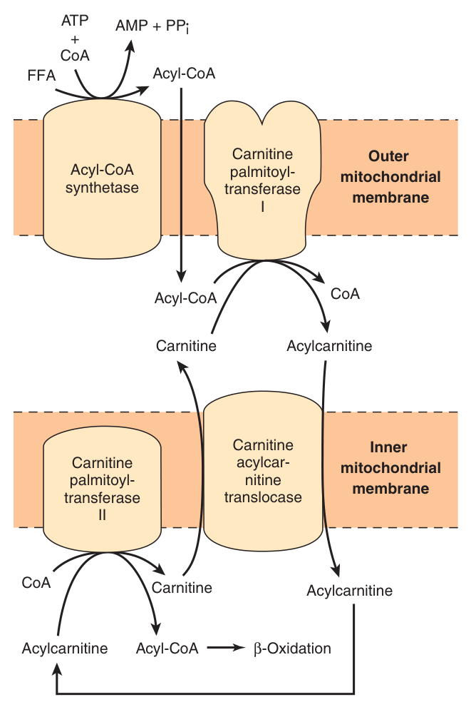
Acyl-CoA has been made in the cytosol. But long-chain acyl-CoA cannot penetrate the inner membrane of mitochondria, and β-oxidation happens in the matrix. Long-chain fatty acids (more than 12 carbons) penetrate the inner mitochondrial membrane as carnitine derivatives.
| # | Enzyme / transporter | What it does |
|---|---|---|
| 1 | Carnitine palmitoyltransferase-I (CPT-I) — outer mitochondrial membrane | Transfers the long-chain acyl group from CoA to carnitine, forming acylcarnitine and releasing CoA |
| 2 | Carnitine-acylcarnitine translocase — inner membrane exchange transporter | Binds acylcarnitine and transports it across the membrane in exchange for carnitine |
| 3 | Carnitine palmitoyltransferase-II (CPT-II) — inside the inner membrane | Transfers the acyl group back to CoA, so acyl-CoA is reformed and carnitine is liberated |
Carnitine is β-hydroxy-γ-trimethylammonium butyrate — widely distributed and particularly abundant in muscle.
Because a shuttle can be regulated and a pore cannot.
Penetration of fatty acyl-CoA into the mitochondrial matrix is rate-limiting for fatty acid oxidation — so by making entry the bottleneck, the cell gets a single point at which to switch the entire pathway on or off. §6 is that switch.
Note also the size cut-off: this applies to fatty acids of more than 12 carbons. Short- and medium-chain fatty acids cross freely — which is why MCAD deficiency (§8) presents so differently from a carnitine transport defect.
- Which fatty acids need the shuttle? → Long-chain, more than 12 carbons
- Name the three components in order → Carnitine palmitoyltransferase-I (outer membrane), carnitine-acylcarnitine translocase, carnitine palmitoyltransferase-II (inner face)
- Where is carnitine especially abundant? → Muscle
- Why does this matter for regulation? → Entry into the matrix is the rate-limiting step for β-oxidation
The four steps of β-oxidation ★★★
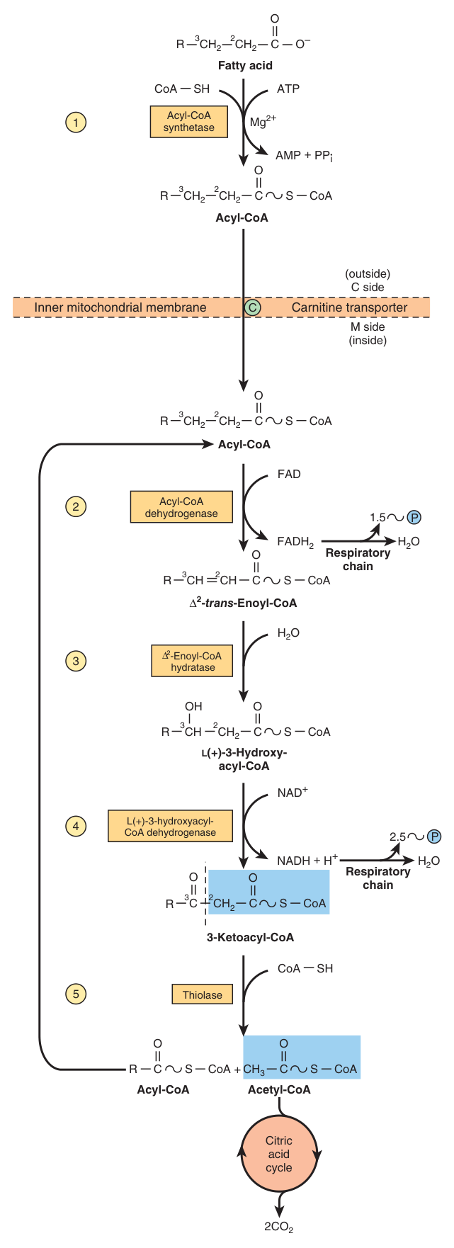
A pathway in which two carbons at a time are cleaved from acyl-CoA molecules, starting at the carboxyl end. The chain is broken between the α(2)- and β(3)-carbon atoms — hence the name β-oxidation. The two-carbon units formed are acetyl-CoA; thus palmitoyl-CoA forms eight acetyl-CoA molecules.
Location: mitochondrial matrix (or inner membrane).
| Step | Reaction | Enzyme | Yield |
|---|---|---|---|
| 1 · Dehydrogenation | Makes a double bond | Acyl-CoA dehydrogenase | FADH₂ |
| 2 · Hydration | Water added across the double bond | Enoyl-CoA hydratase | — |
| 3 · Dehydrogenation | Makes a carbonyl | β-Hydroxyacyl-CoA dehydrogenase | NADH |
| 4 · Thiolysis | Acyl transfer — cleaves off acetyl-CoA | Acyl-CoA acetyltransferase (thiolase) | Acetyl-CoA |
Oxidise, hydrate, oxidise. Compare the last four steps of the citric acid cycle (Unit 11): succinate → fumarate (FADH₂), fumarate → malate (hydration), malate → oxaloacetate (NADH). Identical sequence, identical coenzymes, identical order.
That is not a coincidence — it is the standard chemistry for converting a –CH₂–CH₂– into a –CO–CH₂–, which is what you must do before a carbon–carbon bond can be cleaved. Recognising the pattern means you have effectively already learned this cycle.
Mnemonic if you want one: D-H-D-T — Dehydrogenate, Hydrate, Dehydrogenate, Thiolysis.
- Why is it called β-oxidation? → The chain is broken between the α(2)- and β(3)-carbon atoms
- Name the four steps and their enzymes → Dehydrogenation (acyl-CoA dehydrogenase), hydration (enoyl-CoA hydratase), dehydrogenation (β-hydroxyacyl-CoA dehydrogenase), thiolysis (thiolase)
- What is produced per cycle? → One FADH₂, one NADH and one acetyl-CoA
- How many acetyl-CoA does palmitoyl-CoA give? → Eight
The ATP yield ★★★
A calculation you must be able to reproduce. Work it for palmitate, 16 carbons.
| Step | Working | ATP |
|---|---|---|
| Per cycle | 1 FADH₂ (1.5) + 1 NADH (2.5) | 4 |
| Cycles | 16 carbons → 7 cycles (not 8 — the last cycle yields two acetyl-CoA) → 4 × 7 | 28 |
| Acetyl-CoA | 8 acetyl-CoA × 10 ATP each in the citric acid cycle | 80 |
| Subtotal | 28 + 80 | 108 |
| Activation | Subtract the initial activation (ATP → AMP + PPi) | −2 |
| NET | 106 |
Subtract two, not one. Activation makes AMP + PPi, so ATP is split twice over (§2).
Note also that this deck uses 1.5 and 2.5 ATP — confirming the values settled in Unit 9. And compare the scale: 1 mole of glucose yields 30/32 ATP against palmitate's 106. Fat is a far denser fuel, exactly as Unit 15 §7 predicted.
- How many ATP per cycle of β-oxidation? → 4 — one FADH₂ (1.5) and one NADH (2.5)
- How many cycles for palmitate, and why? → Seven — the last cycle yields two acetyl-CoA, giving eight in total
- Give the full palmitate arithmetic → (7 × 4) + (8 × 10) − 2 = 28 + 80 − 2 = 106 ATP
- Compare with glucose → One mole of glucose gives 30–32 ATP; palmitate gives 106
Regulation ★★★
An acyl-CoA in the cytosol can go in one of two directions: (1) entry into the mitochondrion to be oxidised, or (2) packed into triacylglycerol and sent off to storage in an adipocyte. β-oxidation is only turned on when it is needed, and the switch is at the entry point.
Entry into the mitochondrion, via carnitine, is the major point of regulation for β-oxidation. Penetration of fatty acyl-CoA into the mitochondrial matrix is rate-limiting.
Glucose ↑ → malonyl-CoA ↑ → carnitine acyltransferase I ↓
When glucose is plentiful, acetyl-CoA carboxylase makes malonyl-CoA — the first committed intermediate of fatty acid synthesis (Unit 16). And malonyl-CoA inhibits carnitine palmitoyltransferase-I, closing the door to oxidation.
So the act of starting to make fat simultaneously blocks the pathway that burns it. One molecule enforces reciprocal regulation across two compartments — exactly as fructose 2,6-bisphosphate does for glycolysis and gluconeogenesis in Unit 13.
A second control operates further down: a high [NADH]/[NAD⁺] ratio inhibits β-hydroxyacyl-CoA dehydrogenase, step 3 of the cycle.
The physiological consequence: CPT-I activity is low in the fed state, leading to depression of fatty acid oxidation, and high in starvation, allowing fatty acid oxidation to increase.
- What is the major point of regulation? → Entry into the mitochondrion via the carnitine shuttle — CPT-I
- What inhibits CPT-I, and where does it come from? → Malonyl-CoA, the first committed intermediate of fatty acid synthesis
- Why is that elegant? → Starting to make fat automatically blocks the pathway that burns it
- When is CPT-I activity high? → In starvation; it is low in the fed state
Odd-chain and unsaturated fatty acids ★★
| Situation | What happens |
|---|---|
| Odd number of carbons | β-oxidation proceeds normally until a three-carbon propionyl-CoA remains. This is converted via D-methylmalonyl-CoA → succinyl-CoA, entering the citric acid cycle |
| Unsaturated fatty acids | A modified β-oxidation pathway requiring two additional enzymes: enoyl-CoA isomerase, which converts cis to trans configuration, and 2,4-dienoyl-CoA reductase |
Succinyl-CoA is a citric acid cycle intermediate, and Unit 11 established that all cycle intermediates are potentially glucogenic. So the three terminal carbons of an odd-chain fatty acid can become glucose.
That is why propionate appeared in Unit 13's list of gluconeogenic substrates, and why it matters so much in ruminants. It is the second exception — with glycerol — to “fat cannot make glucose”, and both exceptions are small parts of the molecule.
- What remains after β-oxidation of an odd-chain fatty acid? → Propionyl-CoA
- How does propionyl-CoA enter the cycle? → Via D-methylmalonyl-CoA to succinyl-CoA
- Which two extra enzymes are needed for unsaturated fatty acids? → Enoyl-CoA isomerase (cis to trans) and 2,4-dienoyl-CoA reductase
Peroxisomes and inherited defects ★★
Some β-oxidation takes place in the peroxisomes — of very long chain fatty acids (26 C and above), using the same four enzymatic steps.
If a patient has a high level of very long chain fatty acids (VLCFAs) in the blood, think of a peroxisomal defect first.
In X-linked adrenoleukodystrophy (XALD), peroxisomes fail to oxidise VLCFAs, which accumulate in the blood. It affects young boys under 10 years, causing loss of vision, behavioural disturbances, and death within a few years.
Your lecture poses the therapeutic problem honestly. Restricting the diet — fatty meat, nuts, the waxy coating of fruits and vegetables are high in VLCFAs — is not sufficient, because you MAKE about two-thirds of them yourself as part of fat synthesis. The hypothesis offered is to block the elongating enzymes with shorter-chain fatty acids.
Many people lack the gene for medium-chain acyl-CoA dehydrogenase (MCAD) and are unable to oxidise 6- to 12-carbon fatty acids.
Population: US and northern European. Carrier frequency: 1 in 40. Mortality 25–60% in early childhood.
Syndrome: fat accumulation in the liver, high blood levels of octanoic acid, low blood glucose (hypoglycaemia), sleepiness, vomiting and coma.
Treatment: a low-fat, high-carbohydrate diet, and the patient must eat often to keep the body from using fat reserves. That last instruction is the whole pathophysiology in one sentence: these children are fine until they fast.
- Where are very long chain fatty acids oxidised? → In peroxisomes, by the same four steps
- What should raised blood VLCFAs suggest? → A peroxisomal defect — X-linked adrenoleukodystrophy
- What is MCAD deficiency, and how common? → Inability to oxidise 6- to 12-carbon fatty acids; carrier frequency 1 in 40 in northern European populations
- Why is MCAD deficiency treated with frequent feeding? → To stop the body mobilising fat reserves it cannot oxidise
Ketone bodies ⭐
This was Section II question 4 in the 2019 paper: “What are ketone bodies? Try to describe their metabolic properties.” This section is that answer.
Acetoacetate, D-β-hydroxybutyrate and acetone (which is volatile) are collectively termed ketone bodies.
Ketogenesis occurs when there is a high rate of fatty acid oxidation in the liver, and takes place in liver mitochondria.
Formation
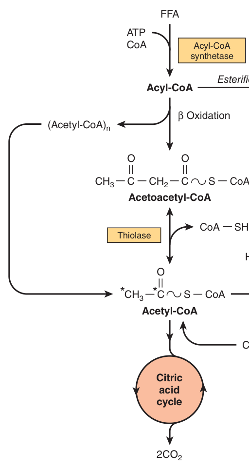
Two acetyl-CoA condense to acetoacetyl-CoA (thiolase); HMG-CoA synthase adds a third acetyl-CoA to give 3-hydroxy-3-methylglutaryl-CoA (HMG-CoA); HMG-CoA lyase cleaves this to acetoacetate. Acetoacetate is reduced to D-β-hydroxybutyrate by D(−)-3-hydroxybutyrate dehydrogenase, or decarboxylates spontaneously to acetone.
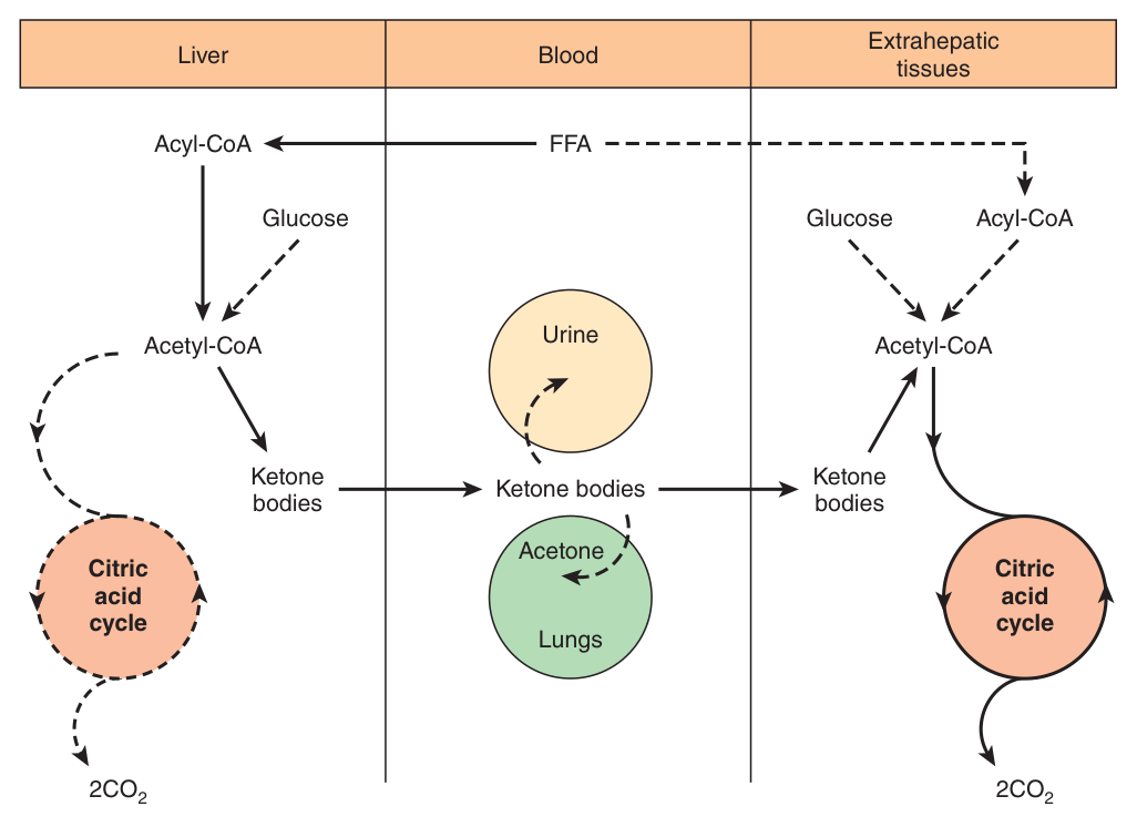
The liver, during starvation, is oxidising fatty acids far faster than its own citric acid cycle can consume the resulting acetyl-CoA — because oxaloacetate is being drained into gluconeogenesis (Unit 13). So it packages the surplus two-carbon units into a water-soluble, transportable form and ships them out.
The elegance is in the solubility. Fatty acids need albumin to travel; ketone bodies dissolve freely in plasma — and, crucially, they cross the blood-brain barrier, which long-chain fatty acids cannot. Ketone bodies are how fat feeds the brain. In prolonged starvation they supply a large fraction of cerebral energy, which is what spares muscle protein from being consumed for gluconeogenesis.
In extrahepatic tissues they are reconverted to acetyl-CoA and oxidised in the citric acid cycle. The liver lacks the enzyme to do this — which is why it never consumes its own product.
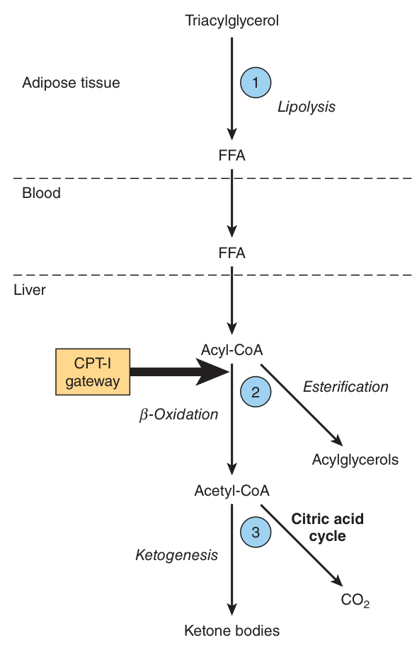
Regulation — three crucial steps
| # | Step | Detail |
|---|---|---|
| 1 | Lipolysis in adipose tissue | Ketosis does not occur in vivo unless there is an increase in the level of circulating FFAs arising from lipolysis of adipose triacylglycerol. The liver extracts about 30% of the FFAs passing through it, in both fed and fasting conditions |
| 2 | CPT-I — the gateway | After uptake, FFAs are either β-oxidised, or esterified to triacylglycerol and phospholipid. CPT-I activity is low in the fed state and high in starvation |
| 3 | Partition of acetyl-CoA | Acetyl-CoA is directed either into the citric acid cycle or into ketogenesis |
Starvation (or dieting). Gluconeogenesis depletes citric acid cycle intermediates — particularly oxaloacetate — so acetyl-CoA is diverted to ketone bodies. This is Unit 11's anaplerotic problem and “fat burns in the flame of carbohydrate”, arriving at last.
Untreated diabetes. Insulin is low → glucose does not enter cells → malonyl-CoA falls → inhibition of carnitine acyltransferase-I is relieved → fatty acid oxidation runs unchecked.
Accumulation of acetyl-CoA → ketone body formation → acidosis → coma or death. The numbers are worth quoting: blood ketone bodies in untreated diabetics reach 90 mg/100 mL against a normal <3 mg/100 mL, with urinary excretion of 5000 mg/24 h against a normal 125 mg/24 h — ketosis.
One practical point: because renal handling varies, measurement of the ketonaemia, not the ketonuria, is the preferred method of assessing the severity of ketosis.
- Name the three ketone bodies → Acetoacetate, D-β-hydroxybutyrate and acetone
- Where are they made? → Liver mitochondria, when fatty acid oxidation is high
- Why does the liver make them? → To export surplus acetyl-CoA as a water-soluble fuel that other organs — including the brain — can use
- Name the three regulatory steps → Lipolysis raising FFA, the CPT-I gateway, and the partition of acetyl-CoA between the cycle and ketogenesis
- Why does starvation cause ketosis? → Gluconeogenesis depletes oxaloacetate, so acetyl-CoA cannot enter the citric acid cycle
- Why does untreated diabetes cause ketosis? → Low insulin lowers malonyl-CoA, relieving inhibition of CPT-I so fatty acid oxidation runs unchecked
- Which is the better measure of severity? → Ketonaemia, not ketonuria
Revision layer
The pathway in one line
TAG → (hormone-sensitive lipase, ATGL) → FFA + glycerol → activation (acyl-CoA synthetase, −2 ATP) → carnitine shuttle (CPT-I, translocase, CPT-II) → β-oxidation (dehydrogenate, hydrate, dehydrogenate, thiolysis) → acetyl-CoA → citric acid cycle, or ketogenesis.
Definitions from this unit — Section I material
| Term | Definition |
|---|---|
| β-oxidation | The pathway in which two carbons at a time are cleaved from acyl-CoA starting at the carboxyl end, the chain being broken between the α(2)- and β(3)-carbon atoms; each cycle comprises dehydrogenation, hydration, dehydrogenation and thiolysis, yielding one FADH₂, one NADH and one acetyl-CoA. It occurs in the mitochondrial matrix |
| The carnitine shuttle | The mechanism by which long-chain fatty acids (over 12 carbons) cross the inner mitochondrial membrane: carnitine palmitoyltransferase-I on the outer membrane transfers the acyl group from CoA to carnitine; carnitine-acylcarnitine translocase exchanges acylcarnitine for carnitine across the inner membrane; and carnitine palmitoyltransferase-II on the inner face restores acyl-CoA. It is the rate-limiting step of fatty acid oxidation |
| Ketone bodies ⭐ | Acetoacetate, D-β-hydroxybutyrate and acetone (volatile), formed in liver mitochondria when there is a high rate of fatty acid oxidation, and exported to other organs as a water-soluble fuel — including to the brain, which cannot use long-chain fatty acids |
| Ketogenesis | The formation of ketone bodies from acetyl-CoA in liver mitochondria, via acetoacetyl-CoA and HMG-CoA (HMG-CoA synthase), with HMG-CoA lyase releasing acetoacetate; regulated at three steps — lipolysis, the CPT-I gateway, and the partition of acetyl-CoA |
| Ketosis | The state of raised blood and urinary ketone bodies arising when acetyl-CoA accumulates faster than the citric acid cycle can consume it — in starvation (oxaloacetate depleted by gluconeogenesis) and in untreated diabetes (low malonyl-CoA relieving CPT-I inhibition); severe ketosis causes acidosis, coma or death |
Numbers worth carrying in
| Item | Value |
|---|---|
| Cost of activation | 2 ATP equivalents (ATP → AMP + PPi) |
| Carnitine shuttle applies to | fatty acids of >12 carbons |
| ATP per cycle | 4 (FADH₂ 1.5 + NADH 2.5) |
| Palmitate | 7 cycles → 8 acetyl-CoA → 106 ATP net |
| Glucose, for comparison | 30–32 ATP |
| Peroxisomal β-oxidation | very long chain, ≥26 carbons |
| MCAD carrier frequency | 1 in 40; mortality 25–60% in early childhood |
| Blood ketone bodies | normal <3 mg/100 mL · untreated diabetes 90 mg/100 mL |
| Urinary ketone excretion | normal 125 mg/24 h · ketosis 5000 mg/24 h |
| Liver FFA extraction | about 30% of what passes through it |
- Trace fat mobilisation from hormone to free fatty acid
- Explain why activation costs two ATP equivalents
- Name the three components of the carnitine shuttle and say why it is regulated
- Give the four steps of β-oxidation with enzymes and yields
- Work the palmitate ATP arithmetic, explaining 7 cycles and −2
- Explain how malonyl-CoA links synthesis and oxidation
- Answer the 2019 question: what are ketone bodies and their metabolic properties
- Contrast the mechanism of ketosis in starvation and in untreated diabetes