Metabolism of Glycogen
What glycogen is ★★★
The major storage carbohydrate in animals, corresponding to starch in plants. It is a branched polymer of α-D-glucose.
It occurs mainly in liver (up to 6%) and muscle (rarely exceeding 1%). However, because of its greater mass, muscle contains about three to four times as much glycogen as liver.
Liver has the higher concentration (6% vs 1%). Muscle has the greater total amount, three to four times more, simply because there is so much more muscle.
Examiners love this because it catches students who memorised one figure without the other. And the two facts point at the two different purposes in §2: the liver's store is concentrated because it is a rapidly mobilised export depot; muscle's is large because it is a private fuel tank.
- Define glycogen → The major storage carbohydrate in animals, a branched polymer of α-D-glucose
- Which tissue has the higher concentration, and which the greater total? → Liver has up to 6%; muscle rarely exceeds 1% but contains 3–4 times as much in total
Liver versus muscle — two different jobs ★★★
| Muscle glycogen | Liver glycogen | |
|---|---|---|
| Purpose | A readily available source of glucose for glycolysis WITHIN THE MUSCLE ITSELF | To store and export glucose to maintain BLOOD glucose between meals |
| Has glucose-6-phosphatase? | NO | YES (also kidney) |
| Can it release free glucose? | No — the glucose stays in the muscle | Yes — exported, raising blood glucose |
| Depletion | With exercise | Almost totally depleted after 12–18 hours of fasting |
Glycogenolysis in either tissue produces glucose 6-phosphate. Phosphorylated sugars cannot cross the plasma membrane. Only the liver (and kidney) possesses the phosphatase that hydrolyses glucose 6-phosphate, yielding free glucose that is exported, leading to an increase in blood glucose concentration.
Muscle has no such enzyme, so its glycogen is a private store — it can fuel that muscle's own glycolysis and nothing else. This single enzymatic difference is why hepatic glycogen governs blood sugar and muscle glycogen governs sprinting, and it recurs in Unit 13 as the last step of gluconeogenesis.
After 12–18 hours of fasting, liver glycogen is almost totally depleted. That number is worth carrying: it is the reason gluconeogenesis (Unit 13) has to exist. Overnight, glycogen covers you. Beyond about a day, the liver must start manufacturing glucose from non-carbohydrate precursors, or the brain fails.
- What is muscle glycogen for? → A readily available source of glucose for glycolysis within the muscle itself
- What is liver glycogen for? → To store and export glucose to maintain blood glucose between meals
- Which enzyme does muscle lack? → Glucose-6-phosphatase — present in liver and kidney only
- How long does liver glycogen last? → It is almost totally depleted after 12–18 hours of fasting
Glycogenesis — building the polymer ★★★
| # | Reaction | Enzyme |
|---|---|---|
| 1 | Glucose → glucose 6-phosphate | Hexokinase in muscle, glucokinase in liver |
| 2 | Glucose 6-phosphate → glucose 1-phosphate | Phosphoglucomutase — the enzyme itself is phosphorylated, and glucose 1,6-bisphosphate is an intermediate |
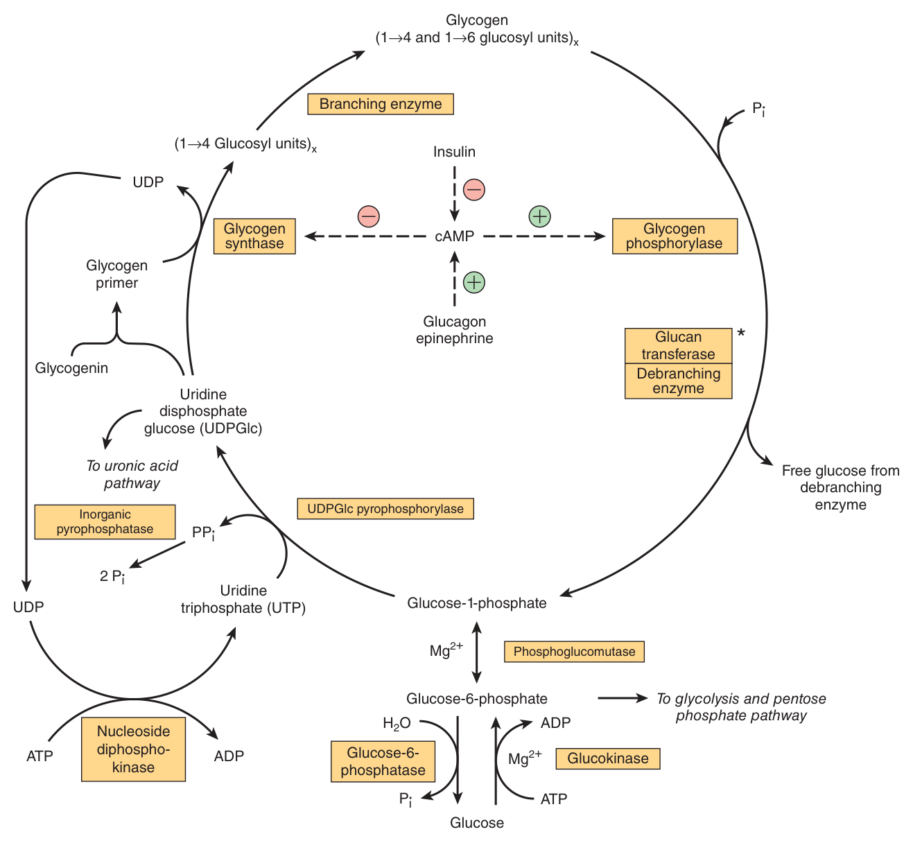
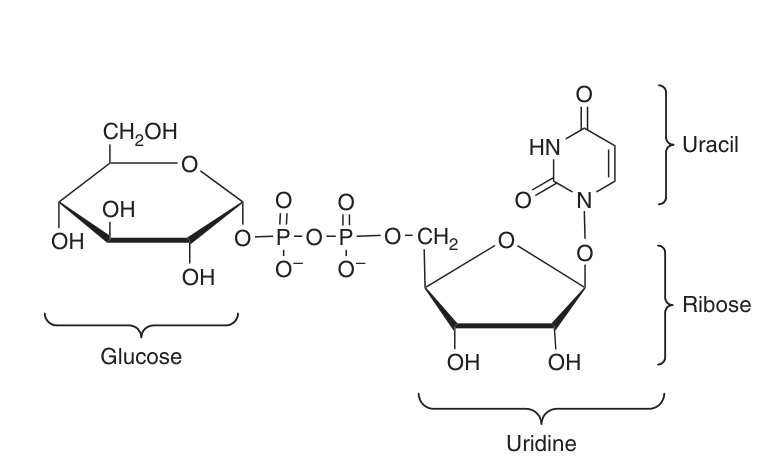
| 3 | Glucose 1-phosphate + UTP → UDP-glucose (UDPGlc) + PPi | UDPGlc pyrophosphorylase |
| 4 | PPi → 2 Pi | Pyrophosphatase — shifting the equilibrium of the main reaction by removing one of its products |
| 5 | UDPGlc + glycogen(n) → glycogen(n+1) + UDP | Glycogen synthase — forms a glycosidic bond between C1 of the activated glucose and C4 of a terminal glucose residue |
Pyrophosphatase does not add anything to the glycogen. Its whole function is to destroy one of the products of step 3, so that step 3 cannot run backwards.
This is precisely the device Unit 8 described for ATP → AMP + PPi: hydrolysing the pyrophosphate (ΔG⁰′ −19.2 kJ/mol) pulls the reaction irreversibly forward. The cell pays an extra high-energy bond to buy directionality. Watch for this pattern again in nucleotide synthesis and DNA replication.
And that primer is itself formed on glycogenin, a 37-kDa protein which is glycosylated on a specific tyrosine residue by UDPGlc. Further glucose residues are attached in the 1→4 position to make a short chain that is then a substrate for glycogen synthase.
So every glycogen granule has a protein at its core. A neat tissue difference worth quoting: in skeletal muscle glycogenin remains attached in the centre of the glycogen molecule, whereas in liver the number of glycogen molecules exceeds the number of glycogenin molecules.
- What is the activated form of glucose for glycogen synthesis? → UDP-glucose (UDPGlc), formed from glucose 1-phosphate and UTP
- Why does pyrophosphatase matter? → It hydrolyses PPi, removing a product and driving the reaction forward
- What bond does glycogen synthase form? → Between C1 of UDPGlc's glucose and C4 of a terminal glycogen residue — a 1→4 linkage
- What is glycogenin? → A 37-kDa protein, glycosylated on a tyrosine residue by UDPGlc, that primes glycogen synthesis
Branching ★★
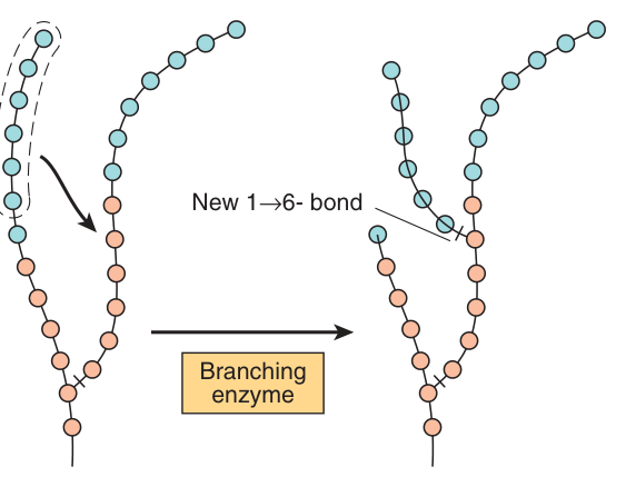
Addition occurs at the non-reducing, outer end of the molecule, so the “branches” of the glycogen “tree” become elongated as successive 1→4 linkages are formed.
When a chain has been lengthened to at least 11 glucose residues, branching enzyme transfers a part of the 1→4 chain (at least six glucose residues) to a neighbouring chain to form a 1→6 linkage, establishing a branch point.
The branches then grow by further 1→4 additions and further branching.
Solubility. A single long unbranched chain would be an insoluble fibre. Branching makes a compact, hydrated granule.
Speed. Phosphorylase can only chew inwards from a non-reducing end. A linear polymer has exactly one such end — so mobilisation would be hopelessly slow. Every branch creates another end to attack simultaneously.
The architecture is optimised for the emergency in §5: you need glucose now, from as many points at once as possible.
- At what chain length does branching occur? → At least 11 glucose residues
- How many residues are transferred? → At least six
- What linkage is formed at a branch point? → 1→6
- Why is glycogen branched? → For solubility, and to create many non-reducing ends for rapid simultaneous mobilisation
Glycogenolysis ★★★
This is Unit 7's principle again: a pathway and its opposite must use different enzymes at the irreversible steps, otherwise they could not be regulated independently and would simply run in a futile cycle. Synthesis uses UDPGlc and glycogen synthase; breakdown uses phosphorolysis by phosphorylase. Different chemistry, different enzymes, separately controlled.
State this in the first line and the rest of your answer has a frame.
| # | Step | Enzyme |
|---|---|---|
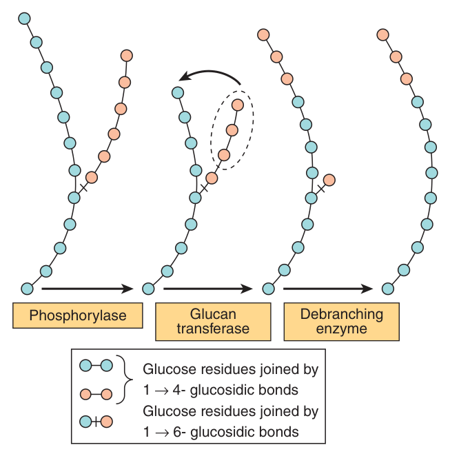
| 1 | Phosphorolytic cleavage of 1→4 linkages by inorganic phosphate, yielding glucose 1-phosphate. Terminal residues are removed sequentially until approximately four glucose residues remain on either side of a 1→6 branch | Glycogen phosphorylase — catalyses the rate-limiting step |
| 2 | Transfer of a trisaccharide unit from one branch to the other, exposing the 1→6 branch point | Transferase (glucan transferase) |
| 3 | Hydrolysis of the 1→6 linkage | Debranching enzyme |
| 4 | Glucose 1-phosphate ⇌ glucose 6-phosphate | Phosphoglucomutase — reversible, so it serves both pathways |
| 5 | Glucose 6-phosphate → free glucose, exported | Glucose-6-phosphatase — LIVER AND KIDNEY ONLY |
Note the word. Phosphorolysis uses inorganic phosphate to break the bond (compare hydrolysis, which uses water). The product is therefore glucose 1-phosphate, already phosphorylated.
That is worth an ATP. If glycogen were hydrolysed to free glucose, the cell would have to spend ATP at hexokinase to phosphorylate it again. By using phosphate instead of water, muscle gets its glucose into glycolysis for free — which is exactly what you want in an emergency.
- Which enzyme catalyses the rate-limiting step? → Glycogen phosphorylase
- What is phosphorolysis, and what is the product? → Cleavage by inorganic phosphate rather than water, yielding glucose 1-phosphate
- Where does phosphorylase stop? → About four glucose residues from a 1→6 branch point
- What two activities are needed at a branch? → A transferase moving a trisaccharide, then debranching enzyme hydrolysing the 1→6 bond
- Why is phosphorolysis energetically preferable? → The product is already phosphorylated, saving the ATP that hexokinase would spend
cAMP and the phosphorylase cascade ★★★
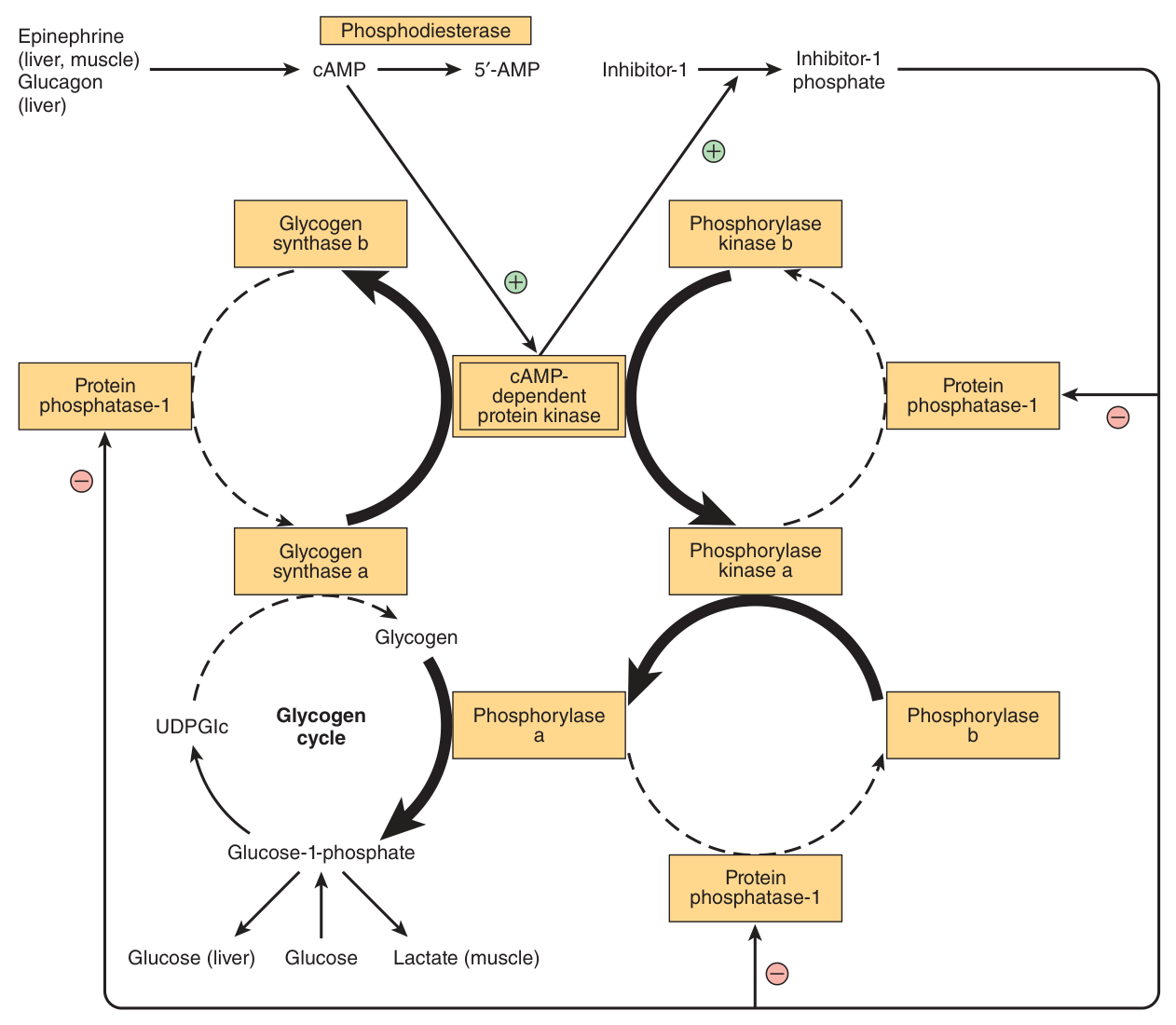
The two principal enzymes — glycogen phosphorylase and glycogen synthase — are regulated by allosteric mechanisms and by covalent modification through reversible phosphorylation and dephosphorylation in response to hormone action. Unit 7's two short-term mechanisms, both operating on the same pathway.
Formed from ATP by adenylyl cyclase at the inner surface of cell membranes, acting as an intracellular second messenger in response to hormones such as epinephrine, norepinephrine and glucagon.
It is hydrolysed by phosphodiesterase, terminating hormone action — and in liver, insulin increases the activity of phosphodiesterase.
The cascade
Epinephrine → ↑cAMP → activates cAMP-dependent protein kinase → which phosphorylates inactive phosphorylase kinase b to active phosphorylase kinase a → which, by a further phosphorylation, activates phosphorylase b to phosphorylase a.
Four steps to turn on one enzyme looks wasteful. It is amplification. One hormone molecule activates one receptor, which makes many cAMP; each cAMP activates a kinase; each kinase phosphorylates many molecules of the next enzyme. By the bottom of the chain, a handful of adrenaline molecules have mobilised a whole liver's worth of glycogen.
Harper's gives the scale in muscle: glycogenolysis increases several hundred-fold immediately after the onset of contraction.
Liver phosphorylase versus muscle phosphorylase
| Liver | Muscle | |
|---|---|---|
| Structure | One of the serine hydroxyls of active phosphorylase a is phosphorylated | A dimer, each monomer containing 1 mol of pyridoxal phosphate (vitamin B₆) |
| Phosphorylase a | Active, phosphorylated | Phosphorylated and active in either the presence OR absence of 5′-AMP. The normal physiologically active form |
| Phosphorylase b | Formed by hydrolytic removal of phosphate by protein phosphatase-1; reactivated by phosphorylase kinase | Dephosphorylated and active ONLY in the presence of 5′-AMP — which rises during exercise, providing fuel for the muscle |
The mechanism is beautifully direct. Muscle phosphorylase kinase has four subunit types, (αβγδ)₄. The α and β subunits contain the serines phosphorylated by cAMP-dependent protein kinase. And the δ subunit binds four Ca²⁺ and is identical to the Ca²⁺-binding protein CALMODULIN.
Binding Ca²⁺ activates the catalytic site of the γ subunit while the molecule remains in the dephosphorylated b configuration — so the enzyme can be switched on by calcium alone, without any hormone at all.
Calmodulin is built into the enzyme. The nerve impulse that tells the muscle to contract simultaneously tells it to start burning glycogen. Compare Unit 11 §9, where Ca²⁺ activates the citric acid cycle for the same reason.
- How is cAMP made and destroyed? → Made from ATP by adenylyl cyclase; hydrolysed by phosphodiesterase, whose activity insulin increases in liver
- Trace the cascade → Epinephrine → ↑cAMP → cAMP-dependent protein kinase → phosphorylase kinase b to a → phosphorylase b to a
- Which form of muscle phosphorylase needs AMP? → Phosphorylase b — active only in the presence of 5′-AMP
- What vitamin is in muscle phosphorylase? → Pyridoxal phosphate (B₆), 1 mol per monomer
- What is the δ subunit of phosphorylase kinase? → Calmodulin — it binds four Ca²⁺
Reciprocal regulation — the exam question ⭐
The 2020/21 paper asked, in Section II: “Please try to explain how insulin and glucagon regulate glycogenesis and glycogenolysis reciprocally.” This section is that answer.
The same phosphorylation event that activates phosphorylase simultaneously inactivates glycogen synthase. One signal, two opposite effects — so the cell can never break down and synthesise glycogen at the same time.
| Glucagon / epinephrine | Insulin | |
|---|---|---|
| Signal | Low blood glucose (glucagon), or stress (epinephrine) | High blood glucose |
| cAMP | ↑ — via adenylyl cyclase | ↓ — in liver, insulin increases phosphodiesterase activity, destroying cAMP |
| Phosphorylation state | Enzymes become phosphorylated | Enzymes become dephosphorylated |
| Glycogen phosphorylase | ACTIVATED (b → a) | INACTIVATED |
| Glycogen synthase | INACTIVATED | ACTIVATED |
| Net effect | Glycogenolysis — glucose released | Glycogenesis — glucose stored |
For glycogen, phosphorylation means BREAKDOWN.
Phosphorylase is active when phosphorylated; synthase is inactive when phosphorylated. So if you can remember which hormone raises cAMP — glucagon and adrenaline, the “we need glucose now” hormones — everything else follows automatically.
Be careful, though: this rule is specific to glycogen. In other pathways phosphorylation activates synthesis. Do not generalise it; anchor it to “adrenaline phosphorylates, and adrenaline wants glucose released.”
Glucagon acts mainly on liver — its job is to raise blood glucose, so it acts where glucose-6-phosphatase exists. Epinephrine acts on muscle as well — but muscle has no phosphatase, so the glucose stays put and fuels the muscle itself.
Same second messenger, same cascade, two different outcomes — decided entirely by which tissue has the enzyme from §2.
- Which hormones raise cAMP? → Glucagon, epinephrine and norepinephrine
- How does insulin lower cAMP in liver? → By increasing phosphodiesterase activity
- What does phosphorylation do to phosphorylase and to synthase? → Activates phosphorylase, inactivates synthase — hence reciprocal control
- Why does glucagon act mainly on liver? → Only liver (and kidney) has glucose-6-phosphatase, so only liver can export glucose to the blood
Glycogen storage diseases ★★
A group of inherited disorders characterised by deficient mobilisation of glycogen or deposition of abnormal forms of glycogen, leading to muscular weakness or even death.
| Disease | Deficient enzyme | Consequence |
|---|---|---|
| Type I — von Gierke's disease | Glucose-6-phosphatase | Liver glycogen accumulates but cannot be exported as glucose — severe fasting hypoglycaemia, hepatomegaly, lactic acidosis |
| Type V — McArdle's syndrome | Muscle glycogen phosphorylase | Muscle glycogen cannot be mobilised — exercise intolerance, cramps, with no rise in blood lactate on exercise |
von Gierke removes the enzyme that lets the liver do its job — so the liver fills with glycogen it cannot release, and the patient is hypoglycaemic despite abundant stores.
McArdle removes the enzyme that lets muscle do its job — so blood glucose is normal but the muscle cannot access its own fuel, and it cramps under load.
Two diseases, two tissues, two purposes. If you understood §2, you can derive both presentations without memorising them.
- Define glycogen storage disease → Inherited disorders with deficient mobilisation of glycogen or deposition of abnormal glycogen
- What is deficient in von Gierke's disease? → Glucose-6-phosphatase — causing fasting hypoglycaemia despite hepatic glycogen accumulation
- What is deficient in McArdle's syndrome? → Muscle glycogen phosphorylase — causing exercise intolerance with no rise in blood lactate
Revision layer
Glycogenesis versus glycogenolysis
| Glycogenesis | Glycogenolysis | |
|---|---|---|
| Activated form of glucose | UDP-glucose (from glucose 1-P + UTP) | — |
| Key enzyme | Glycogen synthase (1→4 bonds) | Glycogen phosphorylase — rate-limiting |
| Chemistry | Glycosidic bond formation | Phosphorolysis, giving glucose 1-phosphate |
| Branch handling | Branching enzyme — moves ≥6 residues to make a 1→6 bond, at chain length ≥11 | Transferase (moves a trisaccharide) + debranching enzyme (hydrolyses 1→6) |
| Primer | Glycogenin, 37 kDa, glycosylated on a tyrosine | — |
| Activated by | Insulin (dephosphorylation) | Glucagon, epinephrine (phosphorylation), and Ca²⁺ in muscle |
Definitions from this unit — Section I material
| Term | Definition |
|---|---|
| Glycogen | The major storage carbohydrate in animals, a branched polymer of α-D-glucose; occurring mainly in liver (up to 6%) and muscle (rarely over 1%, but 3–4 times more in total mass) |
| Glycogenesis | The synthesis of glycogen from glucose, occurring mainly in muscle and liver, via glucose 6-phosphate, glucose 1-phosphate and the activated nucleotide UDP-glucose, with chain extension by glycogen synthase and branch formation by branching enzyme |
| Glycogenolysis | The breakdown of glycogen — not the reverse of glycogenesis but a separate pathway — in which glycogen phosphorylase catalyses the rate-limiting phosphorolytic cleavage of 1→4 linkages to yield glucose 1-phosphate, with debranching enzyme hydrolysing the 1→6 linkages |
| Glycogenin | A 37-kDa protein, glycosylated on a specific tyrosine residue by UDPGlc, which acts as the primer on which the glycogen primer is formed |
| Glycogen storage disease | One of a group of inherited disorders characterised by deficient mobilisation of glycogen or deposition of abnormal forms of glycogen, leading to muscular weakness or even death |
| Second messenger (cAMP) | Cyclic AMP, formed from ATP by adenylyl cyclase at the inner surface of the cell membrane, acting as an intracellular second messenger in response to hormones such as epinephrine, norepinephrine and glucagon, and hydrolysed by phosphodiesterase |
Numbers worth carrying in
| Item | Value |
|---|---|
| Liver glycogen | up to 6% |
| Muscle glycogen | rarely over 1%, but 3–4× more in total |
| Liver glycogen exhausted after | 12–18 hours of fasting |
| Glycogenin | 37 kDa |
| Branching occurs at chain length | ≥ 11 residues; ≥ 6 transferred |
| Phosphorylase stops | about 4 residues from a branch point |
| Phosphorylase kinase | (αβγδ)₄; δ = calmodulin, binds 4 Ca²⁺ |
| Glycogenolysis on contraction | increases several hundred-fold |
- Define glycogen, glycogenesis and glycogenolysis
- Contrast liver and muscle glycogen, and explain the glucose-6-phosphatase difference
- Give the five steps of glycogenesis with enzymes, including the role of pyrophosphatase
- Explain glycogenin and why a primer is needed
- Describe branching and debranching, with the numbers
- Trace the cAMP cascade from epinephrine to phosphorylase a
- Answer the 2020/21 question on reciprocal insulin/glucagon regulation
- Explain how Ca²⁺ and calmodulin synchronise glycogenolysis with contraction
- Give von Gierke and McArdle with their enzymes and presentations