Glycolysis & the Oxidation of Pyruvate
Why glycolysis matters ★★★
The major pathway for glucose metabolism, which occurs in the cytosol of all cells.
It is unique in that it can function either aerobically or anaerobically.
This is the 2019 answer sheet's wording almost exactly — “the major initial pathway for glucose metabolism, occurs in the cytosol of all cells”. Open with it.
Three facts in that definition, and each is doing work. Cytosol — no mitochondrion required, which is why it can run without oxygen. All cells — there is no tissue that does not do this. Either aerobically or anaerobically — this is the property that makes glycolysis unique among the major pathways, and it is what §5 is about.
| Tissue | Why glycolysis matters there |
|---|---|
| Brain | Most tissues have at least some requirement for glucose; in brain the requirement is substantial |
| Erythrocytes | Lack mitochondria, so are completely reliant on glucose and metabolise it by anaerobic glycolysis — always terminating in lactate, even with oxygen present |
| Skeletal muscle | Glycolysis provides ATP in the absence of oxygen, allowing very high work rates when oxygen supply is insufficient, and allowing tissues to survive anoxic episodes |
| Heart muscle | Adapted for aerobic performance; has relatively low glycolytic activity and poor survival under ischaemia |
Skeletal muscle can cope with an interrupted blood supply for a while, because it can make ATP without oxygen. Cardiac muscle largely cannot — its glycolytic capacity is low, so when a coronary artery occludes, the myocardium cannot fall back on anaerobic glycolysis and the cells die.
One line of biochemistry explains the entire clinical difference between a cramp and an infarct.
Glycolysis is also the main pathway for the metabolism of fructose, galactose and other dietary carbohydrates — not just glucose.
- Define glycolysis → The major pathway for glucose metabolism, occurring in the cytosol of all cells; unique in functioning either aerobically or anaerobically
- Why do erythrocytes always produce lactate? → They lack mitochondria, so pyruvate cannot be oxidised further
- Why does heart muscle tolerate ischaemia poorly? → It is adapted for aerobic performance and has relatively low glycolytic activity
The overall equation ★★★
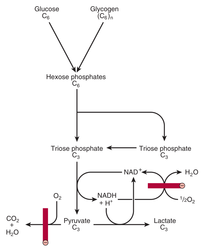
Glycolysis has a shape, and the shape is the mnemonic:
The investment phase (steps 1–5) spends 2 ATP to prepare a six-carbon sugar and split it in two.
The payoff phase (steps 6–10) earns 4 ATP — but note that everything from here on happens twice, because there are now two three-carbon molecules.
Net: 4 − 2 = 2 ATP. The doubling is where most students lose marks: if you forget that the payoff phase runs twice per glucose, every number you compute afterwards will be halved.
- Write the overall equation to lactate → Glucose + 2 ADP + 2 Pi → 2 lactate + 2 ATP + 2 H₂O
- Where are the enzymes? → All in the cytosol
- Why is the net yield 2 and not 4? → Two ATP are invested in the first phase; the payoff phase yields 4 because it runs twice
The investment phase — steps 1 to 5 ★★★
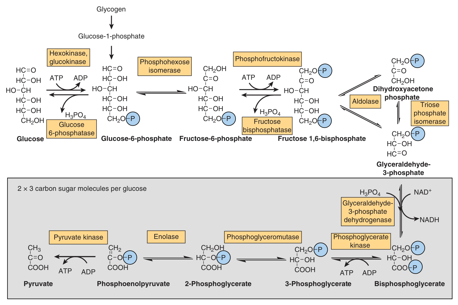
| # | Reaction | Enzyme | Note |
|---|---|---|---|
| 1 | Glucose → glucose 6-phosphate | Hexokinase (or glucokinase) | Uses ATP. ⚠️ IRREVERSIBLE — see the coupling calculation in Unit 8 |
| 2 | Glucose 6-phosphate → fructose 6-phosphate | Phosphohexose isomerase | Freely reversible |
| 3 | Fructose 6-phosphate → fructose 1,6-bisphosphate | Phosphofructokinase-1 (PFK-1) | Uses ATP. ⚠️ IRREVERSIBLE — and the major site of regulation of glycolysis |
| 4 | Fructose 1,6-bisphosphate → glyceraldehyde 3-phosphate + dihydroxyacetone phosphate | Aldolase | The split — one six-carbon sugar becomes two three-carbon ones |
| 5 | Dihydroxyacetone phosphate ⇌ glyceraldehyde 3-phosphate | Phosphotriose isomerase | Converts DHAP into the form the pathway can use — so both halves continue |
Glucokinase is found in liver and pancreatic β islet cells and has a Km very much higher. It therefore only becomes active when blood glucose is high — after a meal.
The two functions that follow:
• In the liver, glucokinase's job is to remove glucose from the blood following a meal, providing glucose 6-phosphate in excess of the requirements for glycolysis — which is then used for glycogen synthesis and lipogenesis.
• In the pancreas, the glucose 6-phosphate formed by glucokinase signals increased glucose availability and leads to the secretion of insulin.
Same reaction, two Km values, two entirely different physiological roles. This is the best example in the whole course of why Km is not an abstraction.
- Name the five enzymes of the investment phase → Hexokinase/glucokinase, phosphohexose isomerase, PFK-1, aldolase, phosphotriose isomerase
- Which two steps consume ATP? → Hexokinase (step 1) and phosphofructokinase-1 (step 3)
- Which is the major site of regulation? → Phosphofructokinase-1
- Where is glucokinase found, and why does its high Km matter? → Liver and pancreatic β cells; it acts only when blood glucose is high, i.e. after a meal
The payoff phase — steps 6 to 10 ★★★
Everything below happens twice per glucose, because step 4 produced two triose phosphates.
| # | Reaction | Enzyme | Note |
|---|---|---|---|
| 6 | Glyceraldehyde 3-phosphate → 1,3-bisphosphoglycerate | Glyceraldehyde 3-phosphate dehydrogenase | NAD⁺ → NADH + H⁺. The only oxidation in glycolysis — and the step that makes NAD⁺ regeneration necessary |
| 7 | 1,3-Bisphosphoglycerate → 3-phosphoglycerate | Phosphoglycerate kinase | MAKES ATP — substrate-level phosphorylation. 2 ATP per glucose |
| 8 | 3-Phosphoglycerate → 2-phosphoglycerate | Phosphoglycerate mutase | 2,3-Bisphosphoglycerate is likely an intermediate |
| 9 | 2-Phosphoglycerate → phosphoenolpyruvate | Enolase | A dehydration. Inhibited by fluoride; requires Mg²⁺ or Mn²⁺ |
| 10 | Phosphoenolpyruvate → pyruvate | Pyruvate kinase | MAKES ATP. ⚠️ IRREVERSIBLE. 2 ATP per glucose |
Go back and look at it. The only glycolytic intermediates above ATP were 1,3-bisphosphoglycerate (−49.3) and phosphoenolpyruvate (−61.9).
And here they are — steps 7 and 10, the only two steps that make ATP. It could not have been otherwise: a compound below ATP in that table has no capacity to phosphorylate ADP. You could have derived which steps make ATP without ever seeing the pathway.
Fluoride. Enolase is inhibited by fluoride, which is why blood samples taken for measurement of glucose are collected into tubes containing fluoride — it stops the erythrocytes in the tube from consuming the glucose you are trying to measure. A laboratory fact with a biochemical reason, and a favourite MCQ.
The cell makes the step one-way by immediately converting the product into something else — the same trick as hydrolysing PPi in Unit 8.
- Which step is the only oxidation? → Step 6, glyceraldehyde 3-phosphate dehydrogenase, producing NADH
- Which two steps make ATP, and how much per glucose? → Phosphoglycerate kinase and pyruvate kinase, 2 ATP each
- What inhibits enolase, and what is the practical use? → Fluoride — blood glucose tubes contain it to stop glycolysis in the sample
- How does arsenate poison glycolysis? → It competes with Pi, giving a product that hydrolyses spontaneously without forming ATP
The fork — lactate or acetyl-CoA ★★★
Pyruvate has been made. What happens next is decided by the availability of oxygen — or, more precisely, by whether NADH can be reoxidised.
| Anaerobic | Aerobic | |
|---|---|---|
| The problem | NADH cannot be reoxidised through the respiratory chain | NADH can be reoxidised normally |
| What happens to pyruvate | Reduced to lactate by lactate dehydrogenase | Transported into mitochondria, oxidatively decarboxylated to acetyl-CoA, then oxidised to CO₂ in the citric acid cycle |
| What happens to the NADH | It is used to reduce pyruvate — which is the point | Reducing equivalents enter mitochondria via the malate-aspartate or glycerophosphate shuttle (Unit 9) |
| ATP yield | 2 per glucose | up to 32 per glucose |
Step 6 — glyceraldehyde-3-phosphate dehydrogenase — consumes NAD⁺. The cell's pool of NAD⁺ is small, so unless it is continuously regenerated, glycolysis stops dead after a few turns. Normally the respiratory chain does the regenerating. Without oxygen it cannot, so the cell uses pyruvate itself as the electron acceptor.
Harper's puts it exactly: “the reoxidation of NADH via lactate formation allows glycolysis to proceed in the absence of oxygen by regenerating sufficient NAD⁺ for another cycle of the reaction catalysed by glyceraldehyde-3-phosphate dehydrogenase.”
Lactate is not a waste product. It is the price of a recycled coenzyme.
Which tissues make lactate
Skeletal muscle, particularly the white fibres, where the rate of work output — and therefore the need for ATP — may exceed the rate at which oxygen can be taken up and utilised. Erythrocytes always, even aerobically. Also brain, gastrointestinal tract, renal medulla, retina and skin. The liver, kidneys and heart normally take up lactate and oxidise it — but produce it under hypoxic conditions.
When lactate production is high — vigorous exercise, septic shock, cancer cachexia — much of it is taken to the liver for gluconeogenesis (Unit 13). That is an energy-expensive process, so it raises the metabolic rate to supply the ATP and GTP required.
The resulting increase in oxygen consumption is seen as the oxygen debt after vigorous exercise. You keep breathing hard after you stop running because your liver is busy turning the lactate back into glucose.
- What determines the fate of pyruvate? → The availability of oxygen — specifically, whether NADH can be reoxidised
- Why is pyruvate reduced to lactate? → To regenerate NAD⁺ so that glyceraldehyde-3-phosphate dehydrogenase can continue
- Name five tissues that normally produce lactate → Skeletal muscle (white fibres), erythrocytes, brain, GI tract, renal medulla, retina, skin
- What is oxygen debt? → The increased oxygen consumption after exercise, needed to supply ATP and GTP for hepatic gluconeogenesis from lactate
The three regulated steps ★★★
Unit 7 told you that regulation targets the rate-limiting, irreversible step. Here is that principle applied. Although most of the reactions of glycolysis are freely reversible, three are markedly exergonic and must therefore be considered physiologically irreversible — and those three are the major sites of regulation.
| Step | Enzyme | Reversed in gluconeogenesis by |
|---|---|---|
| 1 | Hexokinase (and glucokinase) | Glucose 6-phosphatase |
| 3 | Phosphofructokinase-1 | Fructose 1,6-bisphosphatase |
| 10 | Pyruvate kinase | Pyruvate carboxylase and phosphoenolpyruvate carboxykinase |
Gluconeogenesis is not glycolysis run backwards. Seven of the ten steps are freely reversible and can simply be reversed — but three cannot, and each of those three needs a different enzyme to bypass it.
So when you meet gluconeogenesis, you already know its four bypass enzymes. They are in the right-hand column above, and they exist for exactly one reason: the left-hand column is irreversible.
That inhibition can be rapidly relieved by 5′AMP, which is formed as ADP begins to accumulate, signalling the need for an increased rate of glycolysis.
This is Unit 8's adenylate kinase reaction paying off. AMP rises steeply when ATP falls, because the AMP pool is small — so it is a far more sensitive alarm than ATP is a gauge. The most important regulatory enzyme in glycolysis listens to the most sensitive available signal.
One further point worth knowing. Fructose enters glycolysis by phosphorylation to fructose 1-phosphate, and thereby bypasses the main regulatory steps — resulting in the formation of more pyruvate and acetyl-CoA than is required. A pathway that skips its own brakes, which is part of why large fructose loads are metabolically unfavourable.
- Name the three irreversible enzymes → Hexokinase (glucokinase), phosphofructokinase-1, pyruvate kinase
- Why are they the regulatory sites? → They catalyse markedly exergonic, physiologically irreversible reactions
- What relieves ATP inhibition of PFK-1? → 5′AMP, formed as ADP accumulates
- Which four enzymes reverse these steps in gluconeogenesis? → Glucose 6-phosphatase, fructose 1,6-bisphosphatase, pyruvate carboxylase, PEP carboxykinase
- Why is fructose metabolically unusual? → It enters as fructose 1-phosphate and bypasses the main regulatory steps
The 2,3-bisphosphoglycerate shunt ★★
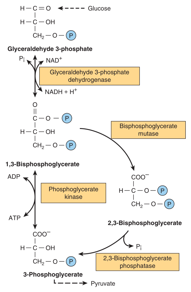
In the erythrocytes of many mammals, the reaction catalysed by phosphoglycerate kinase may be bypassed by a process that effectively dissipates as heat the free energy associated with the high-energy phosphate of 1,3-bisphosphoglycerate.
The route: 1,3-bisphosphoglycerate → 2,3-bisphosphoglycerate (bisphosphoglycerate mutase) → 3-phosphoglycerate (2,3-bisphosphoglycerate phosphatase). The pathway rejoins glycolysis, but no ATP is made at that step.
This looks like a design fault — deliberately skipping an ATP-generating step. But remember what 2,3-BPG does: it binds in the central cavity of haemoglobin, stabilises the T state, and lowers oxygen affinity so that more O₂ is released to the tissues.
The erythrocyte is not a cell that needs much ATP. It is a cell whose job is oxygen delivery. So it sacrifices one ATP per turn to manufacture the molecule that makes it better at its job. Unit 4 and Unit 10 are the same story told from two ends.
It also explains why a low pO₂ promotes BPG synthesis — the shunt is the mechanism.
- Which step does the 2,3-BPG shunt bypass? → Phosphoglycerate kinase — so no ATP is formed
- What are the two enzymes? → Bisphosphoglycerate mutase and 2,3-bisphosphoglycerate phosphatase
- Why does the erythrocyte do this? → 2,3-BPG stabilises the T state of haemoglobin, lowering O₂ affinity and improving tissue delivery
Pyruvate dehydrogenase ★★★
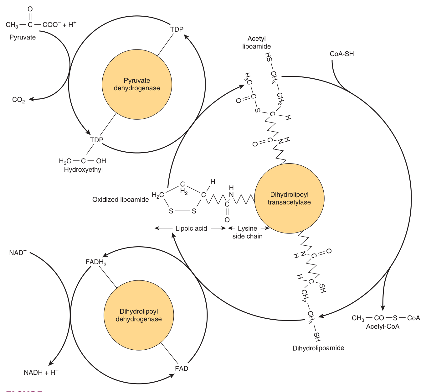
The irreversible route from glycolysis to the citric acid cycle, catalysing the oxidative decarboxylation of pyruvate to acetyl-CoA.
| The three catalytic enzymes | The five cofactors |
|---|---|
| Pyruvate dehydrogenase (PDH) Dihydrolipoamide transacetylase (DLT) Dihydrolipoamide dehydrogenase (DLDH) | TPP (thiamin pyrophosphate) Lipoic acid NAD⁺ FAD CoASH |
Look at what those five cofactors are made of: TPP from thiamin (B₁), FAD from riboflavin (B₂), NAD⁺ from niacin (B₃), CoA from pantothenic acid (B₅), plus lipoic acid.
This single enzyme complex needs four B vitamins. That is why Unit 5's B-vitamin table was worth learning, and it is why thiamin deficiency causes lactic acidosis — §10.
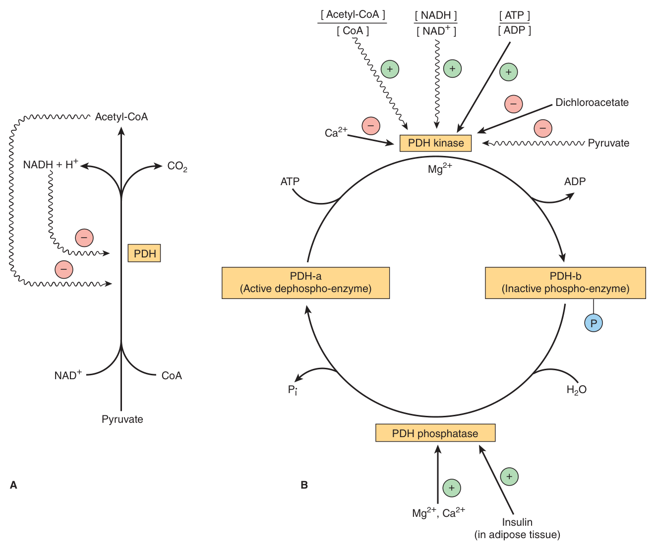
Regulation — two mechanisms, both from Unit 7
| Mechanism | Detail |
|---|---|
| 1 · End-product inhibition | PDH is inhibited by its own products, acetyl-CoA and NADH |
| 2 · Covalent modification | Phosphorylation of three serine residues by a kinase decreases activity; dephosphorylation by a phosphatase increases it |
The kinase — which switches the enzyme off — is activated by increases in the [ATP]/[ADP], [acetyl-CoA]/[CoA] and [NADH]/[NAD⁺] ratios. Read those three ratios: every one of them is a signal that the cell has plenty of energy.
In starvation, free fatty acid concentrations increase, so there is a decrease in the proportion of the enzyme in the active form, leading to a sparing of carbohydrate.
That phrase — sparing of carbohydrate — is the point. PDH is the irreversible gate out of glycolysis: once pyruvate becomes acetyl-CoA, that carbon can never be turned back into glucose. Shutting the gate when fat is available preserves glucose for the brain, which cannot use fatty acids. The switch that protects your brain during a fast is a phosphorylation on three serine residues.
Conversely, in adipose tissue, where glucose provides acetyl-CoA for lipogenesis, the enzyme is activated in response to insulin.
- Name the three enzymes of the PDH complex → Pyruvate dehydrogenase, dihydrolipoamide transacetylase, dihydrolipoamide dehydrogenase
- Name the five cofactors → TPP, lipoic acid, NAD⁺, FAD, CoASH
- What inhibits PDH by end-product inhibition? → Acetyl-CoA and NADH
- What activates the PDH kinase? → Rises in [ATP]/[ADP], [acetyl-CoA]/[CoA] and [NADH]/[NAD⁺]
- Why is PDH inhibited in starvation? → Fatty acid oxidation raises acetyl-CoA and NADH, sparing carbohydrate for the brain
- Which form is active? → The DEPHOSPHORYLATED form (PDH-a)
The ATP balance sheet ★★★
Oxidation of glucose yields up to 32 mol of ATP under aerobic conditions, but only 2 mol when oxygen is absent.
Most of the ATP is formed by oxidative phosphorylation resulting from reoxidation of the reduced coenzymes by the respiratory chain; the remainder is formed by substrate-level phosphorylation.
| Stage | Source | ATP |
|---|---|---|
| Investment | Hexokinase | −1 |
| Phosphofructokinase-1 | −1 | |
| Payoff (×2) | Phosphoglycerate kinase — substrate level | +2 |
| Pyruvate kinase — substrate level | +2 | |
| Net from glycolysis alone | +2 | |
| Oxidative | 2 × NADH from glyceraldehyde-3-P dehydrogenase | via the shuttles |
| 2 × NADH from pyruvate dehydrogenase | respiratory chain | |
| Citric acid cycle, ×2 turns | the bulk of it | |
| TOTAL, aerobic | up to 32 |
Lecture 9 slide 16 stated P:O = 3 and 2, which would give the older figure of 38.
So the department's own material uses the modern numbers in this lecture and the old numbers in the previous one. Combined with Lecture 9 slide 27 conceding “possibly 2.5”, the weight of evidence in your own course materials is on the side of 2.5 / 1.5 and 32 ATP.
Practical advice: quote 32 — it is what this lecturer's slide says and what the textbook says. If a question forces the P:O ratio itself, give your lecturer's figure and note the discrepancy. See Unit 9 §7 for the full comparison.
- Net ATP from glycolysis alone? → 2 per glucose
- Total aerobic yield per glucose? → Up to 32 mol ATP
- Anaerobic yield? → 2 mol — which is why much more glucose must be metabolised anaerobically
- Where does most of the aerobic ATP come from? → Oxidative phosphorylation, from reoxidation of the reduced coenzymes
Clinical aspects ★★
Lactic acidosis from inhibition of pyruvate metabolism
| Cause | Mechanism |
|---|---|
| Arsenite and mercuric ions | React with the –SH groups of lipoic acid and inhibit pyruvate dehydrogenase, allowing pyruvate to accumulate |
| Thiamin deficiency | PDH requires TPP. Nutritionally deprived alcoholics are thiamin-deficient — both from poor diet and because alcohol inhibits thiamin absorption — and may develop potentially fatal pyruvic and lactic acidosis |
| Inherited PDH deficiency | Defects in one or more components of the complex; presents with lactic acidosis, particularly after a glucose load |
Because of the dependence of the brain on glucose as a fuel, these metabolic defects commonly cause neurological disturbances. The brain has a substantial requirement for glucose (§1) and cannot substitute fatty acids — so a block in glucose oxidation hits the brain first and hardest.
Note the practical inference for the alcoholic patient: giving glucose to a thiamin-deficient patient without thiamin drives more pyruvate into a pathway that cannot process it.
Enzyme deficiencies
| Deficiency | Presentation |
|---|---|
| Aldolase A (inherited) | Haemolytic anaemia |
| Pyruvate kinase in erythrocytes | Haemolytic anaemia |
| Muscle phosphofructokinase | Low exercise capacity, particularly on high-carbohydrate diets. Work capacity improves when lipid is available as an alternative fuel — e.g. in starvation, when free fatty acids and ketone bodies are increased |
Look back at §1. The erythrocyte has no mitochondria, so glycolysis is its only source of ATP — a defect anywhere in the pathway leaves it unable to maintain its membrane, and it lyses. Hence haemolytic anaemia.
Skeletal muscle depends on glycolysis for burst activity, so a defect shows up as fatigue and low exercise capacity. Harper's summarises the whole pattern: diseases in which glycolytic enzymes are deficient are seen mainly as haemolytic anaemias, or as fatigue if the defect affects skeletal muscle.
In fast-growing cancer cells, glycolysis proceeds at a higher rate than is required by the citric acid cycle, forming large amounts of pyruvate, which is reduced to lactate and exported. This produces a relatively acidic local environment in the tumour, which may have implications for cancer therapy.
That lactate is then used for gluconeogenesis in the liver — an energy-expensive process responsible for much of the hypermetabolism seen in cancer cachexia. The patient's own liver is spending ATP to recycle the tumour's waste, and the patient wastes away paying for it.
- How do arsenite and mercuric ions inhibit PDH? → They react with the –SH groups of lipoic acid
- Why do alcoholics develop lactic acidosis? → Thiamin deficiency — poor diet plus alcohol inhibiting thiamin absorption — so PDH lacks TPP
- Which deficiencies cause haemolytic anaemia? → Aldolase A and erythrocyte pyruvate kinase
- What is the pattern of glycolytic enzyme disease? → Haemolytic anaemia, or fatigue if skeletal muscle is affected
- Explain cancer cachexia in terms of lactate → Tumours export lactate; the liver spends ATP converting it back to glucose, driving hypermetabolism
Revision layer
“Glycolysis” was set as a Section I definition in the 2019 paper. The definition below is the answer-sheet wording expanded — learn the first sentence exactly.
The major pathway for glucose metabolism, which occurs in the cytosol of all cells.
It converts glucose to pyruvate (aerobically) or lactate (anaerobically), and is unique in being able to function either aerobically or anaerobically. The overall anaerobic equation is glucose + 2 ADP + 2 Pi → 2 lactate + 2 ATP + 2 H₂O. It is regulated at three irreversible steps, catalysed by hexokinase, phosphofructokinase and pyruvate kinase.
The ten steps
| # | Enzyme | ATP / NADH |
|---|---|---|
| 1 | Hexokinase / glucokinase ⚠️ | −1 ATP |
| 2 | Phosphohexose isomerase | |
| 3 | Phosphofructokinase-1 ⚠️ | −1 ATP |
| 4 | Aldolase | splits into 2 trioses |
| 5 | Phosphotriose isomerase | |
| 6 | Glyceraldehyde 3-phosphate dehydrogenase | +2 NADH |
| 7 | Phosphoglycerate kinase | +2 ATP |
| 8 | Phosphoglycerate mutase | |
| 9 | Enolase — inhibited by fluoride | |
| 10 | Pyruvate kinase ⚠️ | +2 ATP |
| 11 | Lactate dehydrogenase (anaerobic only) | −2 NADH |
The three irreversible steps and their gluconeogenic bypasses
| Glycolysis | Gluconeogenesis |
|---|---|
| Hexokinase / glucokinase | Glucose 6-phosphatase |
| Phosphofructokinase-1 | Fructose 1,6-bisphosphatase |
| Pyruvate kinase | Pyruvate carboxylase + PEP carboxykinase |
Definitions from this unit — Section I material
| Term | Definition |
|---|---|
| Glycolysis ⭐ | The major pathway for glucose metabolism, occurring in the cytosol of all cells; unique in functioning either aerobically or anaerobically, yielding pyruvate or lactate respectively |
| Hexokinase vs glucokinase | Hexokinase has a high affinity (low Km) for glucose and is present in most tissues; glucokinase, in liver and pancreatic β cells, has a much higher Km and acts only when blood glucose is high — removing glucose after a meal and signalling insulin secretion |
| The pyruvate dehydrogenase complex | The multienzyme complex catalysing the irreversible oxidative decarboxylation of pyruvate to acetyl-CoA; three catalytic enzymes (PDH, dihydrolipoamide transacetylase, dihydrolipoamide dehydrogenase) and five cofactors (TPP, lipoic acid, NAD⁺, FAD, CoASH) |
| Substrate-level phosphorylation | Formation of ATP by direct transfer of a phosphate group from a substrate — in glycolysis at phosphoglycerate kinase and pyruvate kinase |
| Oxygen debt | The increased oxygen consumption seen after vigorous exercise, resulting from the increased oxidation of metabolic fuels needed to provide ATP and GTP for hepatic gluconeogenesis from lactate |
| The 2,3-bisphosphoglycerate pathway | A bypass of the phosphoglycerate kinase reaction in erythrocytes, dissipating as heat the free energy of the high-energy phosphate of 1,3-bisphosphoglycerate, and generating the 2,3-BPG that lowers haemoglobin's oxygen affinity |
Numbers worth carrying in
| Item | Value |
|---|---|
| Net ATP, anaerobic | 2 per glucose |
| Total ATP, aerobic | up to 32 per glucose |
| ATP invested | 2 — at hexokinase and PFK-1 |
| ATP produced (gross) | 4 — at phosphoglycerate kinase and pyruvate kinase, ×2 trioses |
| NADH produced in glycolysis | 2 — at glyceraldehyde 3-phosphate dehydrogenase |
| PDH complex | 3 enzymes, 5 cofactors, 3 serines phosphorylated |
- Define glycolysis in exam wording and write the overall equation
- List all ten enzymes in order and mark where ATP and NADH change
- Name the three irreversible steps and their four gluconeogenic bypass enzymes
- Contrast hexokinase and glucokinase, including both of glucokinase's roles
- Explain WHY pyruvate is reduced to lactate anaerobically
- Give the PDH complex's three enzymes and five cofactors, and both regulatory mechanisms
- Explain why PDH inhibition in starvation spares carbohydrate
- State the anaerobic and aerobic ATP yields
- Explain fluoride in blood tubes, arsenate poisoning, and the pattern of glycolytic enzyme disease