Respiratory Chain & Oxidative Phosphorylation
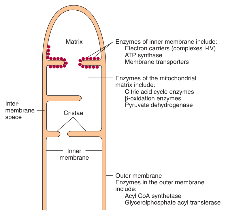
The mitochondrion — and why its membranes differ ★★
Unit 8 ended with a promise: oxidative phosphorylation is the greatest quantitative source of ATP in aerobic organisms. This unit is how. And it begins with an organelle whose entire architecture exists to make one trick possible.
| Compartment | Characteristic contents | Permeability |
|---|---|---|
| Outer membrane | Acyl-CoA synthetase and glycerolphosphate acyltransferase | Permeable to most metabolites |
| Intermembrane space | Adenylyl kinase and creatine kinase | — |
| Inner membrane | The phospholipid cardiolipin, concentrated here together with the enzymes of the respiratory chain. Thrown into many folds, or cristae | SELECTIVELY permeable |
| Matrix | Enzymes of β-oxidation and the citric acid cycle — which produce most of the reducing equivalents | — |
The outer membrane is permeable; the inner membrane is selectively permeable. Everything in this unit depends on that asymmetry.
You are about to learn that mitochondria pump protons out across the inner membrane and let them fall back in through a turbine. That only works if the protons cannot leak back any other way — which is exactly what an impermeable inner membrane provides. Puncture it and the whole machine stops; §9 is a catalogue of poisons that do precisely that.
The cristae are the same idea applied to area: fold the membrane and you fit far more respiratory chain into the same volume.
The compartment markers are examinable in themselves — an enzyme found in the intermembrane space but not the matrix tells a biochemist which compartment a preparation came from. Note that creatine kinase in the intermembrane space is Unit 8's phosphagen system, positioned exactly where ATP emerges.
- Which membrane is selectively permeable? → The inner; the outer is permeable to most metabolites
- Which phospholipid is concentrated in the inner membrane? → Cardiolipin
- Name the intermembrane space marker enzymes → Adenylyl kinase and creatine kinase
- What are cristae, and why? → Folds of the inner membrane, increasing the area available for the respiratory chain
What the respiratory chain is for ★★★
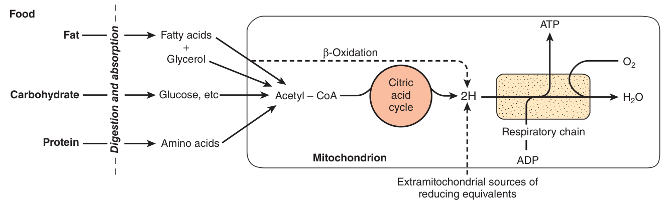
A series of redox carriers, present in the inner mitochondrial membrane, which collect and transport reducing equivalents, directing them to their final reaction with oxygen to form water, and which couple the free energy so liberated to the generation of ATP.
Follow the logic of the whole of metabolism in one sentence. Fat, carbohydrate and protein are digested to fatty acids, glucose and amino acids; these converge on acetyl-CoA; β-oxidation and the citric acid cycle oxidise them, and what they actually produce is not ATP but reducing equivalents (2H). The respiratory chain collects those reducing equivalents and cashes them in.
You could burn glucose in a flame and get 2870 kJ instantly — as heat, uselessly, and once. The cell instead strips off hydrogen atoms two at a time, hands them to carriers, and lets them fall to oxygen down a staircase of carriers rather than off a cliff.
Harper's describes the result precisely: the process is stepwise, efficient and controlled — rather than explosive, inefficient and uncontrolled. Each step releases a manageable packet of energy, and three of those steps release enough to pump a proton. That is the entire design.
- Define the respiratory chain → Redox carriers in the inner mitochondrial membrane that transport reducing equivalents to oxygen, forming water and coupling the free energy to ATP synthesis
- What do β-oxidation and the citric acid cycle actually produce? → Reducing equivalents (2H), not ATP directly
- What is the final electron acceptor? → Molecular oxygen, reduced to water
The carriers ★★★
| Carrier | Nature | Key facts |
|---|---|---|
| NAD⁺ / NADH | Nicotinamide coenzyme | Collects reducing equivalents from most dehydrogenases. Pyruvate and α-ketoglutarate dehydrogenase use complex systems involving lipoate and FAD before passing electrons to NAD; others, e.g. L(+)-3-hydroxyacyl-CoA dehydrogenase, couple directly |
| Flavoproteins (FMN, FAD) | Riboflavin-derived | NADH dehydrogenase is a metalloflavoprotein containing FeS and FMN, tightly bound to the chain, passing reducing equivalents to Q |
| Ubiquinone (Q, coenzyme Q) | Lipid-soluble quinone | n = 10 isoprenoid units in higher animals, hence Q₁₀. Structurally similar to vitamins K and E and to plastoquinone. A MOBILE component, collecting reducing equivalents from the fixed flavoprotein complexes and passing them to the cytochromes |
| Iron-sulfur proteins (Fe-S) | Non-haem iron | Found in Complexes I, II and III. Contain one, two or four Fe atoms linked to inorganic sulfur and/or via cysteine-SH groups. Take part in single-electron transfers, the Fe cycling between Fe²⁺ and Fe³⁺ |
| Cytochromes | Haem proteins | Carry one electron each. Cytochrome c is the only soluble cytochrome and, with Q, is a mobile component connecting the fixed complexes |
Q is lipid-soluble and diffuses within the membrane, connecting Complexes I and II to Complex III. Cytochrome c is water-soluble — the only soluble cytochrome — and moves along the outer face of the inner membrane, connecting Complex III to Complex IV.
Note also the electron-counting problem they solve: Q carries two electrons but the cytochromes carry only one. So the oxidation of one QH₂ is coupled to the reduction of two molecules of cytochrome c — which is exactly what the Q cycle in §4 is for.
- How many isoprenoid units does ubiquinone have in higher animals? → 10 — hence Q₁₀
- Which two carriers are mobile? → Ubiquinone (within the membrane) and cytochrome c (the only soluble cytochrome)
- In which complexes are Fe-S proteins found? → I, II and III
- What does NADH dehydrogenase contain? → FeS and FMN
- How many electrons does Q carry versus a cytochrome? → Two versus one
The four complexes ★★★
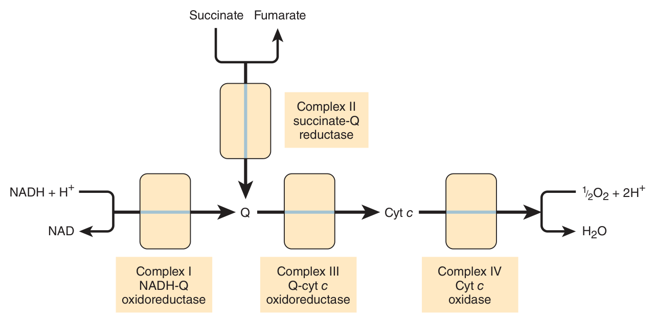
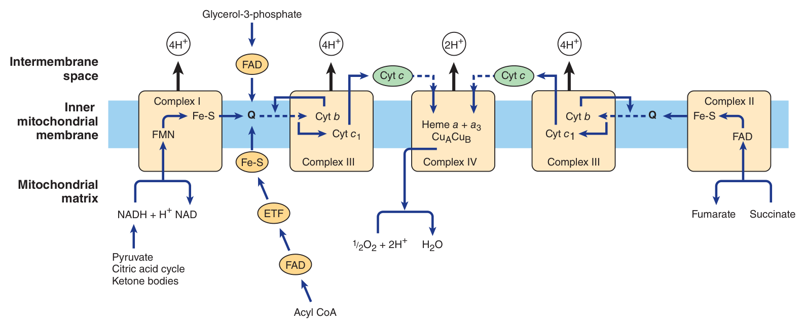
The components are organised as four protein-lipid respiratory chain complexes that span the inner membrane. Learn the four names, what each does, and — crucially — how many protons each pumps.
| Complex | Proper name | Reaction | H⁺ pumped |
|---|---|---|---|
| I | NADH-Q oxidoreductase (NADH:ubiquinone oxidoreductase) | Electrons from NADH → FMN → a series of Fe-S centres → Q. A large L-shaped multisubunit protein | 4 |
| II | Succinate-Q reductase | FADH₂ formed during the conversion of succinate to fumarate in the citric acid cycle; electrons pass via several Fe-S centres to Q | 0 |
| III | Q-cytochrome c oxidoreductase | Electrons from QH₂ to cytochrome c, via cytochromes c₁, bL and bH and a Rieske Fe-S — the Q cycle | 4 |
| IV | Cytochrome c oxidase | 4 reduced cyt c + O₂ + 8H⁺ → 4 oxidised cyt c + 2H₂O + 4H⁺. Involves haems a and a₃ and two copper centres, CuA and CuB | 2 |
NADH drives Complexes I, III and IV: 4 + 4 + 2 = 10 protons.
FADH₂ drives only III and IV: 4 + 2 = 6 protons.
Fewer protons pumped means less proton motive force means less ATP. That is the whole reason for the two different P:O ratios in §7 — and it is a far better answer than quoting the numbers.
Clinically: cytochrome c oxidase is the target of cyanide, carbon monoxide and H₂S — poisons that can totally arrest respiration precisely because this step is irreversible and unbypassable.
One refinement on Complex IV's arithmetic worth noticing: of the eight H⁺ removed from the matrix, four are used to form the two water molecules and four are pumped into the intermembrane space. Per pair of electrons, that is the 2 H⁺ in the table.
- Name the four complexes → I NADH-Q oxidoreductase, II succinate-Q reductase, III Q-cytochrome c oxidoreductase, IV cytochrome c oxidase
- How many protons does each pump per NADH? → I: 4, II: 0, III: 4, IV: 2 — total 10
- Which complex does NOT pump protons? → Complex II
- Which reaction is irreversible? → Complex IV, cytochrome c oxidase — it gives direction to the whole chain
- Which metals are in Complex IV? → Iron in haems a and a₃, plus copper centres CuA and CuB
The chemiosmotic theory ★★★
This is the intellectual centre of the unit, and one of the great ideas of twentieth-century biology. The puzzle it solved: oxidation happens in the membrane, phosphorylation happens at a completely different protein, and for decades nobody could find a chemical intermediate linking them. Peter Mitchell's answer in 1961 was that there is no chemical intermediate — the link is a gradient.
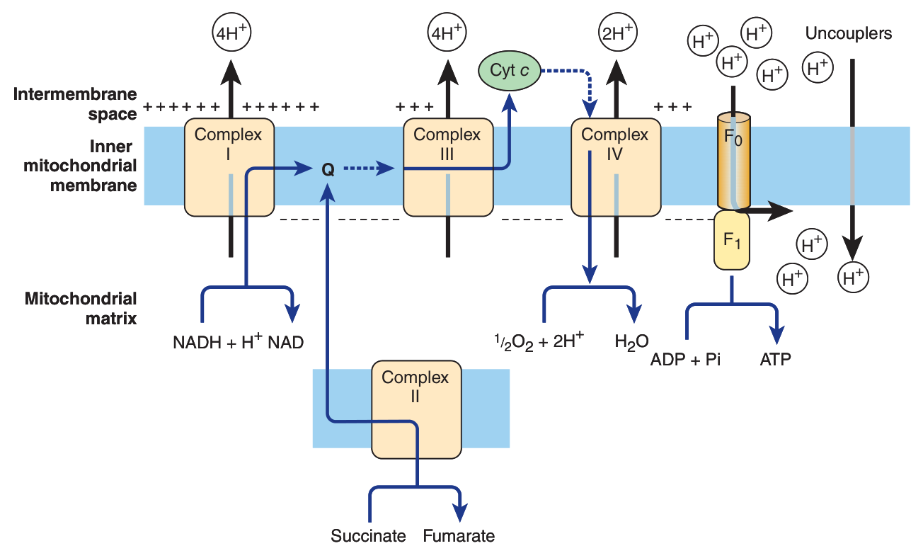
The theory that the energy from oxidation of components in the respiratory chain is coupled to the translocation of protons (H⁺) from the inside to the outside of the inner mitochondrial membrane.
The resulting proton motive force — an electrochemical potential difference, negative on the matrix side — then drives the mechanism of ATP synthesis.
Complexes I, III and IV act as proton pumps. Because the inner membrane is impermeable to ions in general and to protons in particular, the protons accumulate in the intermembrane space.
The respiratory chain is a set of pumps lifting water up behind a dam. It does not make electricity; it makes height. The turbine at the bottom — ATP synthase — makes the electricity as the water falls back through it.
Three things follow, and each is an exam answer:
• The dam must not leak. Hence the impermeable inner membrane; hence uncouplers, which drill holes in it (§9).
• The turbine can be jammed independently of the pumps. Hence oligomycin, which blocks the channel and stops both processes because they are coupled.
• Water cannot be pumped up if the reservoir is already full. Hence respiratory control (§8): with no ADP to consume the gradient, pumping stops.
The Q cycle — how Complex III pumps
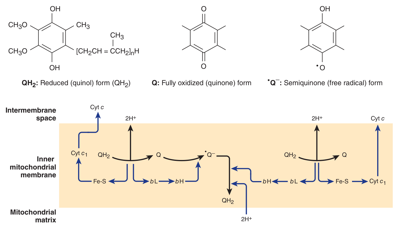
Worth understanding rather than memorising, because it resolves the two-electron / one-electron mismatch of §3. Q exists in three forms: the oxidised quinone, the reduced quinol (QH₂), and the semiquinone free radical, formed transiently.
One turn of the cycle results in the oxidation of 2 QH₂ to Q, releasing 4 H⁺ into the intermembrane space, and the reduction of one Q back to QH₂, taking 2 H⁺ up from the matrix. During oxidation of each QH₂ one electron goes to cytochrome c via the Rieske Fe-S and cytochrome c₁, while the second goes via cytochromes bL and bH to reduce a Q to the semiquinone.
- Who proposed the chemiosmotic theory, and when? → Peter Mitchell, 1961
- State the theory → Oxidation in the respiratory chain is coupled to translocation of protons across the inner membrane; the resulting proton motive force drives ATP synthesis
- Which side is negative? → The matrix side
- Which complexes are proton pumps? → I, III and IV
- Why must the inner membrane be impermeable? → Otherwise protons leak back and the gradient collapses
- What are the three forms of Q? → Quinone (oxidised), quinol QH₂ (reduced), semiquinone (transient free radical)
ATP synthase — a rotary motor ★★★
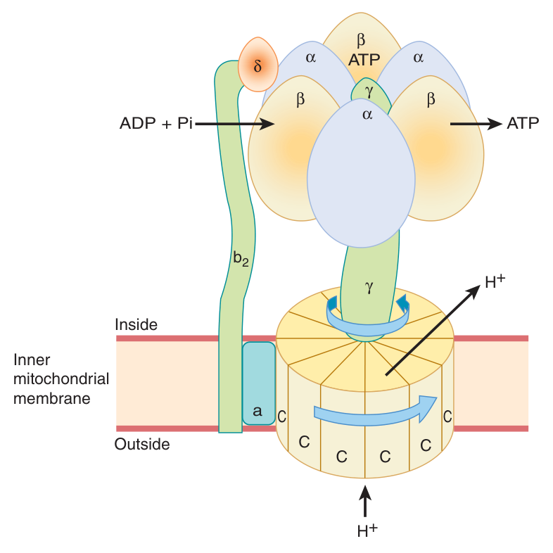
The proton motive force drives a membrane-located ATP synthase that forms ATP from Pi + ADP. It is a genuine rotary engine — one of very few in biology — and the mechanism is examinable in detail.
| Subcomplex | Location | Role |
|---|---|---|
| F₀ | Spans the membrane | Forms the proton channel. A disc of “C” protein subunits; the flow of protons through it causes it to rotate |
| γ-subunit | The “bent axle” | Attached to F₀ and rotating with it; fits inside F₁ |
| F₁ | Projects into the matrix | A ball of three α- and three β-subunits, fixed to the membrane and NOT rotating. Contains the phosphorylation mechanism |
ADP and Pi are taken up sequentially by the β-subunits. As the γ-subunit rotates it squeezes each β-subunit in turn, changing its conformation from one that binds ATP tightly to one that releases ATP and binds ADP and Pi so the next ATP can be formed.
Three ATP molecules are generated per revolution.
The energy is not used to form the bond between ADP and phosphate. ATP forms on the enzyme surface almost spontaneously. The energy of the proton gradient is spent letting go of it — prising the finished ATP off a site that binds it very tightly.
This is why the mechanism is called a binding change mechanism rather than a synthesis mechanism, and it is the detail that separates a memorised answer from an understood one.
- Which subcomplex forms the proton channel? → F₀, spanning the membrane
- Which rotates and which does not? → F₀ and the γ-subunit rotate; F₁ (3α + 3β) is fixed
- How many ATP per revolution? → Three
- What does the energy actually do? → It drives the conformational change that RELEASES the ATP, not the bond formation itself
The P:O ratio — ⚠️ read this carefully
Some phosphorylation happens without the respiratory chain at all. Unit 8 listed it: a net direct capture of two high-energy phosphates in glycolysis, and two more per mole of glucose in the citric acid cycle during the conversion of succinyl-CoA to succinate. All of these occur at the substrate level — they are substrate-level phosphorylation.
The formation of ATP coupled to the oxidation of a substrate via the respiratory chain complexes, as electrons pass to oxygen. The P:O ratio is the number of moles of inorganic phosphate incorporated into ATP per half-mole of O₂ consumed (that is, per pair of electrons).
| Source | Via Complexes I, III, IV (NADH) | Via Complexes II, III, IV (FADH₂) | Efficiency |
|---|---|---|---|
| TMU slide 16 | P:O = 3 | P:O = 2 | 68% |
| Harper's p.131 | P:O = 2.5 | P:O = 1.5 | nearly 90% |
What to do in the exam. Answer with your lecturer's figures — 3 and 2, since that is what the marking key will hold. But if the question asks you to explain rather than state, say that modern proton-counting gives 2.5 and 1.5 and give the reason. Knowing why the numbers changed is worth more than either pair of numbers.
One more figure differs. Since 1 mol of glucose yields approximately 2870 kJ on complete combustion, your slide computes that dehydrogenations plus substrate-level phosphorylations capture 68% of that free energy. Harper's, using the newer ratios, estimates that nearly 90% of the high-energy phosphate from complete oxidation of 1 mol of glucose is obtained via oxidative phosphorylation coupled to the respiratory chain — note that this is a different quantity: a share of the ATP, not a thermodynamic efficiency. Both statements agree on the point that matters: the respiratory chain is responsible for the large majority of ATP formation.
The free energy not captured as high-energy phosphate is liberated as heat. Harper's is careful to say this need not be considered “wasted”, for two reasons: it ensures the respiratory system as a whole is sufficiently exergonic to be removed from equilibrium, allowing continuous unidirectional flow and constant provision of ATP; and it contributes to the maintenance of body temperature.
That second clause is the physiological basis of non-shivering thermogenesis — and it is exactly what an uncoupler does deliberately (§9).
- Define the P:O ratio → Moles of Pi incorporated into ATP per half-mole of O₂ consumed
- Which two processes are substrate-level phosphorylation? → Glycolysis (net 2) and the citric acid cycle at succinyl-CoA → succinate
- What P:O values does your slide give, and what does Harper's give? → Slide 3 and 2; Harper's 2.5 and 1.5 — quote the lecturer's figures but know why they differ
- Why are the modern values non-integer? → 10 H⁺ per NADH and about 4 H⁺ per ATP exported does not divide evenly
- What happens to the energy not captured? → Released as heat — which keeps the system far from equilibrium and maintains body temperature
Respiratory control ★★
The rate of respiration of mitochondria is controlled by the availability of ADP.
This works because oxidation and phosphorylation are tightly coupled: oxidation cannot proceed via the respiratory chain without concomitant phosphorylation of ADP.
Most cells in the resting state are in state 4, in which respiration is limited by ADP availability. When work is performed, ATP is converted to ADP, allowing more respiration to occur, which in turn replenishes the store of ATP. Under certain conditions the concentration of inorganic phosphate can also affect the rate.
As respiration increases — as in exercise — the cell approaches state 3 or state 5, when either the capacity of the respiratory chain becomes saturated, or the pO₂ falls below the Km for cytochrome a₃. There is also the possibility that the ADP/ATP transporter, which facilitates entry of cytosolic ADP and exit of ATP, becomes rate-limiting.
There is no allosteric effector here and no kinase cascade. The regulator is the substrate itself: ADP. Work makes ADP; ADP permits respiration; respiration consumes ADP.
It is the most elegant feedback loop in the body precisely because it needs no machinery — demand automatically creates its own supply signal. Note the Km reference in the paragraph above: even here, Unit 6's kinetics is doing the work.
- What controls the rate of respiration? → The availability of ADP
- Why does that work? → Oxidation and phosphorylation are tightly coupled — oxidation cannot proceed without phosphorylation of ADP
- Which state are resting cells in? → State 4
- Name three things that can limit respiration in exercise → Saturation of the chain, pO₂ below the Km of cytochrome a₃, or the ADP/ATP transporter
Poisons and uncouplers ★★★
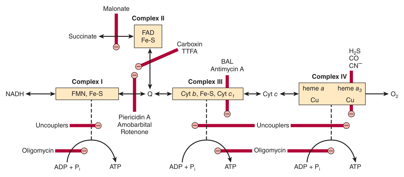
A guaranteed exam topic, and the classification is what earns the marks: inhibitors are either of the respiratory chain, or of oxidative phosphorylation, or uncouplers.
1 · Inhibitors of the respiratory chain
| Agent | Site of action |
|---|---|
| Barbiturates (e.g. amobarbital) | Inhibit NAD-linked dehydrogenases by blocking transfer from FeS to Q — i.e. Complex I. At sufficient dosage they are fatal in vivo |
| Antimycin A and dimercaprol (BAL) | Between cytochrome b and cytochrome c — i.e. Complex III |
| H₂S, carbon monoxide, cyanide | Cytochrome oxidase (Complex IV) — and can therefore totally arrest respiration |
| Malonate | A competitive inhibitor of succinate dehydrogenase — Complex II. This is Unit 6's worked example, reappearing as a respiratory poison |
2 · Inhibitors of oxidative phosphorylation
| Agent | Action |
|---|---|
| Oligomycin | Completely blocks oxidation AND phosphorylation by acting on a step in phosphorylation — specifically it blocks conduction of H⁺ through F₀ |
| Atractyloside | Inhibits the transporter of ADP into and ATP out of the mitochondrion — the adenine nucleotide transporter |
3 · Uncouplers
Compounds that dissociate oxidation in the respiratory chain from phosphorylation. They allow leakage of H⁺ across the membrane, collapsing the electrochemical proton gradient.
They are toxic in vivo, causing respiration to become uncontrolled, since its rate is no longer limited by the concentration of ADP or Pi. The most frequently used is 2,4-dinitrophenol (DNP).
Oligomycin plugs the F₀ channel. Protons cannot return, so the gradient builds until the pumps can no longer push against it — and oxidation stops too. Blocking the turbine backs up the whole dam.
An uncoupler does the reverse: it lets protons leak back without passing through the turbine. The gradient never builds, so the pumps run freely and uncontrollably — oxidation accelerates while ATP synthesis ceases, and the energy comes out as heat.
Oligomycin: both stop. Uncoupler: oxidation speeds up, phosphorylation stops.
DNP was sold as a slimming drug in the 1930s, and the logic was sound: uncouple respiration and you burn fuel without making ATP, so the calories go nowhere. The problem is that all that energy emerges as heat, and respiration is no longer limited by ADP — so it runs flat out. Patients died of hyperthermia. It is still sold illegally and still kills.
The physiological version of the same trick is brown adipose tissue, which uncouples deliberately to generate heat in the newborn.
- Classify the three kinds of agent → Inhibitors of the respiratory chain, inhibitors of oxidative phosphorylation, and uncouplers
- What do cyanide, CO and H₂S inhibit? → Cytochrome oxidase (Complex IV) — totally arresting respiration
- Where do barbiturates act? → Complex I, blocking transfer from FeS to Q
- What does oligomycin do? → Blocks H⁺ conduction through F₀, stopping BOTH oxidation and phosphorylation
- What does atractyloside inhibit? → The adenine nucleotide (ADP/ATP) transporter
- What does an uncoupler do? → Allows H⁺ leakage, collapsing the gradient — oxidation becomes uncontrolled while ATP synthesis stops; DNP is the example
The shuttles ★★
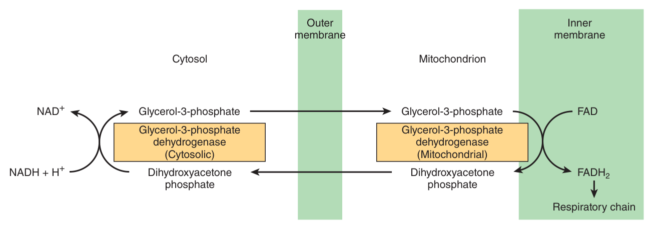
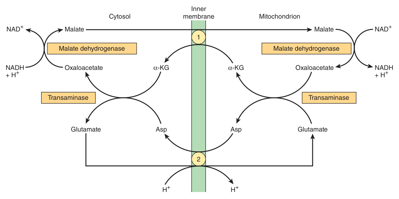
A problem the inner membrane's impermeability creates: NADH generated in the cytosol — by glycolysis — cannot cross into the mitochondrion. Its reducing equivalents must be ferried in by a shuttle.
| Shuttle | How it works | Distribution |
|---|---|---|
| Glycerophosphate shuttle | Transfers reducing equivalents from cytosolic NADH into the mitochondrion via glycerol-3-phosphate, delivering them to a flavoprotein — hence to Q, bypassing Complex I | Present in brain and white muscle; deficient in heart muscle |
| Malate shuttle | Uses the α-ketoglutarate transporter and the glutamate/aspartate transporter (note the proton symport with glutamate); delivers reducing equivalents as NADH inside the matrix | Of more universal utility |
The glycerophosphate shuttle hands electrons to a flavoprotein, so they enter at Q — bypassing Complex I and its four protons. The malate shuttle regenerates NADH inside the matrix, so electrons enter at Complex I and get the full count.
This is why textbooks quote two different ATP yields for glycolysis depending on tissue. It is not a disagreement — it is a genuine biological difference, and the reason is in §4's proton table.
One further transport system worth knowing. The creatine phosphate shuttle augments creatine phosphate's role as an energy buffer by acting as a dynamic system for transferring high-energy phosphate out of mitochondria in active tissues such as heart and skeletal muscle. A mitochondrial isoenzyme of creatine kinase (CKm), located in the intermembrane space, catalyses transfer of high-energy phosphate to creatine from the ATP emerging from the adenine nucleotide transporter — which is exactly why §1 listed creatine kinase as an intermembrane space marker.
Other transporters in the inner membrane, all examinable by name: the phosphate transporter, pyruvate symport, dicarboxylate, tricarboxylate, α-ketoglutarate and adenine nucleotide transporters. Four protons are taken into the mitochondrion for each ATP exported.
- Why are shuttles needed? → Cytosolic NADH cannot cross the impermeable inner membrane
- Name the two shuttles → Glycerophosphate and malate
- Which is more universal? → The malate shuttle; the glycerophosphate shuttle is deficient in heart muscle
- Why do they give different ATP yields? → The glycerophosphate shuttle delivers to a flavoprotein and enters at Q, bypassing Complex I
- How many protons enter per ATP exported? → Four
Revision layer
“Oxidative phosphorylation” was set as a Section I definition in BOTH readable papers — the 2019 and the 2020/21. That makes it one of the two most reliably examined terms in the whole subject. Get the definition below word-perfect.
The definition to memorise
The system in mitochondria that couples respiration to the generation of the high-energy intermediate, ATP.
Electrons pass down the respiratory chain in the inner mitochondrial membrane to their final reaction with oxygen to form water; the free energy released is used by Complexes I, III and IV to translocate protons out across the membrane, and the resulting proton motive force drives ATP synthase to phosphorylate ADP.
The first sentence is the 2019 answer sheet's wording almost exactly — open with it, then add the mechanism.
The four complexes and their protons
| Complex | Name | Entry point | H⁺ pumped |
|---|---|---|---|
| I | NADH-Q oxidoreductase | NADH | 4 |
| II | Succinate-Q reductase | Succinate (FADH₂), glycerol-3-P, acyl-CoA | 0 |
| III | Q-cytochrome c oxidoreductase | QH₂ — the Q cycle | 4 |
| IV | Cytochrome c oxidase | Cytochrome c → O₂ | 2 |
| Route | Total H⁺ | P:O (slide) | P:O (Harper's) |
|---|---|---|---|
| NADH — via I, III, IV | 10 | 3 | 2.5 |
| FADH₂ — via II, III, IV | 6 | 2 | 1.5 |
Inhibitors — the classification is the answer
| Class | Agents | Site |
|---|---|---|
| Respiratory chain | Barbiturates (amobarbital) | Complex I — FeS to Q |
| Antimycin A, dimercaprol (BAL) | Complex III — cyt b to cyt c | |
| Cyanide, CO, H₂S | Complex IV — cytochrome oxidase | |
| Malonate | Complex II — competitive inhibitor of succinate dehydrogenase | |
| Oxidative phosphorylation | Oligomycin | Blocks H⁺ conduction through F₀ — stops BOTH |
| Atractyloside | The ADP/ATP (adenine nucleotide) transporter | |
| Uncouplers | 2,4-dinitrophenol | Allow H⁺ leakage — oxidation uncontrolled, no ATP, energy as heat |
Definitions from this unit — Section I material
| Term | Definition |
|---|---|
| Oxidative phosphorylation ⭐ | The system in mitochondria that couples respiration to the generation of the high-energy intermediate ATP, via proton translocation by Complexes I, III and IV and the action of ATP synthase |
| The respiratory chain | A series of redox carriers in the inner mitochondrial membrane that collect and transport reducing equivalents, directing them to their final reaction with oxygen to form water, and couple the liberated free energy to ATP generation |
| Chemiosmotic theory | Mitchell's theory that the energy from oxidation in the respiratory chain is coupled to translocation of protons from inside to outside the inner mitochondrial membrane, the resulting proton motive force driving ATP synthesis |
| P:O ratio | The number of moles of inorganic phosphate incorporated into ATP per half-mole of oxygen consumed |
| Respiratory control | Control of the rate of mitochondrial respiration by the availability of ADP, arising because oxidation and phosphorylation are tightly coupled |
| Uncoupler | A compound that dissociates oxidation in the respiratory chain from phosphorylation by allowing leakage of protons across the inner membrane, collapsing the electrochemical gradient; respiration becomes uncontrolled and the energy is released as heat |
| Substrate-level phosphorylation | Formation of ATP by direct transfer of a phosphate group from a substrate, independent of the respiratory chain — the net 2 ATP of glycolysis and the 1 ATP of the citric acid cycle at succinate thiokinase |
Numbers worth carrying in
| Item | Value |
|---|---|
| Ubiquinone in higher animals | Q₁₀ — ten isoprenoid units |
| Protons pumped per NADH | 10 (I:4, III:4, IV:2) |
| Protons pumped per FADH₂ | 6 (III:4, IV:2) |
| ATP per revolution of ATP synthase | 3 |
| F₁ composition | three α- and three β-subunits |
| H⁺ taken in per ATP exported | 4 |
| Complete combustion of 1 mol glucose | ≈ 2870 kJ |
| Chemiosmotic theory | Peter Mitchell, 1961 |
| Resting cells | state 4 — ADP-limited |
- Define oxidative phosphorylation in exam wording, then give the mechanism
- Name the four complexes, their substrates and their proton counts
- State the chemiosmotic theory and name its author and year
- Explain why FADH₂ yields less ATP than NADH, in terms of protons
- Describe ATP synthase — F₀, F₁, the γ-axle, the binding change mechanism
- Explain respiratory control and why resting cells are in state 4
- Classify the inhibitors into three groups with examples of each
- Explain why oligomycin stops oxidation but an uncoupler accelerates it
- Name the two shuttles and explain why they give different ATP yields