Enzymes: Mechanism of Action
What an enzyme is
Everything so far has been about protein structure. Now comes the payoff: what structure is for. Section II of Harper's is titled “Enzymes”, and this unit is its foundation.
A biological polymer that catalyses a chemical reaction. With the exception of a few catalytic RNA molecules — ribozymes — the vast majority of enzymes are proteins.
It looks like a footnote and it is a favourite MCQ. The safe formulation is “enzymes are mostly protein catalysts” — because a statement that all enzymes are proteins is false. Ribozymes are RNA, and they are real enzymes. You will meet them again in Unit 24, where RNA splicing is catalysed by RNA itself.
Two thermodynamic points the lecture sets up first. Enzymes work by lowering the activation energy (Ea) — the energy required to start a reaction — so that a far larger proportion of molecules can reach the transition state at body temperature. What they do not do is change ΔG or the position of equilibrium. An enzyme makes a reaction faster; it cannot make an unfavourable reaction favourable.
- Define an enzyme → A biological polymer that catalyses a chemical reaction; mostly proteins, but ribozymes are RNA
- What do enzymes lower? → The activation energy — the energy required to start the reaction
- Do they change the equilibrium? → No — equilibrium is reached faster, but its position is unchanged
The two general properties ★★★
The TMU deck states these as a numbered pair, which means they are meant to be reproduced as a numbered pair.
| Property | The detail that earns the mark |
|---|---|
| 1 · High efficiency | Enzymes enhance the rate of the corresponding non-catalysed reaction by factors of at least 10⁶. Like all catalysts, they are neither consumed nor permanently altered by the reaction, and are not metabolites themselves |
| 2 · High specificity | Specific both for the type of reaction catalysed and for a single substrate or a small set of closely related substrates — and additionally stereospecific |
The property whereby an enzyme is specific both for the type of reaction it catalyses and for a single substrate or a small set of closely related substrates.
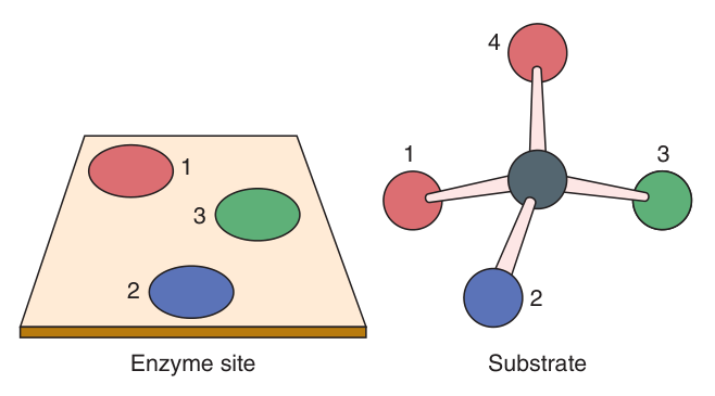
Enzymes are additionally stereospecific: they typically catalyse the reaction of only one stereoisomer of a compound — D- but not L-sugars, L- but not D-amino acids.
So apparently identical atoms become distinguishable once bound, permitting a stereospecific chemical change. This is also why an enzyme can produce a chiral product from a non-chiral substrate — the reduction of pyruvate yields exclusively L-lactate, not a racemic mixture.
Efficiency is impressive; specificity is what makes life possible. Because each enzyme touches only its own substrate, a cell can run hundreds of different reactions in the same small volume at the same time, and control each one independently. Harper's puts it exactly that way. Without specificity a cell would just be a beaker of competing chemistry.
- By what factor do enzymes accelerate reactions? → At least 10⁶
- Define enzyme specificity → Specific for the reaction type AND for one substrate or a small set of closely related ones; also stereospecific
- Which stereoisomers do enzymes act on? → D-sugars but not L; L-amino acids but not D
- How is stereospecificity achieved? → Three-point attachment fixes the substrate's orientation, so apparently identical groups become distinguishable
The six IUB classes ★★★
The International Union of Biochemistry classification is a list to memorise, and there is a specific reason it appears in exams: “protease” is not one of the six, and that fact makes an easy MCQ. Learn the six, and learn what is not on the list.
| # | Class | Reaction catalysed | Example |
|---|---|---|---|
| 1 | Oxidoreductases | Oxidations and reductions | Lactate dehydrogenase |
| 2 | Transferases | Transfer of groups such as methyl or glycosyl from a donor to an acceptor molecule | Aminotransferases (AST, ALT) |
| 3 | Hydrolases | Hydrolytic cleavage of C–C, C–O, C–N, P–O and certain other bonds, including acid anhydride bonds | Trypsin, lipase, amylase |
| 4 | Lyases | Cleavage of C–C, C–O, C–N and other bonds by elimination, leaving double bonds; and the reverse — adding groups to double bonds | Aldolase, fumarase |
| 5 | Isomerases | Geometric or structural changes within a single molecule | Phosphoglucose isomerase |
| 6 | Ligases | Joining together of two molecules, coupled to hydrolysis of a pyrophosphoryl group in ATP or a similar nucleoside triphosphate | DNA ligase, pyruvate carboxylase |
Oxidoreductase — moves electrons. Transferase — moves a group from A to B. Hydrolase — cuts using water. Lyase — cuts without water, leaving a double bond. Isomerase — rearranges one molecule internally. Ligase — joins two molecules, and costs ATP.
The two distinctions that carry marks: hydrolase vs lyase is whether water is used; ligase is the only one that spends ATP.
- Name the six IUB classes → Oxidoreductases, transferases, hydrolases, lyases, isomerases, ligases
- Which class cuts bonds using water? → Hydrolases
- Which class cuts bonds by elimination, leaving a double bond? → Lyases
- Which class requires ATP? → Ligases
- Is protease an IUB class? → No — proteases are hydrolases
Cofactors, coenzymes and prosthetic groups ★★★
This is one of the four TMU study questions, and the reason these accessories exist is worth stating first because it makes the whole section make sense. The twenty amino acid side chains offer only a limited number of functional groups. Non-protein helpers extend the repertoire of catalytic capabilities beyond what those side chains can do — they let an enzyme carry electrons, carry acyl groups, act as a Lewis acid, and so on.
All three terms describe small non-protein components. What distinguishes them is how tightly they are held.
| Term | How it associates | Key facts |
|---|---|---|
| Prosthetic group | Tightly and stably incorporated into the protein's structure, by covalent or non-covalent forces | Examples: pyridoxal phosphate, FMN, FAD, thiamin pyrophosphate, biotin. Metal ions are the commonest type. The roughly one-third of enzymes containing tightly bound Fe, Co, Cu, Mg, Mn or Zn are called metalloenzymes |
| Cofactor | Reversibly / transiently — a dissociable association with the enzyme or with the substrate | Because the binding is transient, cofactors must be present in the surrounding medium for catalysis to occur. Most are metal ions; enzymes needing one are metal-activated enzymes, as distinct from metalloenzymes |
| Coenzyme | Recyclable — binds, carries, and leaves | Serve as substrate shuttles, transporting substrates from one point in the cell to another |
One-third of all enzymes are metalloenzymes — Fe, Co, Cu, Mg, Mn, Zn.
Why coenzymes are called shuttles
Harper's gives the function of a shuttle as twofold, and both halves are quotable. First, they stabilise species too reactive to survive on their own — a hydride ion (NADH) or a hydrogen atom (FADH) would not last long floating free among the water and organic molecules of the cytoplasm. Second, they act as an adaptor or handle that lets a target enzyme recognise and bind a small chemical group — acetate is hard to recognise, acetyl-CoA is easy.
The B-vitamin connection — learn this table
| B vitamin | Coenzyme it becomes | What that coenzyme carries |
|---|---|---|
| Nicotinamide (niacin, B₃) | NAD⁺ and NADP⁺ | Redox — hydride ions |
| Riboflavin (B₂) | FMN and FAD | Redox — hydrogen atoms |
| Pantothenic acid (B₅) | Coenzyme A | Acyl groups |
| Thiamin (B₁) | Thiamin pyrophosphate | Decarboxylation of α-keto acids |
| Folic acid | Folate coenzymes | One-carbon metabolism |
| Cobamide (B₁₂) | Cobamide coenzymes | One-carbon metabolism |
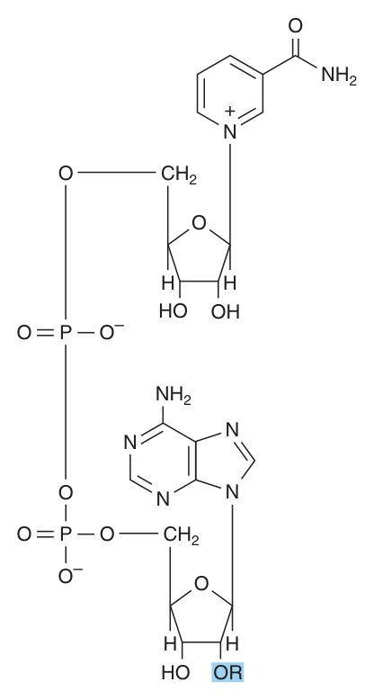
Two structural notes. Many coenzymes contain the adenine, ribose and phosphoryl moieties of AMP or ADP — look at NAD⁺ and you will see AMP sitting inside it. And the difference between NAD⁺ and NADP⁺ is a single group: NADP⁺ carries an extra phosphate (R = PO₃²⁻ where NAD⁺ has R = H). Other shuttles worth naming: methyl groups on folates, glucose on UDP, oligosaccharides on dolichol.
Every B-vitamin deficiency syndrome you will meet in clinical medicine is, at bottom, an enzyme without its coenzyme. Thiamin deficiency (beriberi, Wernicke's) disables the decarboxylation of α-keto acids — which is pyruvate dehydrogenase, Unit 10. Niacin deficiency (pellagra) cripples every NAD-dependent dehydrogenase. Learning this table now means those diseases explain themselves later.
- Distinguish prosthetic group, cofactor and coenzyme → Tightly/stably bound; reversibly and dissociably bound (must be in the medium); a recyclable substrate shuttle
- What is a metalloenzyme? → An enzyme containing a tightly bound metal ion as a prosthetic group — about one-third of all enzymes
- Why do enzymes need these at all? → They extend catalytic capability beyond the limited functional groups of amino acid side chains
- Which vitamin gives NAD/NADP, and which gives FMN/FAD? → Nicotinamide and riboflavin
- Which vitamin gives coenzyme A, and what does it carry? → Pantothenic acid; acyl groups
The active site ★★★
The first of the four TMU study questions is “what is the active site of an enzyme? Describe its characteristics.” That phrasing — describe the characteristics — means a definition alone will not do. Learn the definition plus four features.
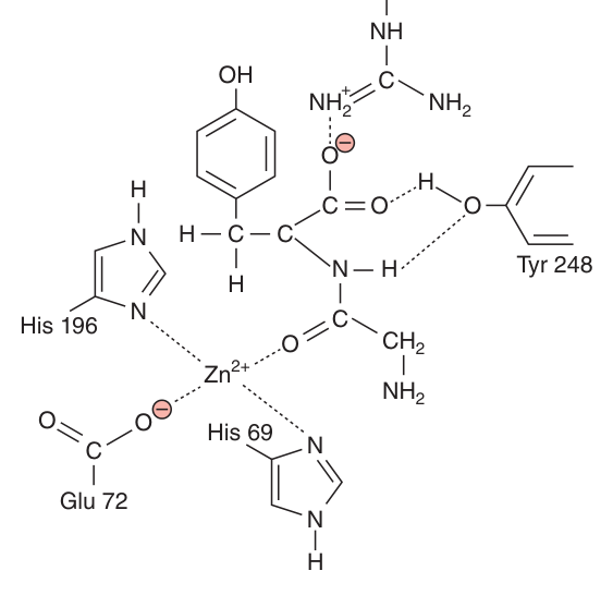
The three-dimensional catalytic centre of an enzyme — a cleft or pocket, usually on the surface, formed by the amino acid residues that bind the substrate and carry out catalysis.
| Characteristic | Detail |
|---|---|
| A small part of the molecule | Only a few residues form it; the rest of the protein positions them and provides the scaffold |
| A three-dimensional entity | The residues are far apart in the primary structure but brought together by folding. This is the point examiners test |
| Located in a cleft or crevice | Substrates are bound in a pocket, shielded from water |
| A distinct microenvironment | The site can have a polarity, hydrophobicity, acidity or alkalinity markedly different from the surrounding cytoplasm |
| Specific, non-covalent binding | The substrate is held by specific interactions — hydrogen bonds, ionic and hydrophobic interactions |
| Optimal alignment | Substrates are brought into close proximity to one another and into optimal alignment with the cofactors, prosthetic groups and side chains that catalyse the change |
All the other options are true — the active site is a three-dimensional catalytic centre, binding involves specific interactions, active sites are often in crevices or clefts, and all active enzymes have one.
- Define the active site → The three-dimensional catalytic centre — a cleft or pocket whose residues bind substrate and perform catalysis
- Are the catalytic residues close together in the primary structure? → NO — they are far apart in sequence, brought together by folding
- Name two features of the microenvironment → It shields substrate from water, and its polarity/acidity can differ markedly from the cytoplasm
Lock-and-key versus induced fit ★★★
Two models, in historical order, and the exam wants both — the second is a correction of the first, so explaining what was wrong with the first is half the answer.
| Model | Proposed by | The idea | Its limitation |
|---|---|---|---|
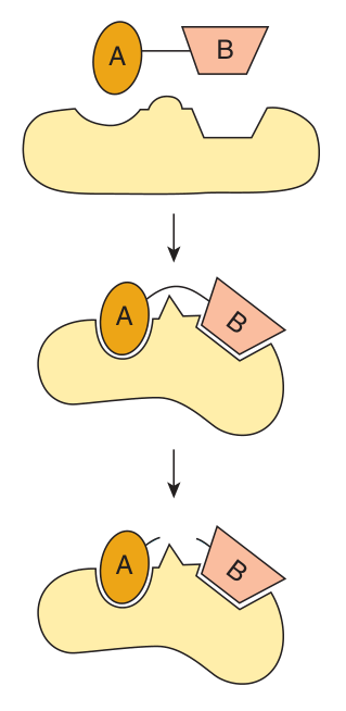
| Lock and key | Emil Fischer | The enzyme's active site is a rigid pocket whose shape is exactly complementary to the substrate, as a lock matches its key. It arose from the observation that substrates make enzymes more resistant to thermal denaturation, implying a stable enzyme-substrate (ES) complex | It failed to account for the dynamic changes that accompany catalysis — a rigid site cannot explain how a bond is strained and broken |
| Induced fit | Daniel Koshland | When a substrate approaches and binds, it induces a conformational change in the enzyme. Enzyme and substrate induce mutual conformational changes in one another, facilitating both recognition and catalysis | The accepted model |
A hand entering a glove. The glove is not a rigid mould; it changes shape around the hand, and the fit only becomes perfect once the hand is inside.
This is not a nicer picture, it is a better explanation. A rigid lock could hold a substrate but could never distort it. A site that closes around its substrate can strain the bond you want to break — which is precisely catalytic mechanism 3 in the next section. Induced fit is what makes catalysis by strain possible.
- Who proposed lock and key, and who induced fit? → Emil Fischer and Daniel Koshland
- What is the ES complex? → The enzyme-substrate complex, whose thermal stability exceeds that of the enzyme alone
- Why did lock and key fail? → It could not account for the dynamic conformational changes that accompany catalysis
- State induced fit in one line → Substrate binding induces a conformational change; enzyme and substrate change each other's shape
The four catalytic mechanisms ★★★
Harper's states it plainly: enzymes use combinations of four general mechanisms to achieve dramatic rate enhancements. This is an excellent short-essay question because the four are genuinely distinct ideas.
| Mechanism | How it works |
|---|---|
| 1 · Catalysis by proximity | For molecules to react they must come within bond-forming distance. Binding substrates at the active site creates a region of high local substrate concentration, with the molecules held in ideal orientation to interact. Worth at least a thousandfold rate enhancement on its own |
| 2 · Acid-base catalysis | Ionisable side chains — and prosthetic groups where present — act as general acids (proton donors) or general bases (proton acceptors). Specific acid/base catalysis = only protons or hydroxide ions participate. General acid/base catalysis = the rate responds to all the acids or bases present |
| 3 · Catalysis by strain | Enzymes catalysing lytic reactions bind the substrate in a conformation that is unfavourable for the bond targeted for cleavage. This strained conformation mimics the transition state, stretching and distorting the bond, weakening it and making it more vulnerable. Linus Pauling first proposed transition-state stabilisation as a general mechanism |
| 4 · Covalent catalysis | Formation of a covalent bond between the enzyme and one or more substrates, creating a transient covalent intermediate. Often follows a “ping-pong” mechanism — the first substrate binds and its product is released before the second substrate binds |
The worked examples the lecture uses
| Enzyme | Mechanism illustrated | The detail |
|---|---|---|
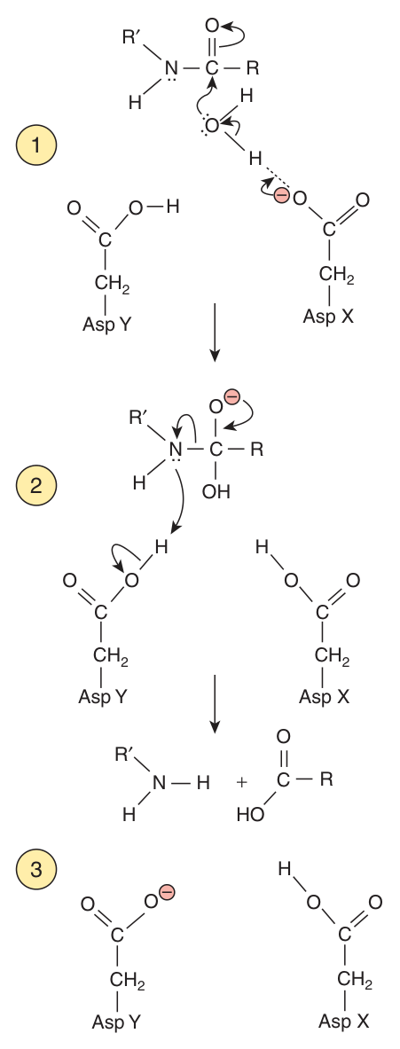
| HIV protease (an aspartic protease) | Acid-base catalysis | Aspartate X acts as a base, abstracting a proton to activate a water molecule; the activated water attacks the peptide bond, forming a transient tetrahedral intermediate; Aspartate Y acts as an acid, donating a proton to the newly formed amino group to facilitate breakdown of the intermediate and release of products. Shuttling the proton from Asp X to Asp Y restores the enzyme |
| Chymotrypsin | Covalent catalysis | A serine protease; forms a covalent acyl-enzyme intermediate |
| Fructose-2,6-bisphosphatase | Covalent catalysis | A regulatory enzyme of gluconeogenesis (Unit 13) that hydrolytically removes the phosphate on carbon 2 of fructose 2,6-bisphosphate |
Knowing that the transition state is strained and mimicked is not academic. Chemists exploit knowledge of the transition state to design transition state analogues — molecules that resemble the strained intermediate rather than the substrate, and therefore bind far more tightly than any substrate could. The HIV protease inhibitors are the most famous clinical example, and they exist because of the mechanism in row 3 of the table above.
- Name the four catalytic mechanisms → Proximity, acid-base catalysis, strain, covalent catalysis
- What does catalysis by proximity achieve? → High local substrate concentration and ideal orientation — at least a thousandfold rate enhancement
- Distinguish specific from general acid-base catalysis → Specific involves only H⁺/OH⁻; general responds to all acids or bases present
- What does catalysis by strain mimic? → The transition state — Pauling's transition-state stabilisation
- What is a ping-pong mechanism? → The first substrate binds and its product is released BEFORE the second substrate binds

Catalytic residues are highly conserved
A short section, but it links this unit to Unit 2's bioinformatics and to Unit 26's gene duplication. Most enzyme families arose through gene duplication: a second copy of a gene is made, the two copies evolve independently, and the result is divergent homologs recognising different substrates but using the same mechanism.
Harper's example is one you already know from Unit 2: chymotrypsin, which cleaves after large hydrophobic residues, and trypsin, which cleaves after basic residues. Same serine-protease mechanism, different substrate preference — two branches from one ancestral gene.
Conserved residues are specific amino acids found in the same relative position in every member of a protein family, from which common ancestry can be inferred. Proteins that diverged from a common ancestor, sharing a large number of conserved residues, are said to be homologous.
Among the most highly conserved residues are those that participate directly in catalysis — which is the point: evolution can tolerate change almost anywhere except the active site.
- How do enzyme families arise? → Through gene duplication, followed by independent evolution of the two copies
- What are conserved residues? → Specific amino acids present in the same relative position in every family member
- Which residues are most conserved of all? → Those that participate directly in catalysis
Isozymes ★★★
Distinct enzyme forms that catalyse the SAME reaction.
They arise through gene duplication, and although the reaction is identical their physical, chemical and immunological properties differ significantly. Differences may include sensitivity to regulatory factors, or substrate affinity — hexokinase and glucokinase are the classic pair — adapting them to specific tissues or circumstances. They also provide a “backup” copy of an essential enzyme.
Lactate dehydrogenase — the worked example
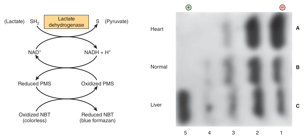
LDH catalyses lactate + NAD⁺ → pyruvate + NADH + H⁺. It is a tetramer built from two monomer types: H (for heart), encoded by LDHB, and M (for muscle), encoded by LDHA. Four subunits drawn from two types gives five possible tetramers — and that is why there are five LDH isozymes.
| Isozyme | Composition | Predominates in |
|---|---|---|
| LDH-1 | HHHH | Heart |
| LDH-2 | HHHM | |
| LDH-3 | HHMM | |
| LDH-4 | HMMM | |
| LDH-5 | MMMM | Liver |
LDH is tissue-specific as a consequence of its quaternary structure — Unit 3 again. The relative proportion of each subunit in a given organ is set by tissue-specific expression of the H and M genes. So the pattern of isozymes in the plasma reports which organ has been damaged. Harper's line is worth quoting: when LDH levels rise in plasma, the tissue of origin can be inferred from the characteristic pattern of LDH isozymes.
In health LDH is largely intracellular. In disease, damaged cells release their own isozyme pattern into the serum.
Myocardial infarction → elevated LDH-1.
Liver disease → elevated LDH-5.
Individual isozymes are separated in the laboratory by electrophoresis and detected with a coupled assay — which works precisely because their physical properties differ.
- Define isozymes → Distinct enzyme forms that catalyse the same reaction, differing in physical, chemical and immunological properties
- What are the LDH subunits? → H (heart, gene LDHB) and M (muscle, gene LDHA)
- Why are there five isozymes? → LDH is a tetramer built from two subunit types
- Which rises in myocardial infarction, and which in liver disease? → LDH-1 and LDH-5
- How are isozymes separated clinically? → By electrophoresis
Enzymes in clinical diagnosis ★★
Enzymes are present in cells in tiny quantities, but each molecule transforms thousands of substrate molecules — an amplification that lets a vanishingly small amount of enzyme announce itself. Under appropriate conditions the rate of the catalysed reaction is proportional to the amount of enzyme present, so activity can be used to infer concentration.
The 340 nm assay — know this one properly
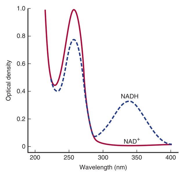
The reduced coenzymes NADH and NADPH absorb light at 340 nm; their oxidised forms NAD(P)⁺ do not.
So when NAD(P)⁺ is reduced, absorbance at 340 nm increases in proportion to the NAD(P)H produced. Conversely, for a dehydrogenase catalysing the oxidation of NAD(P)H, absorbance at 340 nm decreases. In each case the rate of change of absorbance is proportional to the quantity of enzyme present.
280 nm (Unit 1) measures protein, via the aromatic side chains of tryptophan, tyrosine and phenylalanine.
340 nm (here) measures NADH / NADPH, and therefore the activity of a dehydrogenase.
Two different numbers, two entirely different measurements.
Non-functional plasma enzymes
| Serum enzyme | Principal diagnostic use |
|---|---|
| Aspartate aminotransferase (AST / SGOT) | Myocardial infarction |
| Alanine aminotransferase (ALT / SGPT) | Viral hepatitis |
| Amylase · Lipase | Acute pancreatitis |
| Creatine kinase | Muscle disorders and myocardial infarction |
| γ-Glutamyl transferase | Various liver diseases |
| LDH isozyme 5 | Liver disease |
| Alkaline phosphatase (isozymes) | Bone disorders, obstructive liver disease |
| Ceruloplasmin | Hepatolenticular degeneration (Wilson disease) |
| β-Glucocerebrosidase | Gaucher disease |
The first enzymes used to diagnose MI were AST, ALT and LDH. AST and ALT proved less than ideal: they appear in plasma slowly and are not specific to heart muscle. LDH is also slow, but offered tissue specificity through its quaternary structure (the LDH-1 pattern).
Creatine kinase has three isozymes — CK-MM (skeletal muscle), CK-BB (brain), CK-MB (heart) — and CK-MB has a useful diagnostic window: it appears 4–6 hours after an MI, peaks at 24 hours, and returns to baseline by 48–72 hours. CK is still used for skeletal muscle disorders such as Duchenne muscular dystrophy.
Today, however, plasma troponin has replaced CK as the preferred marker for MI. Cardiac troponins I and T rise 2–6 hours after infarction and remain elevated for 4–10 days. Note the caveat: troponin marks all heart muscle damage, not only infarction. The general principle throughout is that an enzyme appearing in plasma only after 12 hours or more is of limited clinical use.
Two further applications the chapter mentions. ELISA exploits enzyme sensitivity to detect proteins that have no catalytic activity of their own: an antibody is covalently linked to a reporter enzyme such as alkaline phosphatase or horseradish peroxidase, and the bound antibody is quantified by adding that enzyme's substrate. And high-throughput screening runs thousands of assays at once in 96-, 384- or 1536-well plates, principally to find inhibitory compounds with potential as drugs — which is where Unit 6 picks up.
- At what wavelength do NADH and NADPH absorb? → 340 nm — their oxidised forms do not
- What does absorbance at 280 nm measure instead? → Protein, via aromatic side chains
- Which enzyme rises in acute pancreatitis? → Amylase and lipase
- Name the three CK isozymes → CK-MM (skeletal muscle), CK-BB (brain), CK-MB (heart)
- What is the preferred marker for MI today? → Cardiac troponins I and T
- What is a reporter enzyme? → An enzyme linked to an antibody in ELISA, e.g. alkaline phosphatase or horseradish peroxidase
Revision layer
Four study questions close this deck — what is the active site and its characteristics? what is enzyme specificity? what are coenzymes, cofactors and prosthetic groups? what are isozymes? All four are Section I definitions, which makes this one of the highest-yield units in the course for the 3-mark questions.
Definitions from this unit — Section I material
| Term | Definition |
|---|---|
| Enzyme | A biological polymer that catalyses a chemical reaction; with the exception of the catalytic RNAs (ribozymes), the vast majority are proteins |
| Enzyme specificity | The property whereby an enzyme is specific both for the type of reaction catalysed and for a single substrate or a small set of closely related substrates; enzymes are additionally stereospecific |
| Active site | The three-dimensional catalytic centre of an enzyme — a cleft or pocket whose residues, far apart in the primary structure but brought together by folding, bind the substrate and carry out catalysis in a microenvironment shielded from water |
| Prosthetic group | A non-protein component tightly and stably incorporated into an enzyme's structure by covalent or non-covalent forces; metal ions are the commonest type |
| Cofactor | A non-protein component that associates reversibly and transiently with the enzyme or substrate, and which must therefore be present in the surrounding medium for catalysis to occur |
| Coenzyme | A recyclable shuttle that transports substrates from one point in the cell to another, stabilising reactive species and acting as a handle for recognition by target enzymes; many are derivatives of B vitamins |
| Isozymes | Distinct enzyme forms that catalyse the same reaction, arising by gene duplication, whose physical, chemical and immunological properties differ significantly |
| Induced fit | Koshland's model, in which substrate binding induces a conformational change in the enzyme; enzyme and substrate induce mutual conformational changes that facilitate recognition and catalysis |
| Conserved residues | Specific amino acids present in the same relative position in every member of a protein family, from which common ancestry is inferred; those participating directly in catalysis are the most highly conserved |
The six IUB classes
| Class | Reaction |
|---|---|
| Oxidoreductases | Oxidations and reductions |
| Transferases | Transfer of a group from donor to acceptor |
| Hydrolases | Hydrolytic cleavage — uses water |
| Lyases | Cleavage by elimination leaving a double bond — no water; also adds to double bonds |
| Isomerases | Geometric or structural change within one molecule |
| Ligases | Joins two molecules — costs ATP |
The four catalytic mechanisms
| Mechanism | One-line summary |
|---|---|
| Proximity | High local concentration and ideal orientation — ≥1000-fold |
| Acid-base catalysis | Side chains act as general acids (proton donors) or bases (proton acceptors) — HIV protease |
| Strain | Substrate bound in a strained conformation mimicking the transition state — Pauling |
| Covalent catalysis | Covalent enzyme-substrate intermediate, often ping-pong — chymotrypsin, fructose-2,6-bisphosphatase |
The B vitamins and their coenzymes
| Vitamin | Coenzyme | Carries |
|---|---|---|
| Nicotinamide | NAD⁺ / NADP⁺ | Hydride — redox |
| Riboflavin | FMN / FAD | Hydrogen atoms — redox |
| Pantothenic acid | Coenzyme A | Acyl groups |
| Thiamin | Thiamin pyrophosphate | Decarboxylation of α-keto acids |
| Folic acid · cobamide | Folate / cobamide coenzymes | One-carbon units |
Numbers and facts worth carrying in
| Item | Value |
|---|---|
| Rate enhancement by an enzyme | at least 10⁶ |
| Rate enhancement by proximity alone | at least 1000-fold |
| Proportion of enzymes that are metalloenzymes | about one-third |
| NADH / NADPH absorbance | 340 nm (oxidised forms do not absorb) |
| Protein absorbance | 280 nm — do not confuse |
| LDH isozymes | 5, from H and M subunits; LDH-1 heart, LDH-5 liver |
| CK-MB after MI | appears 4–6 h · peaks 24 h · baseline by 48–72 h |
| Troponin after MI | rises 2–6 h · elevated 4–10 days |
- Define the active site and give four of its characteristics
- Define enzyme specificity and explain three-point attachment
- Distinguish coenzyme, cofactor and prosthetic group, with an example of each
- Define isozymes and explain the LDH system, including LDH-1 and LDH-5
- Name the six IUB classes and say which uses water and which uses ATP
- Name the four catalytic mechanisms with one example enzyme each
- Contrast lock-and-key with induced fit, naming both proposers
- Explain the 340 nm assay and why it works