Myoglobin & Haemoglobin
Two proteins, two jobs
This unit is where the three previous ones pay off at once. Myoglobin and haemoglobin are the textbook demonstration that structure determines function — two proteins with nearly identical secondary and tertiary structure but completely different physiological roles, and the difference comes down entirely to quaternary structure.
| Myoglobin | Haemoglobin | |
|---|---|---|
| Where | Red skeletal muscle | Erythrocytes only |
| Job | Stores oxygen — a reserve against O₂ deprivation | Transports O₂ to the tissues, and returns CO₂ and protons to the lungs |
| Subunits | Monomeric — one chain | Tetrameric — α₂β₂ in HbA |
| Quaternary structure | None | Yes |
| Cooperativity | None | Yes |
| O₂ binding curve | Hyperbolic | Sigmoid |
Every entry in the right-hand column follows from one thing: haemoglobin has four subunits. Four subunits means they can talk to each other; talking to each other is cooperativity; cooperativity is what makes the curve sigmoid; and a sigmoid curve is what lets a protein load fully in the lungs and unload heavily in the tissues. Myoglobin, with one chain, has no one to talk to — so it gets a hyperbolic curve and can only be a store.
Harper's states the general rule outright: cooperative interactions are an exclusive property of multimeric proteins.
- Where is each protein found? → Myoglobin in red skeletal muscle; haemoglobin only in erythrocytes
- What does each do? → Myoglobin stores oxygen; haemoglobin transports O₂, CO₂ and protons
- Which has quaternary structure? → Haemoglobin (α₂β₂); myoglobin is monomeric and has none
- Why can myoglobin not show cooperativity? → Cooperative interactions are an exclusive property of multimeric proteins
Haem — the prosthetic group ★★★
Neither protein can bind oxygen by itself. Amino acid side chains simply do not bind O₂. The ability comes from a non-protein component — a prosthetic group — and here that group is haem.
A small non-protein molecule that is a permanent part of a protein and is necessary for its function. Some proteins consist of polypeptide alone; others, like myoglobin and haemoglobin, require a prosthetic group.
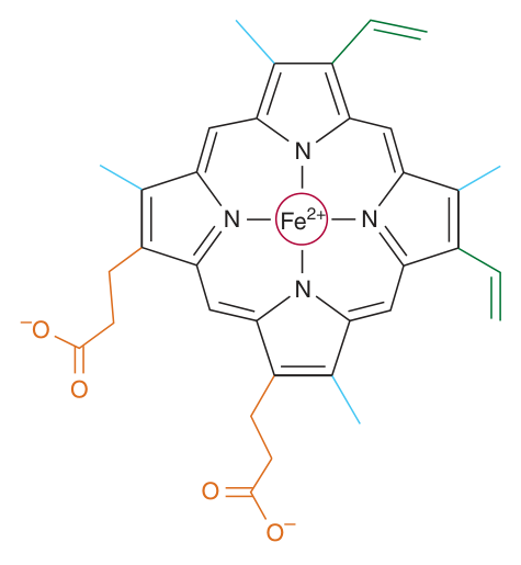
A cyclic tetrapyrrole — four molecules of pyrrole linked by methyne (methylene) bridges — with one atom of ferrous iron (Fe²⁺) at the centre of the planar ring, bonded to all four pyrrole nitrogens.
The substituents at the β-positions are methyl (M), vinyl (V) and propionate (Pr), arranged M, V, M, V, M, Pr, Pr, M. The planar network of conjugated double bonds absorbs visible light — which is why haem, and therefore blood, is deep red.
The six coordination bonds — learn these as a list
| Bond | Partner |
|---|---|
| 1–4 | The four nitrogen atoms of the pyrrole rings of haem |
| 5 | A nitrogen of the imidazole ring of the proximal histidine, His F8 |
| 6 | Oxygen — or left unoccupied. This is the binding site |
The clinical consequence is methaemoglobinaemia, covered in §10.
- What is haem chemically? → A cyclic tetrapyrrole — four pyrroles linked by methyne bridges, with central Fe²⁺
- Name the three substituent types → Methyl, vinyl and propionate
- How many coordination bonds does the iron form, and to what? → Six — four to pyrrole nitrogens, one to His F8, one to O₂ or vacant
- What happens if Fe²⁺ is oxidised to Fe³⁺? → Biological activity is destroyed — methaemoglobin can neither bind nor transport O₂
Myoglobin's structure ★★
Myoglobin is worth knowing in numerical detail because it is the simplest complete worked example of the four orders of structure from Unit 3.
| Order | Myoglobin |
|---|---|
| Primary | A single polypeptide of 153 aminoacyl residues (MW ~17 000) |
| Secondary | An unusually high proportion — about 75% — of residues lie in eight right-handed α-helices of 7–20 residues, named A to H starting from the amino terminal |
| Tertiary | A compact, roughly spherical molecule measuring 4.5 × 3.5 × 2.5 nm. The surface is polar; the interior contains only non-polar residues (Leu, Val, Phe, Met) — with exactly two exceptions |
| Quaternary | None — myoglobin is monomeric |
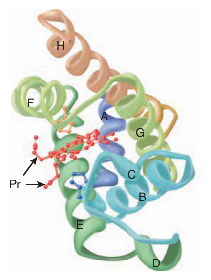
Unit 3 told you the rule: hydrophobic residues inside, polar residues outside. Myoglobin obeys it almost perfectly — almost. The two polar residues buried in the hydrophobic interior are His E7 and His F8, and they are buried precisely because they have a job to do at the haem iron. When a protein breaks its own structural rule, that is where the function is. Look for the exception and you have found the active site.
The naming convention is simple once you see it: E7 = the seventh residue of helix E; F8 = the eighth residue of helix F.
The haem itself sits in a crevice between helices E and F, oriented so that its polar propionate groups face the surface of the globin while the rest sits in the non-polar interior. It is held in that hydrophobic pocket by non-covalent bonding — a point the TMU slides make explicitly, and a common one-mark question.
- How many residues does myoglobin have? → 153
- How many helices, and how are they named? → Eight right-handed α-helices, A to H from the amino terminal
- Which residues are the two exceptions to the hydrophobic interior? → His E7 and His F8
- How is haem held in the pocket? → By non-covalent bonding, in a hydrophobic crevice between helices E and F
His F8 and His E7 — and the movement that starts everything ★★★
| Residue | Name | Role |
|---|---|---|
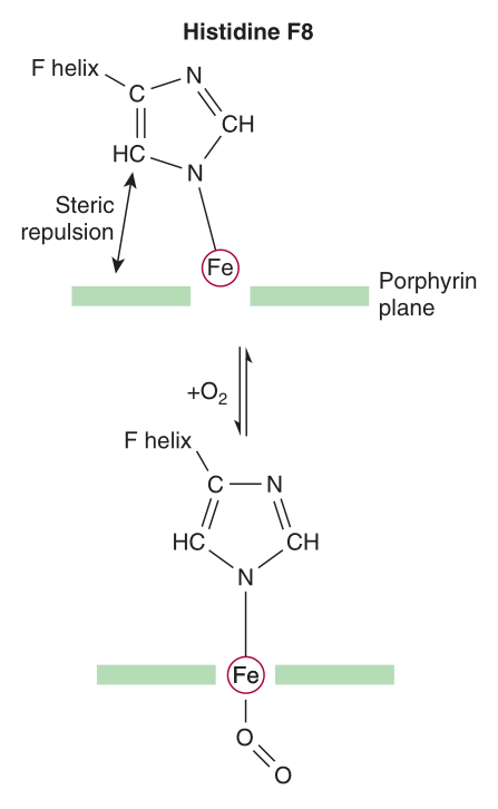
| His F8 | Proximal histidine | Occupies the fifth coordination position of the iron — it is bonded to the iron, anchoring the haem to the protein |
| His E7 | Distal histidine | Lies on the opposite side of the haem ring, not bonded to the iron. It guards the O₂ binding site and sterically hinders CO |
Now the crucial mechanical detail — the single fact from which the whole of cooperativity is built. In deoxymyoglobin the iron does not lie in the plane of the haem ring: it sits about 0.03 nm outside it, pulled towards His F8, so the haem “puckers” slightly.
When O₂ occupies the sixth coordination position, the iron moves into the plane of the ring — to within about 0.01 nm of it. And because the iron is bonded to His F8, His F8 and the residues attached to it are dragged along.
The iron is not free-floating. It is bolted to the protein through His F8. So when a small molecule of oxygen pulls the iron a few hundredths of a nanometre sideways, that movement is transmitted mechanically into the polypeptide.
In myoglobin, with one chain, the movement has nowhere to go and nothing happens. In haemoglobin, with four chains, that same tiny movement ruptures salt bridges between subunits and rotates the whole tetramer — and that is cooperativity. Oxygen binding at one haem is physically communicated to the other three by a chain of levers starting at His F8.
- Which histidine is bonded to the iron? → The proximal histidine, His F8, at the fifth coordination position
- Where is the distal histidine? → His E7, on the opposite side of the haem ring, not bonded to the iron
- Where is the iron in deoxymyoglobin? → About 0.03 nm outside the plane of the haem, towards His F8
- What happens on oxygenation? → The iron moves into the plane, pulling His F8 and its attached residues with it
Why carbon monoxide does not win ★★
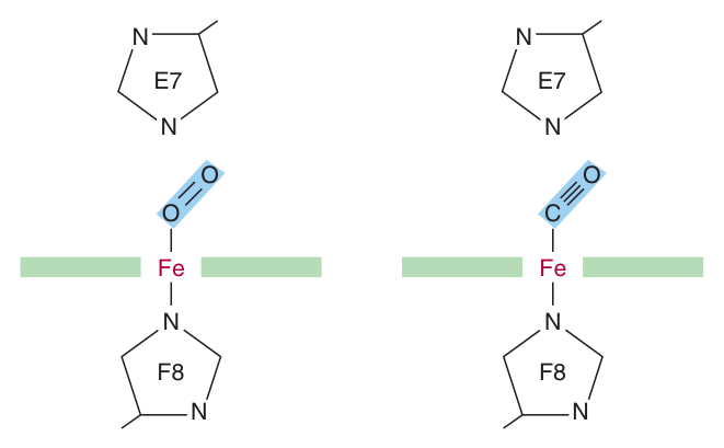
Here is a puzzle Harper's poses and answers, and it makes an excellent short-essay question because it is pure structure-function reasoning. Isolated haem binds carbon monoxide 25 000 times more strongly than oxygen. CO is always present — in the atmosphere, and generated inside cells by the catabolism of haem. So why are we not all permanently poisoned?
Because the protein gets in the way. The apoproteins of myoglobin and haemoglobin create a hindered environment for their gaseous ligands, and the obstruction is His E7, the distal histidine.
| Ligand | Preferred geometry on isolated haem | What the distal histidine does |
|---|---|---|
| O₂ | The O–O bond lies at about 121° to the haem plane, angling the second oxygen away from His E7 | Nothing — O₂ can still adopt its most favourable orientation |
| CO | Fe, C and O all perpendicular to the haem plane, in a straight line | Sterically blocks it. CO is forced to bind at a less favoured angle |
The result is a dramatic reduction in CO's advantage: the haem–CO bond falls from 25 000 times the strength of the haem–O₂ bond to about 200 times. Since O₂ is present in vast excess over CO, oxygen normally wins — although about 1% of myoglobin is typically combined with CO even in health.
Two hundred-fold is a reduction, not an abolition. At raised ambient CO the equilibrium shifts decisively, haemoglobin is occupied by CO, and oxygen carriage collapses. Harper's opens the chapter with the pairing worth remembering: cyanide and carbon monoxide kill because they disrupt the function of two haem proteins — cytochrome oxidase and haemoglobin respectively.
- How much more strongly does isolated haem bind CO than O₂? → 25 000 times
- What reduces this in the intact protein? → The distal histidine E7 sterically blocks CO's preferred perpendicular geometry
- By how much is it reduced? → To about 200-fold
- Which haem protein does cyanide poison? → Cytochrome oxidase (CO poisons haemoglobin)
The two dissociation curves ★★★
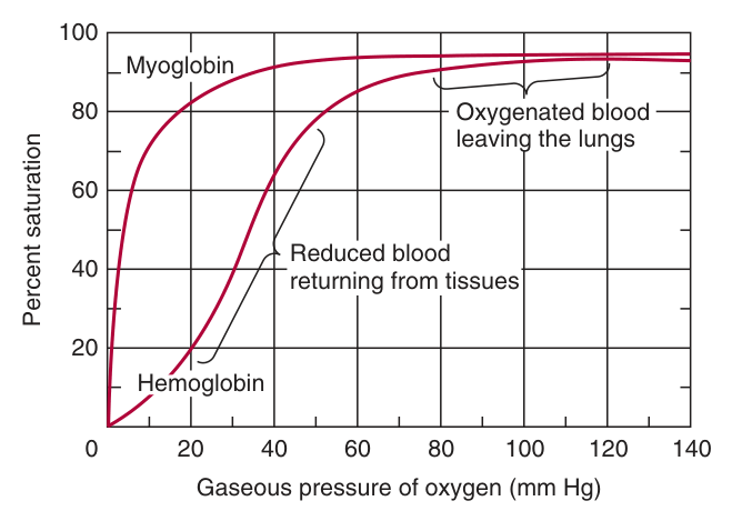
This is the diagram to be able to draw from memory, with the axes labelled and the four physiological pressures marked. Everything about the two proteins' roles can be read off it.
| Site | pO₂ |
|---|---|
| Arterial blood / lung capillary bed | 100 mm Hg |
| Mixed venous blood | 40 mm Hg |
| Capillary of active muscle | 20 mm Hg |
| Minimum required by cytochrome oxidase | 5 mm Hg |
Myoglobin's curve is hyperbolic. It loads O₂ readily at the 100 mm Hg of the lung — but that is not its problem. Its problem is unloading: at 40 mm Hg, or even the 20 mm Hg of active muscle, myoglobin is still nearly saturated and releases only a small fraction of its oxygen. That makes it an ineffective transport vehicle. Only when strenuous exercise drives muscle pO₂ down to about 5 mm Hg does myoglobin let go — and at that point the released O₂ permits mitochondrial ATP synthesis, and muscular activity, to continue. It is an emergency reserve, released exactly when the tissue becomes hypoxic.
Haemoglobin's curve is sigmoid. The shallow start means it holds on to oxygen at high pO₂ while filling up; the steep middle section falls exactly across the physiological range between lung and tissue, so a modest drop in pO₂ produces a large release of oxygen. Association of chains into a tetramer results in far greater oxygen delivery than single chains could ever manage.
The partial pressure of O₂ at which a given haemoglobin is half-saturated — a measure of oxygen affinity.
A high P₅₀ means low affinity (more pressure needed to half-fill it) and therefore better release at the tissues. P₅₀ always exceeds the pO₂ of the peripheral tissues.
HbA: 26 mm Hg. HbF: 20 mm Hg.
But the same property is a liability once the baby is breathing. HbF's higher affinity means it is reluctant to release O₂, so it limits the quantity delivered to the tissues — Harper's calls it “suboptimal postpartum”. This is why the switch to HbA matters.
The molecular reason (a good extra mark): residue H21 of the γ subunit is serine rather than histidine. Serine cannot form a salt bridge, so BPG binds more weakly to HbF; less stabilisation of the T state means higher O₂ affinity. See §9.
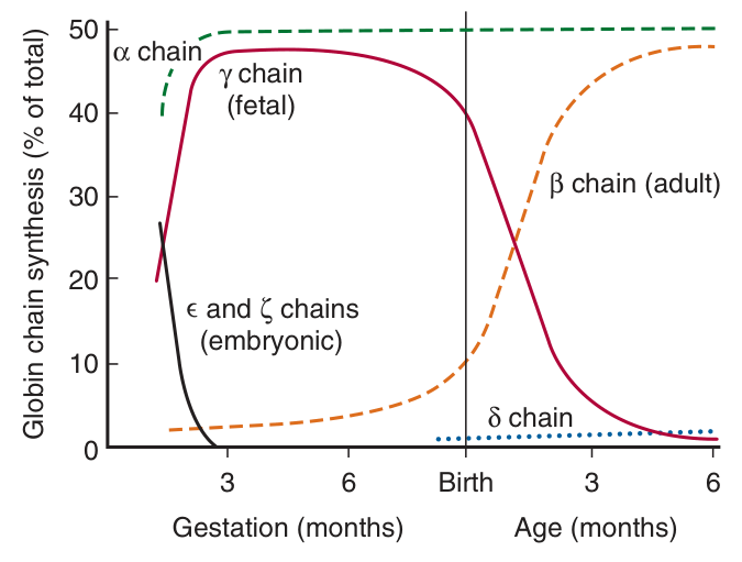
The developmental sequence
| Stage | Haemoglobin |
|---|---|
| Early embryo | ξ₂ε₂ |
| End of the first trimester onwards | HbF = α₂γ₂ — ξ and ε replaced by α and γ |
| Third trimester | β subunit synthesis begins |
| Some weeks after birth | Replacement of γ by β complete — HbA = α₂β₂ |
- Which curve is hyperbolic and which sigmoid? → Myoglobin hyperbolic; haemoglobin sigmoid
- Why is myoglobin a poor transporter? → It releases only a small fraction of its O₂ at tissue pO₂ — it only lets go at about 5 mm Hg
- Define P₅₀ → The partial pressure of O₂ at which a haemoglobin is half-saturated
- Give the P₅₀ of HbA and HbF → 26 and 20 mm Hg
- Does a high P₅₀ mean high or low affinity? → LOW affinity — and therefore better O₂ release
- What is fetal haemoglobin? → HbF = α₂γ₂
Haemoglobin and cooperativity ★★★
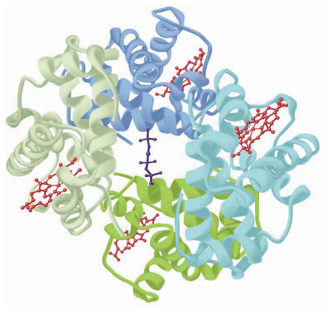
Adult haemoglobin, HbA, is α₂β₂ — a tetramer of two subunit types, binding four molecules of O₂, one per haem. The subunits are held together by non-covalent forces, importantly including salt bridges.
The structural punchline, and a favourite exam sentence: despite having different primary structures, myoglobin and the subunits of haemoglobin have nearly identical secondary and tertiary structures. Both are rich in α-helix, both are highly compact with hydrophilic residues outside and hydrophobic inside, and in both the hydrophobic interior forms the pocket that holds the haem. The β subunit matches myoglobin closely; the α subunit has seven helical regions rather than eight but still resembles it.
Different primary structure. Nearly identical secondary and tertiary structure. Completely different quaternary structure. And it is the quaternary difference that produces every functional difference.
That is a whole essay compressed into three sentences, and it is exactly what the TMU study question — “explain the linkage between protein structure and biologic function” — is asking for.
A molecule of O₂ binds to a haemoglobin tetramer more readily if other O₂ molecules are already bound.
This permits haemoglobin to maximise both the quantity of O₂ loaded at the pO₂ of the lungs and the quantity released at the pO₂ of peripheral tissues. Cooperative interactions are an exclusive property of multimeric proteins and are critically important to aerobic life.
- What is the subunit composition of HbA? → α₂β₂
- How many O₂ molecules per tetramer? → Four — one per haem
- How do Mb and the Hb subunits compare structurally? → Different primary structure, nearly identical secondary and tertiary structure
- Define cooperative binding → O₂ binds a haemoglobin tetramer more readily if other O₂ molecules are already bound
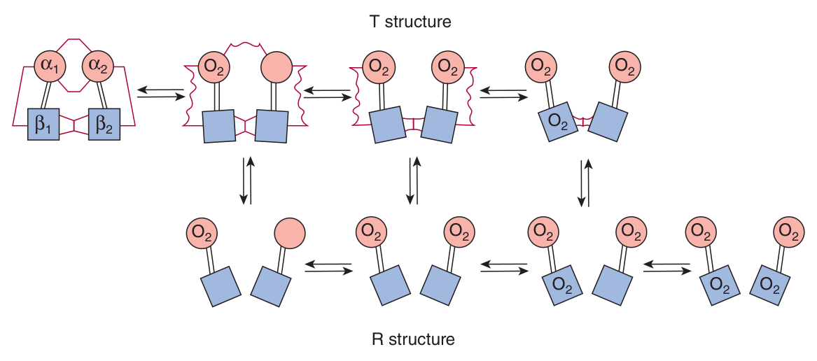
The T and R states — allostery, first appearance ★★★
Now assemble the pieces. §4 gave you the lever: O₂ binding pulls the iron into the haem plane, dragging His F8 with it. In a tetramer that motion has consequences.
| Step | What happens |
|---|---|
| 1 | The first O₂ binds to deoxyHb. The haem iron shifts into the plane of the ring, from about 0.04 nm beyond it |
| 2 | The motion is transmitted to the proximal (F8) histidine and the residues attached to it |
| 3 | This causes the rupture of salt bridges between the carboxyl-terminal residues of all four subunits |
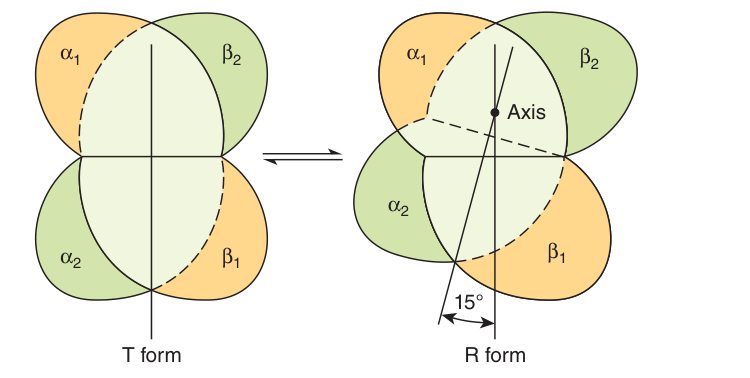
| 4 | One pair of α/β subunits rotates 15° relative to the other, compacting the tetramer |
| 5 | Profound changes in secondary, tertiary and quaternary structure convert the molecule from the T state to the R state |
T (taut) — the low-affinity conformation, held together by salt bridges between subunits; the deoxygenated form.
R (relaxed) — the high-affinity conformation; the oxygenated form.
The same terms are used for the low- and high-affinity conformations of allosteric enzymes.
And here is why this produces cooperativity, stated in one line: the transition significantly increases the affinity of the remaining unoxygenated haems for O₂, because subsequent binding events require the rupture of fewer salt bridges. The first oxygen does the hard work of breaking the bridges; every one after it has an easier job.
The T→R transition does not take place after a fixed number of oxygen molecules have been bound. It becomes progressively more probable as each successive oxygen binds — and salt bridges that have not yet ruptured are progressively weakened.
It is a shifting probability, not a switch that flips at molecule three. The transition is also influenced by protons, CO₂, chloride and BPG: the higher their concentration, the more oxygen must be bound to trigger it. That single sentence contains the whole of §9.
T and R are not haemoglobin-specific jargon. They are the standard vocabulary of allosteric regulation, and Unit 7 (Regulation of Enzyme Activity) uses them throughout. Haemoglobin is the canonical allosteric protein: a ligand binding at one site changes the conformation, and therefore the affinity, of sites elsewhere on the molecule. Learn it here properly and Unit 7 costs you almost nothing.
- What starts the T→R transition? → The first O₂ binding pulls the haem iron into the plane, moving His F8
- What breaks, and what rotates? → Salt bridges between the C-terminal residues of all four subunits break; one α/β pair rotates 15°
- Which state has high affinity? → R (relaxed); T (taut) is the low-affinity deoxy form
- Why does affinity rise as oxygen binds? → Later binding events require the rupture of fewer salt bridges
- Does the transition occur at a fixed number of O₂? → No — it becomes progressively more probable with each successive O₂
The Bohr effect and 2,3-BPG ★★★
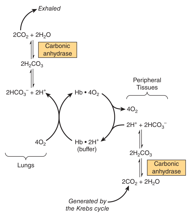
Haemoglobin's second job is to carry the waste products of respiration back to the lungs — and, elegantly, the mechanism for doing so is the same mechanism that improves oxygen delivery. That is the Bohr effect.
How CO₂ is carried
| Form | Share | Detail |
|---|---|---|
| Carbamates | about 15% of venous CO₂ | CO₂ combines with the amino-terminal nitrogens of the chains. This changes the charge on the amino terminals from positive to negative, which favours salt bridge formation between α and β chains — i.e. it stabilises the T state |
| Bicarbonate | most of the rest | CO₂ is hydrated to carbonic acid by carbonic anhydrase in the erythrocyte; at the pH of venous blood H₂CO₃ dissociates into bicarbonate and a proton |
The reciprocal coupling of proton and O₂ binding by haemoglobin.
In the tissues: CO₂ generated by respiration forms carbonic acid, which dissociates to bicarbonate and protons. Deoxyhaemoglobin binds one proton for every two O₂ molecules released, acting as a buffer. The lower pH of peripheral tissues, aided by carbamation, stabilises the T state and so enhances O₂ delivery.
In the lungs: as O₂ binds, protons are released; they combine with bicarbonate to form carbonic acid, which carbonic anhydrase dehydrates to CO₂, which is exhaled. Binding of oxygen thus drives the exhalation of CO₂.
The Bohr effect depends on cooperative interactions between the haems — so the monomeric structure of myoglobin precludes it.
On releasing O₂ the T structure and its salt bridges re-form, which increases the pKa of β-chain His 146 so that it binds protons. Naming His 146 and the pKa shift is what separates a full answer from a general one.
2,3-bisphosphoglycerate
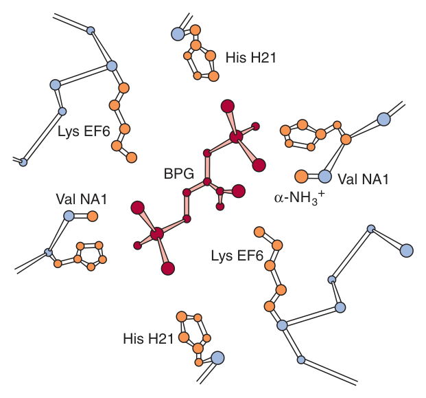
A low pO₂ in peripheral tissues promotes the synthesis of BPG in erythrocytes, from the glycolytic intermediate 1,3-bisphosphoglycerate (a link forward to Unit 10). The tetramer binds one molecule of BPG in the central cavity formed by its four subunits.
The mechanism is a beautiful piece of structural logic. The space between the H helices of the β chains lining that cavity is wide enough to accommodate BPG only when haemoglobin is in the T state. BPG forms salt bridges with three positively charged groups on each β chain — the terminal amino group via Val NA1, and Lys EF6 and His H21. Those are extra salt bridges that must be broken before the R state can form. So BPG stabilises the T state and lowers oxygen affinity.
Your TMU slide 20 says “decreases P₅₀”. That is a mistake. Harper's p.57 reads: “Elevated BPG lowers the affinity of HbA for O₂ (increases P₅₀), which enhances the release of O₂ at peripheral tissues.” The two halves of the slide's own sentence contradict each other — lower affinity cannot mean lower P₅₀.
How to handle it in the exam: the safe answer is to write “elevated BPG lowers the affinity of HbA for oxygen, which enhances release at the tissues”. That is correct, is what the marking key rewards, and sidesteps the contradiction. If you must give a P₅₀ direction, it increases.
Everything that stabilises the T state lowers oxygen affinity and improves delivery to the tissues: protons (low pH), CO₂, chloride and BPG. All four are signals that a tissue is working hard and needs oxygen — and all four act by the same means, adding or preserving the salt bridges that hold the taut state together.
That is not four facts. It is one fact with four inputs.
- How is most CO₂ carried, and how much as carbamate? → Mostly as bicarbonate; carbamates account for about 15% of venous CO₂
- How many protons does deoxyhaemoglobin bind per O₂ released? → One proton for every TWO O₂ molecules released
- Which residue supplies the Bohr protons? → β-chain His 146
- Why can myoglobin not show the Bohr effect? → It is monomeric, and the Bohr effect depends on cooperative interactions
- Where and when does BPG bind? → One molecule in the central cavity, and only in the T state
- What does BPG do to affinity? → Stabilises T, so lowers O₂ affinity and enhances tissue delivery
- Name the four things that stabilise the T state → Protons, CO₂, chloride and BPG
Haemoglobinopathies ★★★
A condition in which a mutation in the gene encoding an α or β subunit compromises the biological function of haemoglobin.
Worth stating the qualifier: of the over 1100 known mutations affecting human haemoglobins (your slide says over 800 — an edition difference), almost all are extremely rare and benign, presenting no clinical abnormality. Only when function is compromised is the term used. Even so, more than 7% of the world's population carry a haemoglobin disorder.
Sickle cell disease — the model molecular disease
This is the second TMU study question — “describe the linkage between primary structure and molecular disease based on sickle cell anaemia” — and it is a past-paper question. The chain of reasoning is what earns the marks, so learn it as a sequence, not a fact.
| Step | What happens |
|---|---|
| 1 · The mutation | In HbS, the non-polar valine replaces the polar surface residue Glu6 of the β subunit. HbA: Val-His-Leu-Thr-Pro-Glu-Glu-Lys → HbS: Val-His-Leu-Thr-Pro-Val-Glu-Lys |
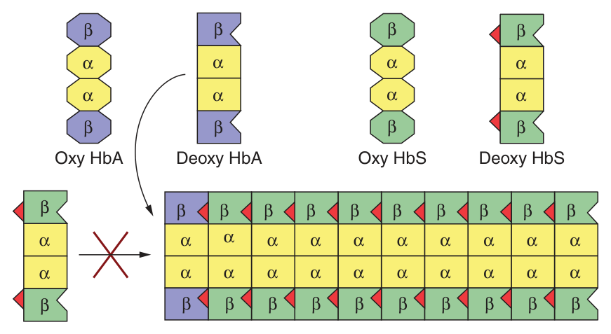
| 2 · The consequence for the surface | This generates a hydrophobic “sticky patch” on the surface of the β subunit — present in both oxyHbS and deoxyHbS |
| 3 · The complementary patch | Both HbA and HbS carry a complementary sticky patch, but it is exposed only in the deoxygenated T state |
| 4 · Polymerisation | So at low pO₂, deoxyHbS polymerises into long, insoluble, twisted helical fibres |
| 5 · The cell | The fibres distort the erythrocyte into the sickle shape, making it vulnerable to lysis in the interstices of the splenic sinusoids — hence the anaemia |
This is precisely why heterozygotes (sickle cell trait) are largely protected: their HbA acts as a chain terminator. It also explains an emerging therapy — inducing HbF expression to inhibit HbS polymerisation, alongside stem cell transplantation and, in future, gene therapy.
The other conditions worth knowing
| Condition | Defect | Consequence |
|---|---|---|
| Methaemoglobinaemia | Haem iron is ferric (Fe³⁺) rather than ferrous | Methaemoglobin can neither bind nor transport O₂. Normally methaemoglobin reductase reduces Fe³⁺ back to Fe²⁺. It arises from oxidation by agents such as sulfonamides, from hereditary haemoglobin M, or from reduced methaemoglobin reductase activity |
| Haemoglobin M | His F8 replaced by tyrosine | The iron forms a tight ionic complex with the phenolate anion of tyrosine, stabilising Fe³⁺. In α-chain variants the R–T equilibrium favours T: oxygen affinity is reduced and the Bohr effect is absent. β-chain variants still show R–T switching, so the Bohr effect is present |
| Haemoglobin Chesapeake | A mutation favouring the R state | Increased O₂ affinity, so the haemoglobin fails to deliver adequate O₂ to the tissues. The resulting tissue hypoxia causes polycythaemia |
| Thalassaemias | Partial or total absence of one or more α or β chains | α-thalassaemia or β-thalassaemia. A superscript indicates complete absence (α⁰, β⁰) or reduced synthesis (α⁻, β⁻). Over 750 mutations known, but only three are common. Apart from marrow transplantation, treatment is symptomatic |
| Myoglobinuria | Massive crush injury to skeletal muscle | Released myoglobin appears in the urine, with renal damage. Myoglobin is also detectable in plasma after myocardial infarction, though serum enzyme assay is a more sensitive index |
Notice what the two named point mutations do. HbS changes a surface residue and the protein still works perfectly as an oxygen carrier — the disease comes from stickiness, not from impaired binding. HbM changes His F8, a residue bonded to the iron itself, and destroys oxygen binding outright.
Same kind of mutation, entirely different mechanism, because of where the residue sits. That is the linkage between primary structure and disease, and it is the sentence to close a sickle-cell essay with.
- What is the HbS mutation? → Valine replaces glutamate at position 6 of the β chain
- What does it create? → A hydrophobic sticky patch on the β subunit surface, present in both oxy and deoxy forms
- When is the complementary patch exposed? → Only in the deoxygenated T state — hence polymerisation at low pO₂
- Why does HbA stop fibre growth? → It lacks the second sticky patch, so it terminates the polymer
- What is wrong in methaemoglobinaemia? → The haem iron is ferric (Fe³⁺) and can neither bind nor transport O₂
- What is the defect in haemoglobin M? → His F8 replaced by tyrosine, stabilising Fe³⁺
- What are the thalassaemias? → Partial or total absence of one or more α or β chains
Revision layer
Two study questions close this deck, and both have appeared on papers: “Describe the structure of Hb and Mb and explain the linkage between protein structure and biologic function” and “Describe the linkage between primary structure and molecular disease based on sickle cell anaemia”. The tables below are those two answers.
Myoglobin versus haemoglobin — the master comparison
| Myoglobin | Haemoglobin | |
|---|---|---|
| Location | Red skeletal muscle | Erythrocytes only |
| Function | O₂ storage | O₂ transport, plus CO₂ and H⁺ |
| Primary structure | 153 residues, one chain | Two chain types, α₂β₂ (HbA) |
| Secondary structure | 8 right-handed α-helices, A–H, ~75% helical | Nearly identical; α subunit has 7 helical regions |
| Tertiary structure | Compact sphere 4.5 × 3.5 × 2.5 nm, polar surface, non-polar interior | Nearly identical to myoglobin |
| Quaternary structure | None | Tetramer, subunits joined by non-covalent forces incl. salt bridges |
| Haem groups | One | Four — one per subunit |
| Cooperativity | No | Yes |
| O₂ curve | Hyperbolic | Sigmoid |
| Bohr effect | No — precluded by monomeric structure | Yes |
| Binds BPG | No | Yes — one molecule, T state only |
The sickle cell chain of reasoning — five steps
| Step | |
|---|---|
| 1 | Point mutation: Val replaces Glu6 of the β chain — a non-polar residue where a polar one was |
| 2 | Creates a hydrophobic sticky patch on the β subunit surface, in both oxy and deoxy HbS |
| 3 | The complementary patch, present on both HbA and HbS, is exposed only in the T state |
| 4 | At low pO₂, deoxyHbS polymerises into long insoluble twisted fibres |
| 5 | Fibres distort the erythrocyte into a sickle, which lyses in the splenic sinusoids → anaemia |
Definitions from this unit — Section I material
| Term | Definition |
|---|---|
| Prosthetic group | A small non-protein molecule that is a permanent part of a protein and is necessary for its function |
| Haem | A cyclic tetrapyrrole of four pyrrole rings linked by methyne bridges, with one atom of ferrous iron at the centre of the planar ring bonded to all four pyrrole nitrogens; substituents are methyl, vinyl and propionate |
| Cooperative binding | A molecule of O₂ binds to a haemoglobin tetramer more readily if other O₂ molecules are already bound; an exclusive property of multimeric proteins |
| P₅₀ | The partial pressure of oxygen at which a given haemoglobin reaches half-saturation; a high P₅₀ indicates low affinity. HbA 26 mm Hg, HbF 20 mm Hg |
| T and R states | T (taut) is the low-affinity, deoxygenated conformation stabilised by salt bridges; R (relaxed) is the high-affinity, oxygenated conformation |
| The Bohr effect | The reciprocal coupling of proton and oxygen binding by haemoglobin: protons and CO₂ stabilise the T state and enhance O₂ delivery in the tissues, while oxygen binding in the lungs releases protons and drives CO₂ exhalation. It depends on cooperativity and is therefore absent in myoglobin |
| Haemoglobinopathy | A condition in which a mutation in the gene encoding an α or β subunit compromises the biological function of haemoglobin |
| Methaemoglobin | Haemoglobin in which the haem iron is ferric (Fe³⁺) rather than ferrous, and which can therefore neither bind nor transport oxygen |
Numbers worth carrying in
| Figure | Value |
|---|---|
| Myoglobin | 153 residues · MW ~17 000 · 8 helices A–H · ~75% helical · 4.5 × 3.5 × 2.5 nm |
| Haem coordination bonds | 6 — four pyrrole N, one His F8, one O₂ |
| Iron displacement, deoxy | ~0.03 nm out of plane (0.04 nm in Hb) |
| CO affinity, isolated haem | 25 000× that of O₂ — reduced to ~200× in the protein |
| Myoglobin normally bound to CO | about 1% |
| pO₂: artery / vein / active muscle / cytochrome oxidase minimum | 100 / 40 / 20 / 5 mm Hg |
| P₅₀ of HbA and HbF | 26 and 20 mm Hg |
| Subunit rotation on T→R | 15° |
| Protons bound per O₂ released | 1 H⁺ per 2 O₂ |
| CO₂ carried as carbamate | about 15% of venous CO₂ |
| BPG bound per tetramer | one molecule, in the central cavity, T state only |
| Known Hb mutations | over 1100 (slide: over 800) — almost all rare and benign |
- Draw both dissociation curves with the four physiological pO₂ values marked
- Compare Mb and Hb across all four orders of structure and every functional consequence
- Describe the six coordination bonds of haem iron and name the two histidines
- Explain cooperativity mechanically, from iron movement to 15° rotation
- Define P₅₀ and explain why HbF's is lower — including the Ser H21 reason
- Explain the Bohr effect in both directions, naming His 146
- Explain how BPG works, and state correctly what it does to affinity and P₅₀
- Give the five-step sickle cell chain and explain why HbA terminates the fibre