Bones of Limbs & Joints
Shoulder Girdle — Clavicle & Scapula
The clavicle is the only horizontal long bone in the body and the most commonly fractured bone — its middle third is mechanically the weakest point where the curvature changes direction, and every FOOSH transmits force straight to this junction. The scapula, by contrast, is wrapped in muscle and fractures only with massive direct trauma; its clinical importance lies entirely in its landmarks — the suprascapular notch being the classic exam trap: the suprascapular nerve goes through, the suprascapular artery goes over. Together, clavicle and scapula form the shoulder girdle that suspends the entire upper limb from the axial skeleton, connected centrally by the sternoclavicular joint — the only bony articulation between the limb and the trunk.
The clavicle is the only horizontal long bone in the body, connecting the axial skeleton (sternum) to the appendicular skeleton (scapula). It is the first bone to ossify (intramembranous, 5th week of gestation) but last to fuse (medial physis fuses ~25 years).
| Feature | Details |
|---|---|
| Shape | S-shaped; medial 2/3 convex anteriorly; lateral 1/3 concave anteriorly |
| Medial end | Rounded; sternoclavicular joint — only joint connecting upper limb to axial skeleton |
| Lateral end | Flat; acromioclavicular joint; coracoid process below (coracoclavicular ligaments: conoid + trapezoid) |
| Inferior surface | Subclavian groove (subclavian muscle); costal tuberosity (costoclavicular ligament) |
| Common fracture site | Middle 1/3 (at the curvature, weakest point) — most commonly fractured bone in the body |
Fall on outstretched hand (FOOSH) or direct blow to shoulder. Middle 1/3 fracture: medial fragment pulled superiorly by sternocleidomastoid; lateral fragment depressed by weight of limb + deltoid. Subclavian vessels and brachial plexus at risk with severe displacement.
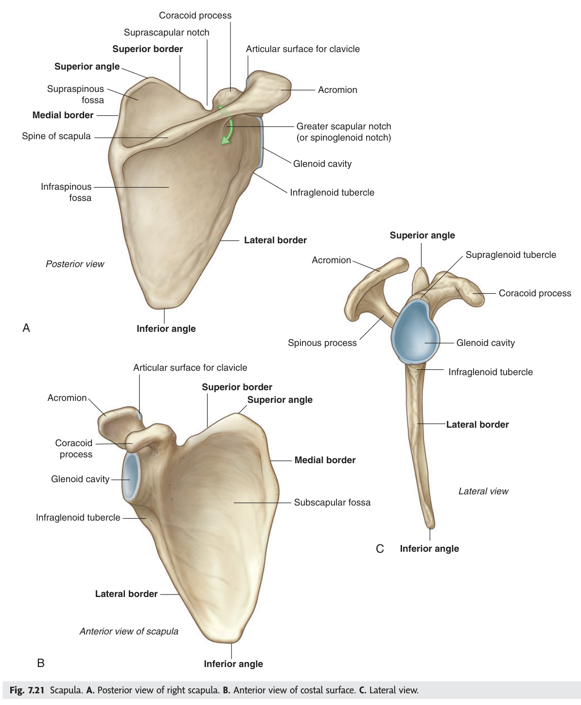
A flat triangular bone lying on the posterior thorax over ribs 2–7. Provides the socket for the shoulder joint and multiple muscle attachments.
| Landmark | Location & Clinical Significance |
|---|---|
| Spine | Posterior ridge dividing supraspinous and infraspinous fossae; continuous with acromion laterally |
| Acromion | Lateral end of spine; articulates with clavicle (AC joint); overhangs supraspinatus (impingement) |
| Coracoid process | Anterior hook-shaped projection; attachment for pectoralis minor, short head biceps, coracobrachialis |
| Glenoid cavity | Lateral angle; shallow socket deepened by fibrocartilaginous glenoid labrum |
| Supraspinous fossa | Superior to spine; supraspinatus muscle |
| Infraspinous fossa | Inferior to spine; infraspinatus muscle |
| Subscapular fossa | Anterior (costal) surface; subscapularis muscle |
| Suprascapular notch | Superior border; suprascapular nerve passes through (artery passes over ligament) |
- Q: Most common site and mechanism of clavicle fracture? — Middle 1/3 (weakest point at curvature change); FOOSH or direct blow. Medial fragment pulled up by SCM, lateral drops by gravity.
- Q: What joint is the only bony connection between upper limb and axial skeleton? — Sternoclavicular joint (medial clavicle–sternum).
- Q: Suprascapular notch rule? — Nerve passes THROUGH (under the superior transverse scapular ligament); artery passes OVER the ligament.
- Q: Name the four muscles attaching to the coracoid process. — Pectoralis minor, short head of biceps brachii, coracobrachialis (+ coracohumeral ligament).
- Q: Which AC joint grade tears both the AC and coracoclavicular ligaments? — Grade III (complete separation, step deformity, clavicle elevated).
Arm & Forearm — Humerus, Radius & Ulna
The humerus is a textbook of nerve anatomy — each level carries a different nerve injury signature. At the surgical neck, the axillary nerve wraps around; a fracture here destroys deltoid and causes regimental badge numbness. In the spiral groove of the mid-shaft, the radial nerve runs with profunda brachii — mid-shaft fracture means wrist drop. Behind the medial epicondyle distally, the ulnar nerve runs unprotected; medial epicondyle fractures (common in children) injure it. Colles' fracture at the distal radius (dorsal displacement, dinner-fork deformity) is the canonical elderly-fall injury, while scaphoid fractures from FOOSH in young adults are dangerous because of delayed diagnosis and avascular necrosis risk from the distal-entering blood supply.
| Region | Landmark | Clinical Significance |
|---|---|---|
| Proximal | Head | Articulates with glenoid cavity; directed superiorly, medially, posteriorly |
| Anatomical neck | Groove between head and tubercles; rarely fractured | |
| Greater tubercle | SITS tendons: supraspinatus (top), infraspinatus (middle), teres minor (bottom) — rotator cuff | |
| Lesser tubercle | Subscapularis tendon; anterolateral | |
| Shaft | Intertubercular (bicipital) groove | Long head of biceps tendon; medial = lesser tubercle, lateral = greater tubercle |
| Shaft | Deltoid tuberosity | Lateral; deltoid muscle insertion (C-shaped) |
| Shaft | Radial (spiral) groove | Posterolateral; radial nerve + profunda brachii artery — mid-shaft fracture → wrist drop |
| Distal | Capitellum | Lateral; articulates with radial head |
| Trochlea | Medial; articulates with trochlear notch of ulna (spool-shaped) | |
| Lateral epicondyle | Extensor muscle origin; lateral epicondylitis = tennis elbow | |
| Medial epicondyle | Flexor muscle origin; ulnar nerve passes behind — medial epicondyle fracture → ulnar nerve injury |
Surgical neck fracture (proximal, common in elderly): axillary nerve injury → loss of deltoid + sensation over 'regimental badge' area (lateral upper arm). Mid-shaft fracture: radial nerve in spiral groove → wrist drop (loss of wrist + finger extension). Supracondylar fracture (most common in children): anterior interosseous nerve injury → "OK sign" loss (FDP index + FPL weakness).
| Bone | Feature | Notes |
|---|---|---|
| Radius | Head | Proximal; disc-shaped; rotates in radial notch of ulna for pronation/supination |
| Radial tuberosity | Biceps brachii tendon attachment (bicipitoradial bursa) | |
| Styloid process | Distal, lateral; extends ~1 cm further distally than ulnar styloid — key X-ray landmark | |
| Distal articular surface | Articulates with scaphoid (lateral) + lunate (medial); tilted 11° anteriorly + 23° ulnarly | |
| Ulna | Olecranon | Proximal; forms the prominence of the elbow; triceps insertion |
| Coronoid process | Anterior projection; trochlear notch between olecranon + coronoid (articulates with trochlea) | |
| Radial notch | Lateral; proximal radioulnar joint (for radial head rotation) | |
| Head (distal) | Lateral; distal radioulnar joint; ulnar styloid medially |
Colles' fracture: FOOSH → distal radius fractures with dorsal displacement → "dinner fork deformity" on lateral X-ray. Most common fracture in elderly women. Smith's fracture: fall on flexed wrist → volar displacement (reverse Colles'). Scaphoid fracture: FOOSH in young adults → anatomical snuffbox tenderness → risk of avascular necrosis of proximal pole (blood supply enters distally). Plain X-ray often negative — if suspected, MRI or bone scan.
- Q: Fracture at radial spiral groove → which nerve → what deficit? — Radial nerve + profunda brachii artery → wrist drop (loss of wrist and finger extension).
- Q: Fracture at surgical neck of humerus → which nerve → what deficit? — Axillary nerve → paralysed deltoid, numbness over regimental badge area (lateral upper arm).
- Q: Colles' vs Smith's fracture — what distinguishes them? — Colles' = dorsal displacement (dinner fork); Smith's = volar displacement (reverse Colles'). Both involve distal radius.
- Q: Why does scaphoid fracture risk avascular necrosis? — Blood supply enters distally and flows proximally; fracture at the waist interrupts flow to the proximal pole.
- Q: Which radial landmark is normally further distal than the ulnar styloid, and by how much? — Radial styloid extends ~1 cm further distally than ulnar styloid (key X-ray sign of Colles' fracture when reversed).
Hand — Carpals, Metacarpals & Phalanges
Eight carpal bones sit in two rows forming the wrist, and the two that matter most clinically are the scaphoid and the lunate. The scaphoid is the most commonly fractured carpal — FOOSH in a young adult, anatomical snuffbox tenderness, often a normal initial X-ray — and its proximal pole is at risk of avascular necrosis because the blood supply enters distally and runs proximally, making it vulnerable to fracture-line interruption. The lunate is the most commonly dislocated carpal and threatens the median nerve in the carpal tunnel when it shifts anteriorly. The carpal tunnel contains the median nerve and nine flexor tendons but not the ulnar nerve — which is why carpal tunnel syndrome spares the little finger and half the ring finger.
Eight small bones in two rows, allowing the complex movements of the wrist. All have rough surfaces for ligament attachment; covered with hyaline cartilage on articular surfaces.
| Row | Bone (lateral → medial) | Key Fact |
|---|---|---|
| Proximal | Scaphoid | Boat-shaped; articulates with radius; most commonly fractured carpal; avascular necrosis risk |
| Lunate | Crescent-shaped; articulates with radius medially; most commonly dislocated carpal | |
| Triquetrum | Pyramid-shaped; articulates with articular disc distally | |
| Pisiform | Smallest; sesamoid in FCU tendon; overlies triquetrum anteriorly | |
| Distal | Trapezium | Articulates with 1st metacarpal (saddle joint — thumb) |
| Trapezoid | Smallest of distal row; articulates with 2nd metacarpal | |
| Capitate | Largest carpal; head articulates with lunate; central keystone of wrist | |
| Hamate | Hook of hamate projects anteriorly → medial wall of carpal tunnel; articulates with 4th + 5th metacarpals |
"Some Lovers Try Positions That They Cannot Handle"
Scaphoid · Lunate · Triquetrum · Pisiform — Trapezium · Trapezoid · Capitate · Hamate
Five metacarpals (I–V) connect the carpals to the phalanges. 14 phalanges: proximal + middle + distal for each finger (3 × 4 = 12) plus proximal + distal for the thumb (2). Total: 27 bones per hand.
| Feature | Details |
|---|---|
| 1st metacarpal | Shortest + most mobile; articulates with trapezium via saddle joint (thumb opposition) |
| Knuckles | Metacarpal heads form knuckles; cartilage-covered for MCP joints |
| Thumb phalanges | Proximal + distal only (2) — no middle phalanx |
| Bennett's fracture | Intra-articular fracture-dislocation at base of 1st metacarpal (axial load on thumb) |
| Mallet finger | Avulsion of extensor tendon from distal phalanx → cannot extend DIP joint |
- Q: Which carpal is most commonly fractured and why is it dangerous? — Scaphoid; blood supply enters distally → fracture at waist interrupts proximal flow → AVN of proximal pole. X-ray often negative; need MRI.
- Q: Which carpal is most commonly dislocated and what structure does it threaten? — Lunate; anterior dislocation presses on the carpal tunnel → acute median nerve compression.
- Q: What are the 10 contents of the carpal tunnel? — Median nerve + 4 × FDS tendons + 4 × FDP tendons + 1 × FPL tendon. The ulnar nerve is NOT in the tunnel (it runs in Guyon's canal).
- Q: Mnemonic for the 8 carpal bones in order? — "Some Lovers Try Positions That They Cannot Handle" (Scaphoid, Lunate, Triquetrum, Pisiform — Trapezium, Trapezoid, Capitate, Hamate).
- Q: Bennett's fracture — which bone and what mechanism? — Intra-articular fracture-dislocation at the base of the 1st metacarpal; axial load on a partially flexed thumb.
Upper Limb Joints ★
The shoulder is the most mobile and least stable joint in the body — a deliberate evolutionary trade-off that gives the arm extraordinary range at the cost of dislocation liability. The rotator cuff (SITS: supraspinatus, infraspinatus, teres minor, subscapularis) compensates by pulling the humeral head into the glenoid, but the inferior capsule has no cuff reinforcement, which is why anterior-inferior dislocations account for ~95% of cases. The elbow's carrying angle (15–20° valgus) explains why throwing athletes tear the medial collateral ligament — every throw generates valgus stress on the medial joint — and at the wrist, the carpal tunnel's anatomy explains why so many systemic conditions (pregnancy, hypothyroidism, RA) eventually present with median nerve compression.
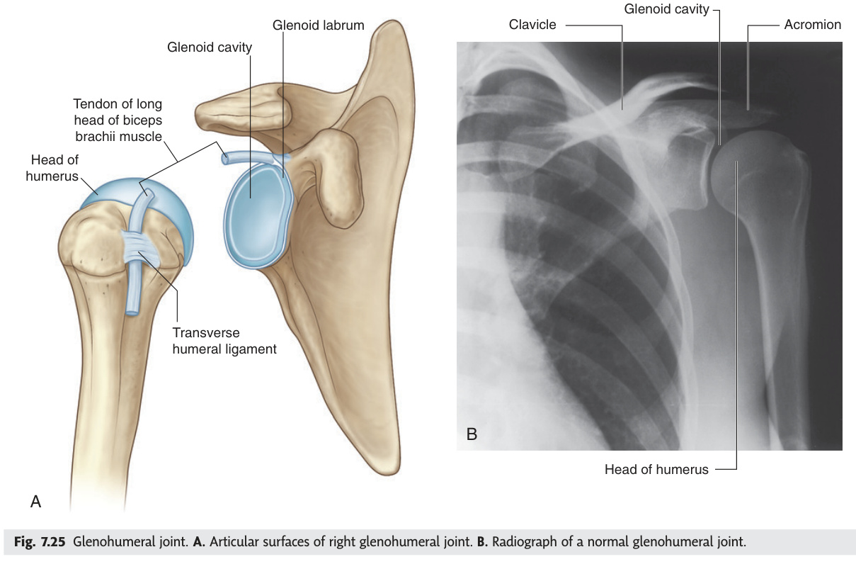
Ball-and-socket synovial joint between the head of the humerus and the glenoid cavity of the scapula. The most mobile and least stable joint in the body.
| Feature | Details |
|---|---|
| Type | Ball-and-socket (multiaxial) — flexion/extension, abduction/adduction, medial/lateral rotation, circumduction |
| Glenoid labrum | Fibrocartilaginous rim deepening the socket by ~50%. Bankart lesion = labral tear with anterior dislocation |
| Rotator cuff (SITS) | Supraspinatus (initiates abduction 0–15°) · Infraspinatus (ER) · Teres minor (ER) · Subscapularis (IR) |
| Weakest point | Inferior capsule — no rotator cuff reinforcement → anterior-inferior dislocation most common (~95%) |
| Stability | Provided by rotator cuff, glenoid labrum, coracohumeral ligament, glenohumeral ligaments |
Anterior dislocation: arm held in slight abduction and ER; flattening of deltoid ("squaring of shoulder"); axillary nerve at risk → test deltoid + skin over regimental badge area. Bankart lesion (labrum) + Hill-Sachs lesion (posterolateral humeral head). Painful arc 60–120°: supraspinatus impinges under acromion. Supraspinatus tear: most commonly torn rotator cuff tendon; inability to initiate abduction.
| Muscle | Origin | Insertion on Greater/Lesser Tubercle | Action | Nerve Supply |
|---|---|---|---|---|
| Supraspinatus | Supraspinous fossa | Greater tubercle — superior facet | Initiates abduction 0–15° (deltoid continues); holds head in socket | Suprascapular n. (C5, C6) |
| Infraspinatus | Infraspinous fossa | Greater tubercle — middle facet | External rotation (ER); posterior capsule support | Suprascapular n. (C5, C6) |
| Teres minor | Lateral scapular border | Greater tubercle — inferior facet | External rotation; weak adduction | Axillary n. (C5, C6) |
| Subscapularis | Subscapular fossa (anterior) | Lesser tubercle | Internal rotation (IR); adduction; anterior capsule support | Upper + lower subscapular nn. (C5, C6) |
Grade II: AC ligament torn + coracoclavicular (CC) ligament intact — mild separation (clavicle rides up slightly).
Grade III: Both AC + CC ligaments torn — complete separation; clavicle elevated. Step deformity. Conservative management for most Grade III.
Grades IV–VI: Clavicle displaced posteriorly/inferiorly — surgical repair.
| Feature | Details |
|---|---|
| Type | Compound hinge joint — 3 articulations in one capsule: humeroulnar, humeroradial, proximal radioulnar |
| Movements | Flexion/extension (humeroulnar + humeroradial) + pronation/supination (proximal radioulnar) |
| Carrying angle | 15–20° valgus in full extension; more in females. Cubitus valgus (increased) → tardy ulnar nerve palsy |
| MCL | Medial (ulnar) collateral ligament; anterior band resists valgus; torn in throwing athletes (UCL reconstruction) |
| LCL | Lateral (radial) collateral ligament + annular ligament of radial head |
Condyloid (ellipsoid) synovial joint between the distal end of the radius (and articular disc) and the proximal row of carpals (scaphoid + lunate). The ulna does not directly contact the carpals.
Carpal Tunnel — an osseofibrous tunnel on the anterior wrist:
| Boundary | Structure |
|---|---|
| Roof | Flexor retinaculum (attached to pisiform, hamate hook, scaphoid tubercle, trapezium ridge) |
| Floor | Concave arch of carpal bones |
| Contents (10) | 4 × FDS tendons + 4 × FDP tendons + 1 × FPL tendon + median nerve |
| NOT in tunnel | Flexor carpi radialis (in its own tunnel) · Ulnar nerve + artery (in Guyon's canal) |
Median nerve compression under the flexor retinaculum. Causes: repetitive use, pregnancy, hypothyroidism, RA, obesity. Features: pain and tingling in lateral 3½ fingers (median nerve territory), nocturnal worsening, thenar wasting (abductor pollicis brevis weakness), positive Tinel's test (tap over carpal tunnel) and Phalen's test (sustained wrist flexion). Treatment: splinting, steroid injection, surgical decompression.
- Q: Why is the shoulder the most commonly dislocated joint? — Inferior capsule has no rotator cuff reinforcement → anterior-inferior dislocation (~95%); glenoid is shallow and contacts only ~25% of the humeral head at any moment.
- Q: SITS — four rotator cuff muscles and their actions? — Supraspinatus (initiates abduction 0–15°), Infraspinatus (ER), Teres minor (ER), Subscapularis (IR). All stabilise the head in the glenoid.
- Q: What is the carrying angle and what increases it? — 15–20° valgus at the elbow in full extension; larger in females. Cubitus valgus → tardy ulnar nerve palsy. Cubitus varus ("gunstock deformity") follows malunited supracondylar fracture.
- Q: Lateral vs medial epicondyle — which is tennis elbow, which has the ulnar nerve? — Lateral epicondyle = tennis elbow (extensor origin). Medial epicondyle = golfer's elbow; ulnar nerve runs in groove behind the medial epicondyle.
- Q: Painful arc syndrome — degrees and which tendon? — Pain between 60–120° of shoulder abduction; supraspinatus tendon impinges under the acromion/coracoacromial arch.
Pelvic Girdle — Hip Bone & Pelvis
The pelvis transmits the entire body weight from the axial skeleton to the lower limbs through the sacroiliac joints and the hip sockets, and the hip bone — formed by the fusion of ilium, ischium, and pubis at the acetabulum — is the structural cornerstone of this system. The ischial spine is a critical surgical landmark: it separates the greater from the lesser sciatic foramen, the pudendal nerve hooks around it to reach the perineum (giving the injection site for pudendal nerve block), and its prominence narrows the pelvic outlet in males. Female pelvic anatomy (wider subpubic angle >90°, rounder inlet, less prominent ischial spines) is directly shaped by obstetric selection — these dimensions define whether a baby can pass.
The hip bone is formed by the fusion of three bones — ilium, ischium and pubis — at the Y-shaped triradiate cartilage in the acetabulum. Fusion is complete by age 16–18.
| Component | Key Landmarks |
|---|---|
| Ilium (superior) | Iliac crest (L4 level) · ASIS (inguinal ligament) · AIIS (rectus femoris) · PSIS · Greater sciatic notch · Iliac fossa (iliacus) · Auricular surface (SI joint) |
| Ischium (posteroinferior) | Ischial tuberosity (hamstring origin, "sitting bone") · Ischial spine (sacrospinous lig, separates greater/lesser sciatic foramina) · Lesser sciatic notch |
| Pubis (anteroinferior) | Body · Superior + inferior rami · Pubic crest · Pubic tubercle (inguinal ligament medial end) · Pectineal line |
| Acetabulum | Cup-shaped socket facing inferolaterally; lunate (articular) surface + acetabular fossa (ligamentum teres) + acetabular notch |
| Feature | Female Pelvis | Male Pelvis |
|---|---|---|
| Overall shape | Gynaecoid (oval inlet) | Android (heart-shaped inlet) |
| Subpubic angle | >90° (wide) | <90° (narrow) |
| Pelvic inlet | Rounded / oval | Heart-shaped (sacral promontory prominent) |
| Ischial spines | Less prominent | Prominent (narrows outlet) |
| Sacrum | Short, wide, curved | Long, narrow, straight |
| Overall | Wider, shallower (obstetric) | Narrower, deeper |
- Q: Three bones that fuse to form the hip bone — where do they meet? — Ilium, ischium, pubis; fuse at the Y-shaped triradiate cartilage in the acetabulum by age 16–18.
- Q: What landmark separates the greater from the lesser sciatic foramen? — Ischial spine (sacrospinous ligament). The pudendal nerve exits the greater foramen, hooks around the ischial spine, and re-enters via the lesser foramen to reach the perineum.
- Q: Subpubic angle — female vs male? — Female >90° (wide, obstetric); male <90° (narrow). Wider angle allows the fetal head to pass through the pelvic outlet.
- Q: At what vertebral level is the iliac crest and why does it matter? — L4 level — landmark for lumbar puncture (L3/4 or L4/5 interspaces, below the cord which ends at L1/2).
- Q: What attaches to the ASIS and the pubic tubercle? — ASIS = lateral end of inguinal ligament; pubic tubercle = medial end. Both also serve as muscle origins (sartorius/TFL at ASIS).
Thigh & Leg — Femur, Patella, Tibia & Fibula
The femoral neck is the most surgically significant region of the lower limb skeleton — an intracapsular fracture here disrupts the medial circumflex femoral artery's retinacular vessels, cutting off blood supply to the femoral head and risking avascular necrosis. The key clinical decision is Garden classification: undisplaced (I and II) → fix; displaced (III and IV) → replace. At the fibular neck, the common peroneal nerve makes a superficial bend around the bone and is the most commonly injured peripheral nerve in the leg — any cast, fracture, or prolonged leg-crossing at this level causes foot drop. Compartment syndrome of the leg (anterior compartment most vulnerable) is the orthopaedic emergency that demands emergency fasciotomy when pain on passive stretch is out of proportion — presence of a pulse does not exclude it.
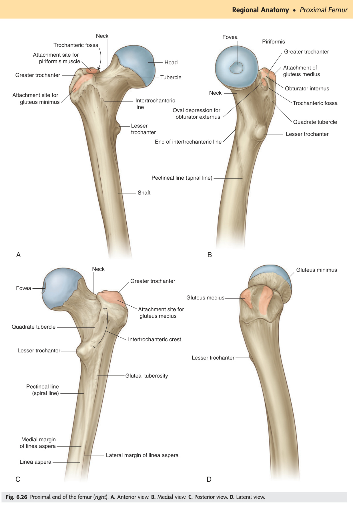
The longest and strongest bone in the body.
| Region | Landmark | Clinical Significance |
|---|---|---|
| Proximal | Head | 2/3 sphere; fovea capitis = ligamentum teres (carries obturator artery to head) |
| Neck | Angle of inclination ~126°; coxa vara <120°, coxa valga >135° | |
| Greater trochanter | Lateral; gluteus medius/minimus, piriformis, obturator internus/externus, gemelli | |
| Lesser trochanter | Posteromedial; iliopsoas (iliacus + psoas) insertion | |
| Shaft | Linea aspera | Posterior ridge; attachment for vastii, adductors, short head biceps femoris |
| Distal | Medial + lateral condyles | Articular surfaces for knee; medial condyle has adductor tubercle (adductor magnus) |
| Intercondylar notch | Between condyles (posterior); ACL + PCL attachment | |
| Patellar groove | Anterior; patellar tracking surface |
Subcapital fracture (intracapsular): disrupts the medial circumflex femoral artery (main blood supply to femoral head, via retinacular vessels) → avascular necrosis of femoral head. Limb: shortened + externally rotated. Treatment: hemiarthroplasty in elderly. Intertrochanteric fracture (extracapsular): below the capsule, blood supply intact → fix with dynamic hip screw (DHS). Elderly osteoporotic women.
Garden II: Complete, undisplaced — trabeculae interrupted but no shift; stable.
Garden III: Complete, partially displaced — femoral head rotated into varus; unstable.
Garden IV: Complete, fully displaced — no trabecular alignment; unstable.
Management rule:
• Garden I + II (undisplaced) → internal fixation with cannulated screws (preserve the femoral head; blood supply may be intact).
• Garden III + IV (displaced) → hemiarthroplasty (elderly, low-demand) or total hip replacement (active patients under 70). Displaced fractures carry ≥30% AVN risk — head replacement avoids this.
Memory: Garden I–II = Fix; Garden III–IV = Replace.
| Bone | Feature | Notes |
|---|---|---|
| Patella | Largest sesamoid | Embedded in quadriceps tendon; protects knee anteriorly; patellar ligament = distal continuation to tibial tuberosity |
| Tibia | Condyles | Medial + lateral; tibial plateau; bears ~5/6 of body weight |
| Tibial tuberosity | Anterior; patellar ligament (quadriceps tendon) insertion; Osgood-Schlatter in adolescents | |
| Medial border | Subcutaneous (shin); common site of tibial stress fractures | |
| Medial malleolus | Distal medial projection; shorter than lateral malleolus | |
| Fibula | Head + neck | Common peroneal (fibular) nerve wraps around fibular neck — fracture → foot drop (loss of dorsiflexion + eversion) |
| Body | Carries ~1/6 body weight; attachment for peroneal + extensor muscles | |
| Lateral malleolus | Extends ~1 cm further distally than medial malleolus — important for ankle stability |
Definition: Raised pressure within a closed osteofascial compartment compromising perfusion to muscles and nerves within that compartment.
Common causes: Tibial shaft fracture (anterior compartment most at risk), supracondylar humerus fracture, crush injuries, circumferential burns, over-tight plaster cast.
The 5 Ps (classic features — in order of appearance):
• Pain — out of proportion to the injury; classically increased by passive stretch of muscles in the compartment (most sensitive early sign).
• Pressure — compartment feels tense/wooden on palpation.
• Paraesthesia — numbness/tingling in the nerve distribution within the compartment.
• Paralysis — weakness of muscles in the affected compartment.
• Pallor / Pulselessness — late, pre-terminal signs; presence of pulses does NOT exclude compartment syndrome.
Diagnosis: Clinical + compartment pressure measurement. Fasciotomy indicated if pressure >30 mmHg or within 30 mmHg of diastolic blood pressure (delta pressure <30 mmHg).
Treatment: Emergency fasciotomy — release all 4 compartments of the leg (anterior, lateral, superficial posterior, deep posterior).
Volkmann's ischaemic contracture: Late consequence of untreated forearm compartment syndrome. Ischaemic necrosis of deep flexors (FDP, FPL) → fibrosis → flexion contracture of wrist + fingers, intrinsic-minus hand posture. Prevention = early fasciotomy.
- Q: Garden classification — which grades fix vs replace? — Garden I & II (undisplaced) → internal fixation with cannulated screws; Garden III & IV (displaced) → hemiarthroplasty or THR. Rule: Fix I–II, Replace III–IV.
- Q: Which artery supplies the femoral head and how is it damaged? — Medial circumflex femoral artery (via retinacular vessels along femoral neck). Intracapsular fracture tears these → AVN of femoral head.
- Q: Nerve at the fibular neck — injury causes what deficit? — Common peroneal (fibular) nerve → foot drop (loss of dorsiflexion + eversion); high-stepping gait.
- Q: First sign of compartment syndrome? — Pain on passive stretch of muscles in the compartment — precedes pulselessness. Presence of pulses does NOT exclude compartment syndrome.
- Q: Trendelenburg test — what is a positive result and what does it mean? — Contralateral pelvis drops when standing on affected leg → weak ipsilateral gluteus medius (or superior gluteal nerve injury). The pelvis tilts toward the unsupported side.
Foot — Tarsals, Metatarsals, Phalanges & Arches
The foot's three arches — medial longitudinal (the most important and the most often fallen), lateral longitudinal, and transverse — are active systems maintained dynamically by ligaments and muscles, not passive bony structures. The talus is unique in having no muscle tendon attachments; it is entirely shaped and positioned by ligament and joint forces. Tibialis posterior is the key dynamic supporter of the medial arch — its degeneration in adults causes acquired flat foot and a characteristic progressive valgus deformity. The 5th metatarsal is the site of two distinct and commonly confused fractures: the avulsion fracture at the tuberosity base (Dancer's fracture — peroneus brevis traction, heals well) and the Jones fracture at the diaphysis just distal to it (stress fracture, poor blood supply, high nonunion rate).
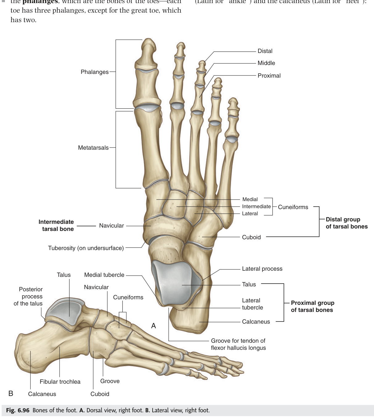
| Bone | Key Facts |
|---|---|
| Calcaneus | Largest tarsal; heel bone; Achilles tendon attaches posteriorly; sustentaculum tali supports talus; peroneal trochlea laterally |
| Talus | Transmits body weight from tibia/fibula to foot; no muscle tendons attach to it; body + neck + head |
| Navicular | Boat-shaped; medial; between talus and cuneiforms; tibialis posterior inserts on tuberosity |
| Cuboid | Lateral; between calcaneus and 4th/5th metatarsals; peroneus longus tendon grooves its plantar surface |
| Cuneiforms (3) | Medial, intermediate, lateral; wedge-shaped; support transverse arch; medial = largest, intermediate = smallest |
Metatarsals (5) and Phalanges (14): same count as hand; great toe has 2 phalanges, others 3 each.
5th metatarsal base avulsion fracture (Dancer's fracture): forced inversion avulses peroneus brevis tendon. Jones fracture: stress fracture of 5th metatarsal diaphysis just distal to tuberosity — poor blood supply → delayed union. Lisfranc injury: tarsometatarsal dislocation/fracture, often missed on X-ray.
| Arch | Bones | Keystone | Supports |
|---|---|---|---|
| Medial longitudinal | Calcaneus → talus → navicular → 3 cuneiforms → 1st–3rd metatarsals | Talus | Plantar fascia, spring ligament (calcaneonavicular), tibialis posterior, FHL |
| Lateral longitudinal | Calcaneus → cuboid → 4th–5th metatarsals | Cuboid | Long plantar ligament, plantar fascia |
| Transverse | Cuneiforms + cuboid (at mid-foot); metatarsal heads (at forefoot) | Intermediate cuneiform | Peroneus longus (key dynamic support) |
- Q: Which tarsal bone has no muscle tendons attaching to it? — Talus; it transmits body weight entirely through ligament and joint forces.
- Q: Keystone of the medial longitudinal arch? — Talus. Dynamic support = tibialis posterior (degeneration → acquired flat foot). Static support = spring (plantar calcaneonavicular) ligament + plantar fascia.
- Q: Dancer's fracture vs Jones fracture — same bone, different location? — Both at 5th metatarsal. Dancer's = avulsion of tuberosity base by peroneus brevis (heals well). Jones = stress fracture of diaphysis just distal to tuberosity (poor blood supply → high nonunion rate).
- Q: Which muscle is the key dynamic stabiliser of the transverse arch? — Peroneus longus; its tendon crosses the plantar surface and cinches the arch transversely.
- Q: Pes cavus — which neurological conditions are associated? — Charcot-Marie-Tooth disease (hereditary motor/sensory neuropathy) and Friedreich's ataxia; always investigate high arch in a young patient for neurological cause.
Lower Limb Joints ★
The hip is the most stable joint in the body (deepest ball-and-socket, reinforced by the Y-shaped iliofemoral ligament — the strongest single ligament in the body), while the knee is the most complex (three articulations in one capsule, paired menisci, four major ligaments). Knee ligament anatomy decodes acute injuries: the unhappy triad (ACL + MCL + medial meniscus) follows valgus-rotation because the MCL physically attaches to the medial meniscus, making them conjoint casualties. At the ankle, ATFL is always the first lateral ligament torn in inversion sprains — the most anterior and weakest of the three lateral components — and stability is maximum in dorsiflexion when the wider anterior dome of the talus is wedged in the mortice, minimum in plantarflexion when the narrow posterior dome is loose.
Ball-and-socket synovial joint between the head of the femur and the acetabulum. The most stable joint in the body, designed for weight-bearing with a large ROM.
| Feature | Details |
|---|---|
| Ligaments | Iliofemoral (Y-ligament, strongest = prevents hyperextension) · Pubofemoral (prevents excessive abduction) · Ischiofemoral (prevents hyperextension + IR) |
| Ligamentum teres | Intracapsular, extrasynovial; carries obturator artery to femoral head; weak mechanical role |
| Acetabular labrum | Fibrocartilaginous rim deepening socket; completed inferiorly by transverse acetabular ligament |
| Blood supply to head | Mainly medial circumflex femoral artery (retinacular vessels); ligamentum teres = minor. Disrupted by intracapsular fractures → AVN |
Posterior dislocation (most common, ~90%): dashboard injury (flexed adducted hip). Limb: flexed, adducted, internally rotated, shortened. Sciatic nerve injury in 10–20% (specifically the common peroneal division → foot drop). Avascular necrosis risk increases with time to reduction (reduce within 6 hours).
| Condition / Test | Details |
|---|---|
| Trendelenburg test | Patient stands on one leg; positive = contralateral pelvis DROPS (weak ipsilateral gluteus medius). Hip tilts toward the unsupported side. Causes: superior gluteal nerve injury (L4–S1), hip OA, coxa vara, post-THR, CDH. Trendelenburg gait = waddling gait. |
| DDH (Developmental Dysplasia of Hip) | Shallow acetabulum or frank hip dislocation from birth. F:M = 6:1; left hip > right; risk factors: breech, first-born, oligohydramnios, family history. Ortolani's test (reduction click) + Barlow's test (dislocation click) in neonates. Ultrasound screening high-risk infants. Treat with Pavlik harness (0–6 months). Missed DDH → limp + leg length discrepancy in childhood. |
| Perthes Disease | Idiopathic AVN of femoral head in children 4–8 years. M > F (4:1). Painless limp → restriction of IR + abduction. X-ray stages: increased density → fragmentation → reossification → remodelling. Treat conservatively (containment in abduction) or surgery if femoral head at risk. Prognosis worse if >8 years at onset. |
| SUFE (Slipped Upper Femoral Epiphysis) | Proximal femoral epiphysis slips postero-inferiorly through the growth plate. Obese adolescent boys 10–14 years (also hypothyroid, growth hormone excess). Presents with hip/knee pain, restricted IR. X-ray (lateral / frog-leg view): "ice cream falling off cone." Treat: pin in situ (surgical emergency). |
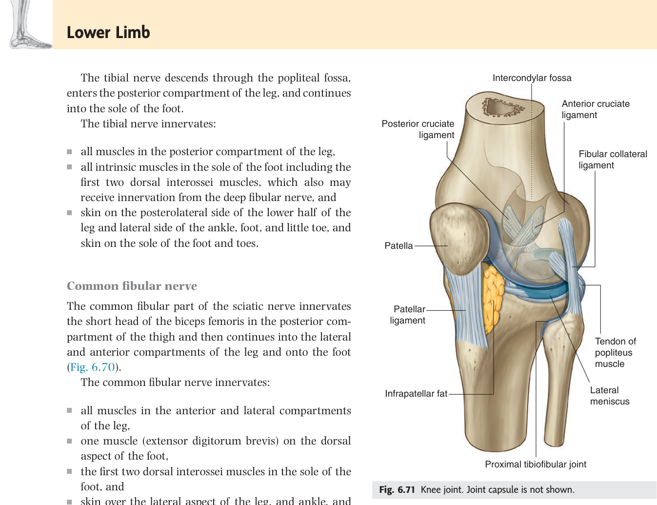
The largest and most complex joint in the body. A modified hinge joint comprising three articulations in one capsule: medial tibiofemoral, lateral tibiofemoral, and patellofemoral.
| Structure | Details |
|---|---|
| Menisci | Medial (C-shaped, firmly attached to MCL — more often injured) · Lateral (more circular/O-shaped, loosely attached — more mobile) |
| MCL | Medial (tibial) collateral ligament; broad flat band; attached to medial meniscus; resists valgus stress |
| LCL | Lateral (fibular) collateral ligament; cord-like; NOT attached to lateral meniscus; resists varus stress |
| ACL | Anterior cruciate ligament; lateral femoral condyle → anterior tibial eminence; prevents anterior tibial slide on femur; tested by Lachman's + anterior drawer |
| PCL | Posterior cruciate ligament; medial femoral condyle → posterior tibial eminence; prevents posterior tibial slide; stronger than ACL |
| Locking mechanism | Terminal extension: femur laterally rotates on tibia ("screw home"); popliteus unlocks for flexion |
Valgus stress + rotation (e.g., American football clip injury): ACL + MCL + medial meniscus injured together. MCL and medial meniscus are both attached — so MCL injury pulls the medial meniscus with it. Presents with immediate haemarthrosis (ACL + vessel), valgus laxity (MCL), joint line tenderness + McMurray's test (medial meniscus). Treat: MCL heals conservatively; ACL reconstruction with hamstring/patellar tendon graft.
| Condition | Details |
|---|---|
| Prepatellar bursitis | "Housemaid's knee" — bursa anterior to the patella, between patella and skin. Occupation: prolonged kneeling (carpet layers, gardeners). Tender fluctuant swelling directly over the patella. Risk of infection → aspirate + culture. Septic bursitis: Staphylococcus aureus most common. |
| Infrapatellar bursitis | "Clergyman's knee" — bursa between patellar ligament and tibia; kneeling upright (prayer position). Pain below the patella over the tibial tuberosity area. |
| Baker's (popliteal) cyst | Distension of the semimembranosus-gastrocnemius bursa in the popliteal fossa. Always associated with intra-articular pathology (RA, OA, meniscal tear). Joint fluid communicates one-way through a valve-like mechanism. Mass in popliteal fossa, transilluminates. Rupture → sudden severe calf pain + swelling mimicking DVT (ultrasonography distinguishes). Treat the underlying joint disease. |
| Osgood-Schlatter disease | Apophysitis of the tibial tuberosity. Boys 10–15 years (active); traction stress from quadriceps via patellar ligament at tibial tuberosity growth plate. Painful bony prominence; worsened by running/jumping. X-ray: fragmentation of tibial tuberosity. Resolves with skeletal maturity; treat with activity modification + physiotherapy. |
| Patellar dislocation | Usually lateral (lateral pull of quadriceps vector + shallow trochlear groove). Young females, valgus knee, patella alta. Medial retinaculum + MPFL (medial patellofemoral ligament) tear. Spontaneous reduction common. Recurrent dislocation → MPFL reconstruction or tibial tuberosity transfer. |
Hinge (ginglymus) synovial joint. The body of the talus sits in a mortice formed by the medial malleolus (tibia) + lateral malleolus (fibula) + inferior articular surface of the tibia.
| Ligament | Components | Mechanism of Injury |
|---|---|---|
| Lateral ligament | ATFL (anterior talofibular) · CFL (calcaneofibular) · PTFL (posterior talofibular, strongest) | Inversion + plantarflexion; ATFL torn first (~85% of sprains) |
| Deltoid ligament | Fan-shaped; very strong; superficial + deep parts; connects medial malleolus to talus/calcaneus/navicular | Eversion injury (rare alone); often fracture of lateral malleolus instead |
| Stability | Most stable in dorsiflexion (wider anterior talus wedged in mortice). Most vulnerable in plantarflexion (narrower posterior talus = loose mortice) | |
85% are lateral (inversion injury). ATFL is torn first, then CFL. Ottawa ankle rules: X-ray if bony tenderness at posterior 6 cm of fibula/tibia OR inability to weight-bear. Pott's fracture = trimalleolar fracture (medial + lateral + posterior malleolus). The lateral malleolus extends ~1 cm further distally than the medial malleolus — important X-ray landmark.
| Structure | Details |
|---|---|
| Subtalar (talocalcaneal) joint | Between inferior talus and superior calcaneus. Responsible for inversion and eversion of the hindfoot — this does NOT occur at the talocrural joint. Three articular facets connected by interosseous talocalcaneal ligament (sinus tarsi). Post-calcaneal fracture subtalar arthritis → subtalar arthrodesis. |
| Tibiofibular syndesmosis | Distal fibula anchored to tibia by anterior + posterior tibiofibular ligaments + interosseous membrane. "High ankle sprain" — tender 2–3 cm above mortice; squeeze test / external rotation stress positive. Wide mortice on X-ray (tibiofibular gap >5 mm; tibiofibular overlap <10 mm on AP). Unstable → surgical fixation. |
| Maisonneuve fracture | Proximal fibula fracture + medial ankle injury (deltoid tear or medial malleolus fracture) + syndesmotic disruption. Force transmits proximally through the interosseous membrane. Key: if medial ankle injury + wide mortice without lateral malleolus fracture → X-ray full fibula. Treat with syndesmotic screw/tightrope + fix medial side. |
- Q: Strongest ligament in the body — which joint, what does it prevent? — Iliofemoral (Y-ligament) of the hip joint; prevents hyperextension of the hip. The hip is the most stable joint in the body.
- Q: Unhappy triad — which three structures and what mechanism? — ACL + MCL + medial meniscus; valgus force + external rotation (e.g., football clip). MCL attaches to the medial meniscus, so MCL injury drags the meniscus with it.
- Q: ACL vs PCL — which prevents anterior tibial slide? — ACL. Tested by Lachman's test and anterior drawer. PCL (stronger) prevents posterior tibial slide — tested by posterior drawer.
- Q: Ankle stability — when is the mortice most and least stable? — Most stable in dorsiflexion (wider anterior talus wedged in mortice); least stable in plantarflexion (narrow posterior dome loose → injury risk).
- Q: Which ankle ligament tears first in an inversion sprain? — ATFL (anterior talofibular ligament) — most anterior, weakest of the three lateral ligaments. Next = CFL; PTFL is the strongest and rarely torn.
Ready to test yourself on Unit 03?
Practice MCQs, True/False statements, and essay questions — timed and scored.