Special Senses
General Sensory Receptor Principles
- A stimulus produces a graded receptor (generator) potential; if it reaches threshold it triggers APs in the afferent.
- Four coded attributes: modality (labelled-line / adequate stimulus), intensity (AP frequency + number of receptors — Weber–Fechner law), location (receptive field), duration.
- Adaptation: tonic receptors (slow-adapting, e.g. pain, proprioception) signal continuously; phasic receptors (fast-adapting, e.g. touch/Pacinian) signal change.
Vision — Optics & Accommodation
The eye is two things: a precision camera and a piece of the brain that pokes out through the orbit. The camera part has to focus light from across the room and from a book held 30 cm away onto the same retinal layer, while letting in the right amount of light. The biological brain part — the retina — then has to convert that light into the electrical signals the cortex can read. Almost every clinical problem in ophthalmology is a failure at one of these two halves: cataracts cloud the lens (camera failure); glaucoma damages the optic nerve fibres (data-transmission failure); macular degeneration kills photoreceptors (brain failure).
The eye is a 3-layer sphere ~24 mm in diameter:
- Outer (fibrous): opaque sclera + transparent cornea (avascular, supplied by aqueous humour & tears).
- Middle (vascular = uvea): choroid (nutritive, pigmented), ciliary body (secretes aqueous humour, contains ciliary muscle & zonules), iris (contains the pupillary sphincter and dilator).
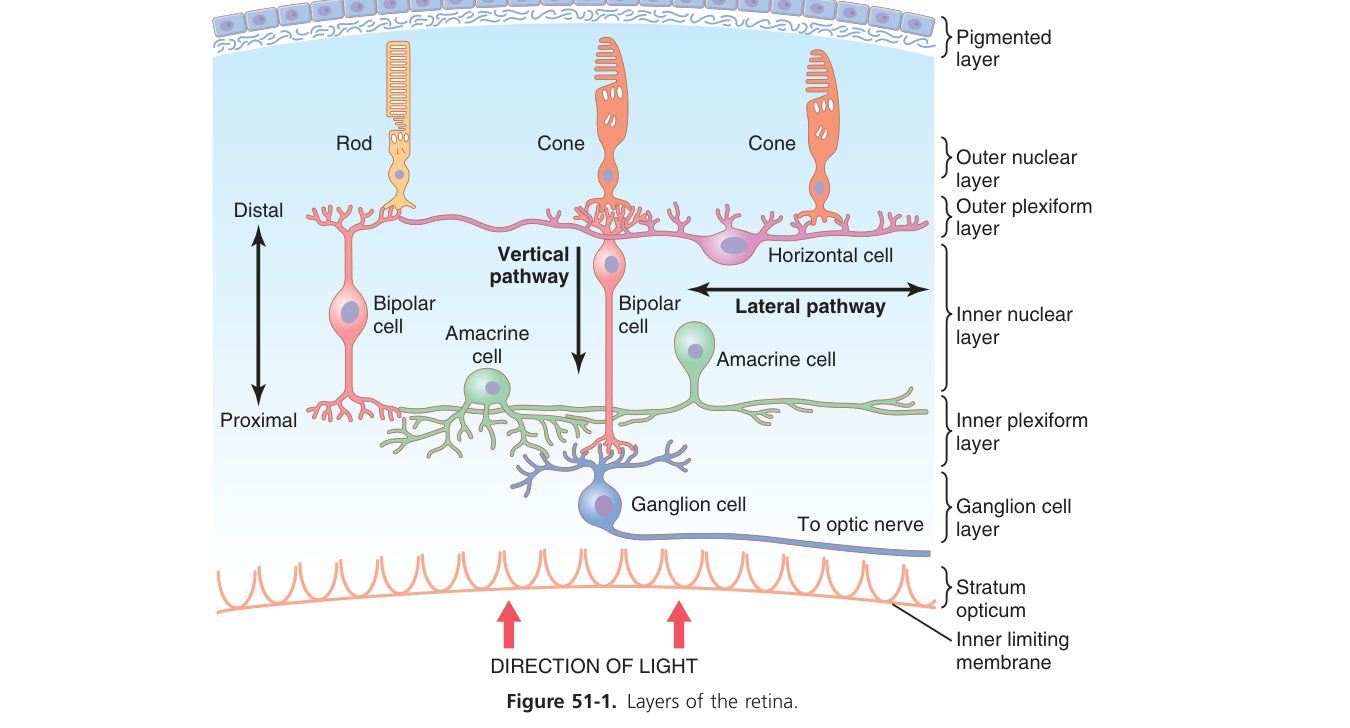
- Inner (neural): retina — 10 layers, with photoreceptors closest to the pigment epithelium (light must cross the inner layers first — the "inverted" retina). Fovea = thinnest retina, pure cones, highest acuity. Optic disc = exit of optic nerve = blind spot (no photoreceptors).
The aqueous humour is produced by the ciliary body → flows through the pupil → drains via the trabecular meshwork → canal of Schlemm. Obstruction → raised intra-ocular pressure (glaucoma).
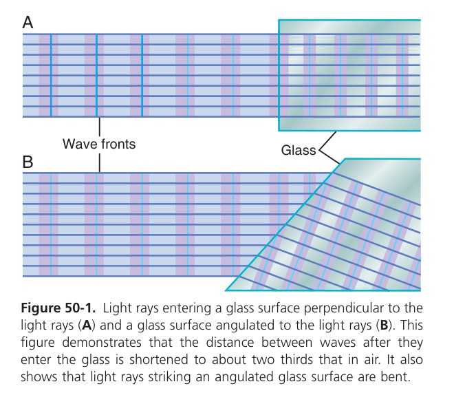
Light is refracted mostly by the cornea (~40 D, fixed) and finely focused by the lens (~20 D, variable) onto the retina. Total eye refractive power ~59 D in the relaxed (far-vision) state.
For a near object: parasympathetic (CN III, Edinger–Westphal) stimulation → ciliary muscle contracts → suspensory zonules slacken → the elastic lens becomes more rounded (↑refractive power). For a far object: ciliary relaxes → zonules pulled taut → lens flattens. The near reflex triad = accommodation + convergence + pupillary constriction (miosis).
Light in either eye → CN II → pretectal nucleus → bilateral Edinger–Westphal nuclei → CN III → pupillary sphincter (M₃) → miosis in both eyes (direct + consensual response).
| Error | Image falls | Cause | Correction |
|---|---|---|---|
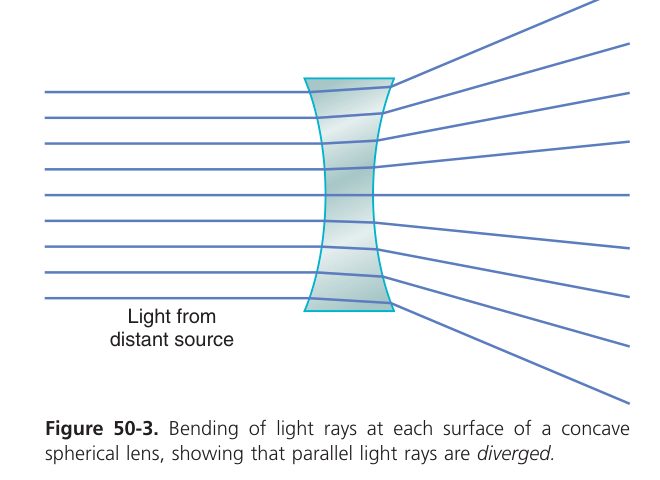
| Myopia (near-sighted) | In front of retina | Eyeball too long / too much power | Concave (diverging) lens |
| Hyperopia (far-sighted) | Behind retina | Eyeball too short / too little power | Convex (converging) lens |
| Astigmatism | Unequal focus | Irregular corneal curvature | Cylindrical lens |
| Presbyopia | Near blur with age | Lens loses elasticity (↓accommodation) | Convex reading lens |
Vision — Phototransduction & the Retina
The retina has two completely different photoreceptor types because the brain needs to see in vastly different lighting conditions, from starlight to bright sun. Rods are exquisitely sensitive (a single photon can fire one), made for dim light — but they don’t distinguish colour and they’re too sluggish for fast scenes. They’re scattered across the peripheral retina; that’s why you see faint stars best by looking slightly to the side. Cones are insensitive (need ~100 photons) but fast, sharp, and come in three colour types (red, green, blue). They’re packed into the central fovea — the only spot of true high-acuity vision; this is why you cannot read peripheral text without moving your eyes. At the optic disc, where the optic nerve exits, there are no photoreceptors at all — that’s your blind spot, which the brain fills in seamlessly so you never notice.
| Feature | Rods | Cones |
|---|---|---|
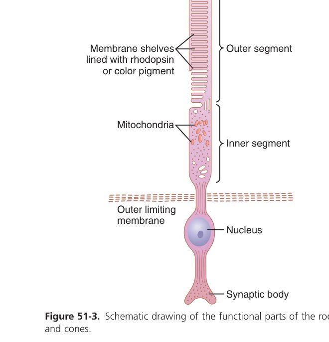
| Pigment | Rhodopsin | 3 opsins (red/green/blue) |
| Function | Dim light (scotopic), no colour | Bright light (photopic), colour |
| Sensitivity / acuity | High sensitivity, low acuity | Low sensitivity, high acuity |
| Location | Peripheral retina | Concentrated at the fovea |
Photoreceptors are unusual: they hyperpolarise in response to light.
- In darkness: high cGMP keeps Na⁺ channels open (the "dark current") → the cell is depolarised and steadily releases glutamate.
- In light: photon → rhodopsin (opsin + 11-cis retinal) isomerises retinal to all-trans → activates transducin (G-protein) → phosphodiesterase → ↓cGMP → Na⁺ channels close → cell hyperpolarises → less glutamate released → signal to bipolar & ganglion cells.
- Dark adaptation (entering a dark cinema): bright-light bleaching has emptied retinal stores; in dark, rhodopsin regenerates from 11-cis retinal & opsin → sensitivity rises ~10,000-fold over ~30 min. Cones recover in 5–7 min, rods over 20–30 min.
- Light adaptation (stepping into sunlight): rapid bleaching of rhodopsin + pupillary constriction + shift from rod to cone vision → sensitivity drops in seconds.
- Vitamin A (retinol) is the precursor of retinal — deficiency impairs rhodopsin regeneration → night blindness (nyctalopia), then xerophthalmia.
The visual pathway is the most clinically useful piece of anatomy in neurology, because lesions at each level produce a characteristic field defect that localises the problem. Light from the right visual field hits the left half of each retina; signals leave the eye through the optic nerve, the two nerves meet at the optic chiasm where nasal fibres (carrying signals from temporal field) cross, then continue as the optic tract to the lateral geniculate nucleus of the thalamus, optic radiation, and primary visual cortex (V1) in the occipital lobe. Each step has a signature lesion: blind one eye? optic nerve. Bitemporal hemianopia (lose the outer halves of both fields)? chiasm — classic pituitary tumour. Right homonymous hemianopia (lose right half of both fields)? left optic tract or beyond.
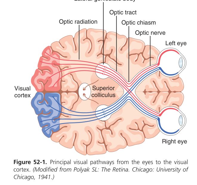
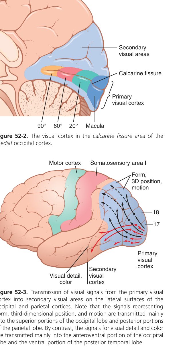
Retina → optic nerve → optic chiasm (nasal-retina fibres cross) → optic tract → lateral geniculate nucleus (LGN, thalamus) → optic radiation → primary visual cortex (V1, occipital lobe, calcarine sulcus).
Image inversion rules: the temporal half of the visual field projects to the nasal retina (which crosses); the nasal half projects to the temporal retina (uncrossed). Right visual field → left hemisphere.
| Lesion site | Field defect | Classic cause |
|---|---|---|
| Optic nerve | Ipsilateral monocular blindness | Optic neuritis, trauma |
| Optic chiasm (centre) | Bitemporal hemianopia | Pituitary adenoma, craniopharyngioma |
| Optic tract | Contralateral homonymous hemianopia | Vascular |
| Meyer loop (temporal) | Contralateral superior quadrantanopia ("pie in the sky") | Temporal lesion |
| Parietal radiation | Contralateral inferior quadrantanopia ("pie on the floor") | Parietal lesion |
| Visual cortex (PCA) | Contralateral homonymous hemianopia with macular sparing | Posterior cerebral artery stroke (occipital pole receives MCA collateral) |
Cones contain one of three opsins maximally absorbing at ~437 nm (blue / S), ~533 nm (green / M), ~564 nm (red / L) — Young–Helmholtz trichromacy. Downstream the signal is recoded into opponent channels (red-green, blue-yellow, light-dark; Hering).
Red-green colour blindness = X-linked cone-opsin defect (commoner in males). Protanopia = loss of red cones; deuteranopia = green cones. Total colour blindness (achromatopsia) is rare. Cataract = lens opacity (ageing). Glaucoma = raised IOP → optic-disc cupping. Diabetic retinopathy & macular degeneration are leading causes of acquired blindness.
Hearing — Sound Conduction
Sound = longitudinal pressure waves in air. Frequency (Hz) is perceived as pitch; amplitude (dB SPL) as loudness. Human hearing range ~20–20,000 Hz, most sensitive at 1–4 kHz (speech). The decibel scale is logarithmic: each 10 dB = 10× intensity; whisper ~30 dB, conversation ~60 dB, traffic ~80 dB, jet ~120 dB (pain threshold).
Sound is a pressure wave in air, and the ear’s problem is converting it into electrical signals despite a fundamental impedance mismatch — air is light, fluid is heavy. If you sent sound straight from air to fluid, ~99.9% of the energy would reflect back. The middle ear solves this with two clever tricks. First, the tympanic membrane is large (~85 mm²) and the oval window is small (~3 mm²); pressure (force/area) is amplified ~17-fold. Second, the three ossicles work as a lever giving another ~1.3-fold gain. Result: ~22-fold (~25 dB) pressure gain, exactly what’s needed to drive cochlear fluid efficiently. Lose the middle ear (otosclerosis, otitis media) and you get conductive deafness. Damage the cochlea or auditory nerve and you get sensorineural deafness. The Rinne and Weber tests at the bedside discriminate the two in seconds.
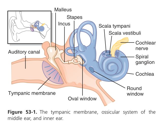
- Outer ear (pinna + external auditory canal) funnels & resonates sound to the tympanic membrane, which vibrates.
- Middle ear ossicles — malleus → incus → stapes — transmit vibration from tympanum to the oval window. Tensor tympani (CN V) and stapedius (CN VII) reflexively dampen ossicles for loud sounds.
- Eustachian tube equalises middle-ear pressure with atmosphere (opens on yawn/swallow).
- Impedance matching: sound passing air → fluid would normally lose ~99.9% of energy. Two mechanisms compensate: the area ratio of tympanum:oval window (~17:1) and the lever action of the ossicles (~1.3:1), giving a pressure gain of ~22-fold (~25 dB).
Hearing — Cochlear Transduction
The cochlea is a snail-shaped, fluid-filled spiral that does pitch discrimination by physical position. The basilar membrane — the floor of the organ of Corti — runs the full length, but it’s narrow and stiff at the base, wide and floppy at the apex. High-pitched sounds resonate at the base; low-pitched at the apex; in between, in between — the famous tonotopic map (G. von Békésy got the Nobel Prize for it). Sitting on the basilar membrane are the hair cells, whose stereocilia bend when basilar membrane moves; that bending opens mechanically-gated channels, K⁺ flows in from the K⁺-rich endolymph, the hair cell depolarises, glutamate is released onto the spiral ganglion, the auditory nerve fires. From there: cochlear nuclei → superior olive (where bilateral comparison lets you localise sound in space) → inferior colliculus → medial geniculate → auditory cortex.
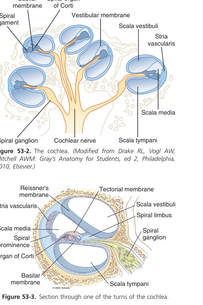
The cochlea is a coiled fluid-filled tube (~2.5 turns) divided by membranes into three scalae:
- Scala vestibuli (perilymph, high Na⁺) — receives oval-window input.
- Scala media (cochlear duct) — contains endolymph (uniquely high K⁺, low Na⁺, secreted by the stria vascularis; produces a +80 mV endocochlear potential).
- Scala tympani (perilymph) — ends at the round window.
The scala media sits on the basilar membrane, which carries the organ of Corti: one row of inner hair cells (IHC) (~3,500, the sensory transducers, drive 95% of CN VIII afferents) and three rows of outer hair cells (OHC) (~12,000, the cochlear amplifier — electromotile via prestin).
- Oval-window push → travelling wave propagates along the basilar membrane.
- Basilar membrane displaces → hair-cell stereocilia bend against the overlying tectorial membrane.
- Bending in the excitatory direction opens tip-link mechanically-gated K⁺ channels → K⁺ enters from the high-K⁺ endolymph (down the +80 mV gradient).
- Hair cell depolarises → voltage-gated Ca²⁺ opens at the basolateral surface → releases glutamate → afferent AP in spiral ganglion → CN VIII.
- OHCs use prestin to shorten/lengthen with each cycle, mechanically amplifying basilar motion ~100-fold (the cochlear amplifier).
Tonotopic (place) coding: the base (narrow, stiff) responds to high frequencies; the apex (wide, floppy) to low frequencies — so pitch maps to position along the basilar membrane (von Békésy travelling wave). Low frequencies also use volley/temporal coding (phase locking of afferents).
Auditory pathway: hair cell → spiral ganglion → CN VIII → cochlear nuclei → superior olivary complex (bilateral — sound localisation) → lateral lemniscus → inferior colliculus → medial geniculate nucleus (thalamus) → primary auditory cortex (Heschl gyrus, BA 41/42). Because of bilateral relay, unilateral cortical lesions cause minimal deafness.
| Type | Site | Rinne (air vs bone) | Weber (lateralisation) |
|---|---|---|---|
| Conductive | Outer/middle ear (wax, otitis media, otosclerosis) | BC > AC in affected ear (Rinne negative) | Lateralises to affected (deaf) ear |
| Sensorineural | Cochlea or CN VIII (noise, presbycusis, aminoglycosides) | AC > BC in affected ear (Rinne positive but reduced) | Lateralises to normal (better) ear |
Presbycusis (age-related): bilateral high-frequency sensorineural loss (basal hair cells fail first). Noise-induced hearing loss: classic 4-kHz notch on audiogram. Aminoglycosides (gentamicin) and cisplatin damage OHCs. Acoustic neuroma (vestibular schwannoma) compresses CN VIII at the cerebellopontine angle.
Vestibular System (Balance)
- Semicircular canals (3, mutually perpendicular — horizontal, anterior, posterior) detect angular (rotational) acceleration: head rotation makes endolymph lag and bend the cupula → deflects hair-cell stereocilia.
- Otolith organs — utricle (horizontal acceleration, head tilt) & saccule (vertical acceleration). Hair-cell stereocilia are embedded in an otolith-laden otolithic membrane; gravity / acceleration shears it across hair cells.
- Excitation = stereocilia bent toward the kinocilium → depolarisation → ↑CN VIII firing; bending away → hyperpolarisation → ↓firing. Resting discharge is tonic, so direction can be encoded by ↑/↓ from baseline.
- Vestibulo-ocular reflex (VOR): head turn left → eyes move right at equal speed → image stays fixed on retina. Pathway: SCC → vestibular nuclei → CN III/IV/VI nuclei.
- Vestibular nystagmus: rotation produces slow eye drift opposite head movement, then fast resetting saccade in the direction of rotation. The fast component names the nystagmus direction.
- Vestibulospinal reflexes: maintain posture against gravity.
BPPV (benign paroxysmal positional vertigo) — otoliths dislodged into a semicircular canal → brief positional vertigo, treated by Epley repositioning. Ménière disease — endolymphatic hydrops → vertigo + sensorineural hearing loss + tinnitus + ear fullness. Motion sickness — sensory mismatch (vestibular says moving, eyes say still).
Taste & Smell (Chemosensation)
Taste is a chemical sense built on five basic modalities — sweet, sour, salty, bitter, umami — each handled by different receptor mechanisms in taste-bud cells on the tongue. Salty senses Na⁺ via direct entry through ENaC channels; sour senses H⁺ via channel block; sweet, bitter, umami all use G-protein-coupled receptors. The taste information funnels through three cranial nerves (VII anterior 2/3, IX posterior 1/3, X for epiglottis) to the nucleus of the solitary tract, then thalamus, then insular cortex. Most of what we call “flavour” is actually smell — the volatiles released from food in the mouth that travel back to the olfactory epithelium. That’s why food tastes bland when you have a cold.
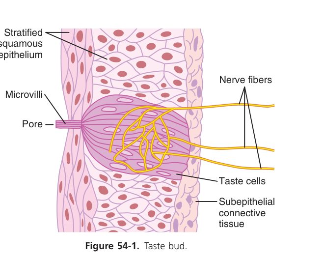
Five basic tastes: sweet, sour, salty, bitter, umami. Taste buds (~10,000) sit in fungiform (anterior tongue), foliate & circumvallate (posterior) papillae — not on filiform.
| Taste | Stimulus | Transduction |
|---|---|---|
| Salty | Na⁺ | Na⁺ entry via ENaC → depolarise |
| Sour | H⁺ | H⁺ blocks K⁺ channels (± PKD2L1) → depolarise |
| Sweet / Umami / Bitter | Sugars / amino acids (glutamate) / alkaloids & toxins | GPCR (T1R / T2R) → PLC → IP₃ → Ca²⁺ → transmitter |
Afferents: anterior 2/3 of tongue via chorda tympani of CN VII; posterior 1/3 via CN IX; epiglottis via CN X. All converge on the nucleus of the solitary tract → thalamus (VPM) → gustatory cortex (insula).
Olfaction is unique among senses because it bypasses the thalamus entirely. Odorant molecules dissolve in mucus on the olfactory epithelium high in the nose, bind G-protein-coupled receptors on olfactory receptor neurons (there are ~400 receptor types — we can discriminate ~10,000 odours by combinatorial coding), and the signal goes straight to the olfactory bulb and on to the piriform cortex and limbic system. This is the anatomical basis for smell’s extraordinary power over memory and emotion — the “Proustian” rush of a forgotten time triggered by a familiar smell happens because the pathway lands in the hippocampus and amygdala before conscious awareness ever sees it.
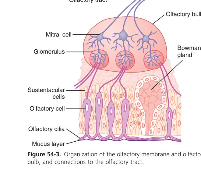
- Olfactory receptor neurons in the olfactory epithelium of the upper nasal cavity express ~400 G-protein-coupled odorant receptors. Each neuron expresses one receptor type; activation → Gₒₒₙ → adenylyl cyclase → cAMP → opens CNG channels → Na⁺/Ca²⁺ influx → depolarise.
- Axons traverse the cribriform plate → synapse in olfactory bulb glomeruli → mitral cells → olfactory tract → piriform & entorhinal cortex (the only sensory pathway that does not obligately relay in the thalamus).
- Olfactory neurons are among the few neurons that regenerate from basal stem cells throughout life.
Anosmia = loss of smell — head trauma can shear olfactory axons at the cribriform plate; URTIs (incl. COVID-19) may damage olfactory epithelium; early anosmia can herald Parkinson or Alzheimer disease. Loss of smell impairs flavour perception (taste seems lost).
Special senses complete
Vision, hearing, vestibular & chemosensation mastered. Next: Metabolism & Body Temperature.