Metabolism & Body Temperature
Energy Metabolism & the Respiratory Quotient
The whole reason you breathe and the whole reason you eat is to make ATP. Every cellular process that requires energy — muscle contraction, neuronal firing, active transport, protein synthesis — ultimately runs on ATP, and ATP is produced by oxidising fuel. The body keeps three fuel stores ready: glycogen (small, short-term, mostly in liver and muscle, ~400 g total), fat (massive, long-term, ~15 kg in an adult of normal weight, providing energy for weeks of starvation), and protein (mostly muscle, dipped into only in extreme deprivation). The interesting thing about fat is its energy density: at 9.3 kcal/g it carries more than twice the energy per gram of carbohydrate or protein. That is why evolution chose fat as the body’s main long-term energy store — you couldn’t carry around the equivalent weight of glycogen.
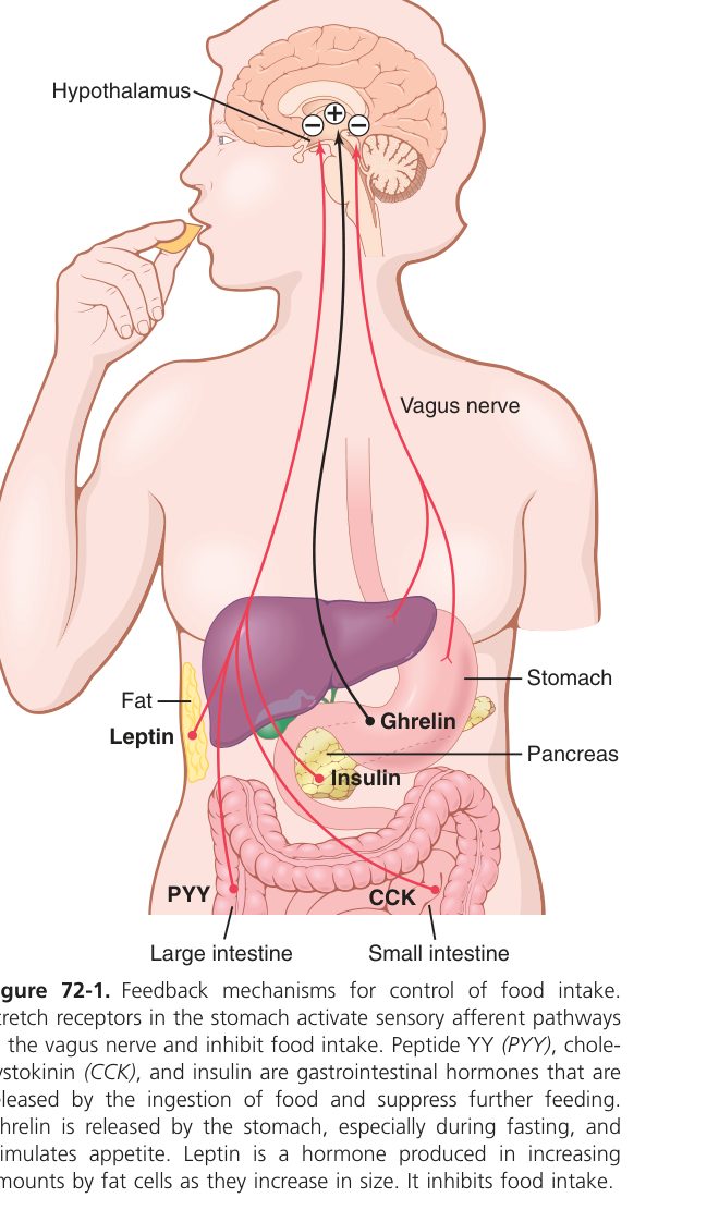
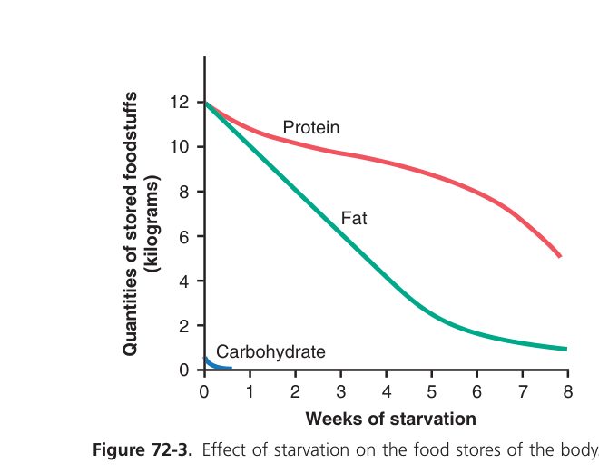
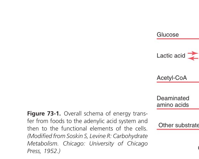
Energy metabolism = the release, storage, transfer and utilisation of energy that accompanies the chemical reactions of metabolism. Metabolic rate = the amount of energy liberated per unit time.
- Immediate fuel = glucose/ATP; the main long-term energy store of the body is FAT (energy-dense, 9.3 kcal/g) — glycogen stores are small.
- Caloric values: carbohydrate & protein ~4.1 kcal/g; fat ~9.3 kcal/g.
Respiratory quotient (RQ) = CO₂ produced ÷ O₂ consumed during metabolism of a foodstuff.
| Foodstuff | RQ |
|---|---|
| Carbohydrate | 1.00 |
| Protein | 0.80 |
| Fat | 0.70 |
| Mixed diet | ~0.85 |
Specific dynamic action = the extra heat produced after eating, as the body processes the food. It is highest for protein (~30%), much less for carbohydrate & fat.
Basal Metabolic Rate (BMR)
Even when you do absolutely nothing — lie still, don’t talk, don’t digest, don’t shiver — your body still burns about 1500-1800 kcal per day, just keeping itself alive: heart pumping, lungs breathing, brain working, ion gradients maintained, body temperature held at 37°C. This irreducible minimum is the basal metabolic rate (BMR), and it’s a useful clinical fingerprint of your overall metabolic state. Because BMR depends on body size, it’s reported per square metre of body surface area (~40 kcal/m²/h in a young man). The main determinant of BMR is thyroid hormone — raise it and BMR climbs (hyperthyroidism: hot, sweaty, restless, weight loss despite eating more); lower it and BMR falls (hypothyroidism: cold, tired, slow, weight gain despite eating less). BMR is also pushed up by catecholamines (fever, exercise), and is naturally higher in children (growth) and lower in elderly.
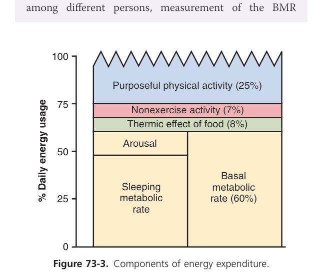
Basal metabolic rate (BMR) = the metabolic rate (energy expenditure per unit time) measured under standard basal conditions — the minimum energy to keep the body alive at rest.
Basal conditions required:
- Awake but physically & mentally at rest, lying down (not standing).
- Post-absorptive — 12–14 hours after the last meal (no SDA).
- Thermoneutral environment (~20–25°C, quiet).
- Normal body temperature.
BMR is normally expressed per unit body surface area (kcal/m²/h or kJ/m²/h) rather than per kg, because heat exchange & metabolism scale with surface area. Adult ♂ ~40 kcal/m²/h, ♀ ~37; a 1.7 m² man ≈ 1600–1800 kcal/day.
| Factor | Effect |
|---|---|
| Surface area | BMR ∝ body surface area (the most reliable normaliser) |
| Sex | ♂ > ♀ by ~5–10% (more lean mass, less fat) |
| Age | Highest in infancy/childhood (growth); falls in adulthood |
| Thyroid hormone | The main hormonal determinant; ↑in hyperthyroidism, ↓in hypothyroidism (BMR is a classic test) |
| Catecholamines, GH, testosterone | Raise BMR |
| Body temperature (fever) | ↑ ~13% per °C rise |
| Environmental temperature | BMR rises in cold (people in cold climates ~higher BMR) |
| Sleep / starvation | Lower BMR (energy conservation) |
| Pregnancy / lactation | Raise BMR |
- Direct calorimetry: measures the heat lost by a subject in an insulated chamber. Accurate but cumbersome — rarely used clinically.
- Indirect calorimetry: measures O₂ consumption (and CO₂ production) and converts using the energy equivalent of O₂ (~4.825 kcal/L O₂ on a mixed diet, depending on RQ). Most common.
- The Benedict–Roth spirometer (closed-circuit) and modern open-circuit metabolic carts both use indirect calorimetry.
Body Temperature
A swimmer pulled from icy water has a hypothermic skin but his organs are still close to 37°C — for a while. That’s the core-shell distinction. The body protects the temperature of the core (brain, thorax, abdomen) ferociously, because enzymes work in a narrow temperature window and severe cooling or heating denatures them. The shell (skin, limbs) is allowed to wander — it acts as a buffer between the harsh outside world and the protected core. In the cold, the shell cools first (you go pale, fingers numb) before the core is touched; in the heat, the shell sweats and vasodilates so heat can leave before the core climbs. Rectal temperature most closely matches core; oral is ~0.5°C lower; axillary lower still. Core temperature has a daily rhythm: lowest at ~4 am, highest at ~6 pm, ~1°C swing.
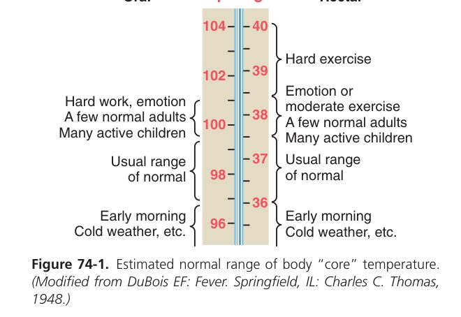
- Core temperature (deep organs — brain, thorax, abdomen) is kept nearly constant ~37°C; this is the clinically relevant "body temperature".
- Shell temperature (skin, limbs) varies with the environment and acts as a heat buffer/radiator.
- Measurement sites: rectal (~37.1–37.5°C, closest to core) > oral (~37°C) > axillary (~36.5°C).
Heat Production & Heat Loss
- At rest: the liver (and other viscera) is the main heat source.
- In cold / during activity: skeletal-muscle shivering is the chief way to increase heat production.
- Also: hormones (thyroxine, adrenaline → non-shivering thermogenesis), and the specific dynamic action of food.
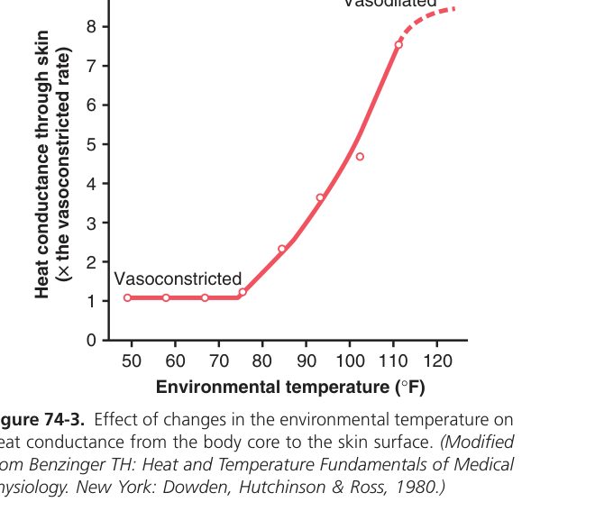
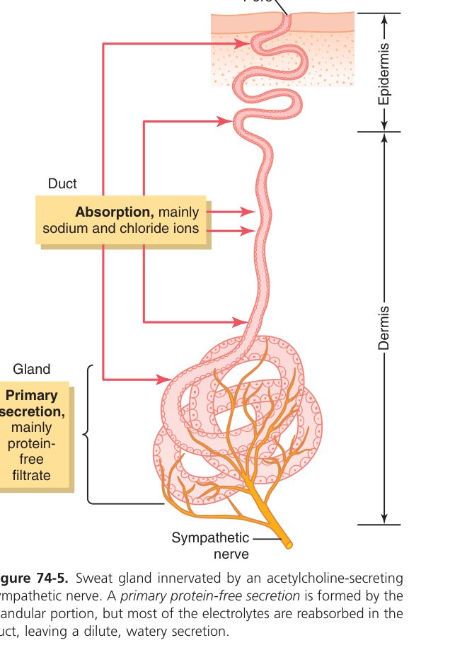
Heat leaves the body by four routes — radiation (~60% at rest, infrared given off to cooler surroundings), conduction (small, by direct contact), convection (heat carried away by moving air or water; rises with wind), and evaporation (sweat). Three of these depend on a temperature gradient between skin and environment, which means when the room is hotter than your skin (~33°C), they go into reverse and you start gaining heat. At that point evaporation becomes the only route left — which is why sweating is your last defence against hyperthermia. A litre of sweat that fully evaporates carries away ~580 kcal, equivalent to about a third of your daily BMR. The catch: that evaporation only happens if the air is dry enough to accept it. In high humidity (e.g. tropical climates) sweat drips off without evaporating, and core temperature climbs — heat stroke risk soars.
| Route | Mechanism | Note |
|---|---|---|
| Radiation | Infrared emission to cooler surroundings | Main route at rest in a cool room |
| Conduction | Direct contact with cooler objects | Small |
| Convection | Heat carried away by moving air/water | Increases with wind |
| Evaporation (sweat) | Latent heat of vaporisation of water | The ONLY route when ambient temp ≥ skin temp (hot environment) |
Thermoregulation & Fever
The body keeps temperature within ~0.5°C of 37°C because the preoptic area of the hypothalamus works as a thermostat. Neurons there directly sense blood temperature and compare it with an internal set point. If body temp climbs above 37°C, the hypothalamus dispatches heat-loss orders: skin vasodilation, sweating, decreased heat production. If body temp falls below 37°C, the opposite happens: cutaneous vasoconstriction (you go pale), piloerection (goosebumps, useful in furred mammals), shivering (large skeletal muscle contractions that produce heat as a byproduct), and behavioural changes (huddle, put on a jumper). Fever is not a broken thermostat — it’s a thermostat reset to a higher set point by pyrogens (mainly prostaglandin E₂ in the hypothalamus, made in response to inflammatory cytokines like IL-1 and IL-6). The body then defends the new (higher) temperature exactly as if it were normal — you feel cold and shiver as it climbs. NSAIDs (aspirin, paracetamol) block PGE₂ synthesis, resetting the thermostat back to 37°C.
The thermoregulatory centre sits in the preoptic area of the (anterior) hypothalamus — neurons there respond directly to blood temperature and compare it with a set point (~37°C), driving negative feedback:
| Situation | Response |
|---|---|
| Body temp > set point (too hot) | ↑ heat loss (skin vasodilation, sweating) + ↓ heat production |
| Body temp < set point (too cold) | ↓ heat loss (vasoconstriction, piloerection) + ↑ heat production (shivering) |
Pyrogens (from infection/inflammation, via prostaglandin E₂) raise the hypothalamic set point above 37°C. Now the actual body temperature is below the new set point, so the body behaves as if cold:
- Onset (chill phase): vasoconstriction + shivering → heat is generated & conserved → temperature climbs to the new (higher) set point — the patient feels cold despite a rising fever.
- Plateau: temperature held at the elevated set point.
- Defervescence (crisis): when the set point falls back to normal (recovery or antipyretic), body temp is now above it → vasodilation + sweating → temperature falls.
| Condition | Mechanism | Signs |
|---|---|---|
| Hyperthermia / heat stroke (>40°C) | Heat gain exceeds loss; set point is normal but the system is overwhelmed (hot environment, exertion, drugs, poor sweating) | Hot dry skin, confusion, collapse; multi-organ failure if untreated. Treat with rapid external cooling. |
| Malignant hyperthermia | RyR mutation triggered by anaesthetics → runaway muscle Ca²⁺ release | Rigidity, fever, acidosis — antidote dantrolene |
| Neuroleptic malignant syndrome | Anti-dopaminergics raise the set point + rigidity | Fever + rigidity + altered consciousness + autonomic instability |
| Hypothermia (<35°C) | Heat loss exceeds gain (immersion, exposure, the elderly) | Shivering → ceases <30°C; bradycardia, J-wave on ECG, depressed consciousness |
- To heat: over days/weeks → sweat begins at a lower core temperature, sweat volume rises (up to 2 L/h), and sweat Na⁺ content falls (aldosterone) → salt conserved.
- To cold: increased non-shivering thermogenesis (brown fat in infants; thyroid up-regulation in adults), peripheral vasoconstriction, lower thermal comfort temperature.
- To altitude (covered in Unit 5): hyperventilation, ↑2,3-DPG, ↑EPO → polycythaemia, ↑capillary density.
Antipyretics (aspirin, paracetamol) inhibit hypothalamic prostaglandin synthesis → reset the elevated set point — effective in fever but useless in heat stroke (where the set point is already normal). For heat stroke, cool the patient externally.
Carbohydrate, Fat & Protein Metabolism
- Glycolysis — cytosolic; 1 glucose → 2 pyruvate + 2 ATP + 2 NADH (anaerobic: pyruvate → lactate). Rate-limited by phosphofructokinase-1 (inhibited by ATP/citrate, activated by AMP/F2,6-BP).
- TCA cycle & oxidative phosphorylation — mitochondrial; complete oxidation yields ~30–32 ATP per glucose.
- Glycogenesis (liver, muscle) stores glucose as glycogen (insulin-driven). Glycogenolysis (glucagon, adrenaline) releases it. Liver glycogen ~100 g, muscle ~400 g.
- Gluconeogenesis (liver, renal cortex) makes glucose from lactate, glycerol & gluconeogenic amino acids in starvation. Driven by glucagon, cortisol.
Normal fasting plasma glucose ~70–100 mg/dL (3.9–5.6 mmol/L). Brain & RBCs depend almost exclusively on glucose; the brain uses ~120 g/day.
- Lipolysis in adipose → free fatty acids (FFA) + glycerol; activated by adrenaline, glucagon, cortisol; inhibited by insulin.
- β-oxidation (mitochondrial) yields ~106 ATP per palmitate (16C) — fat is the densest fuel.
- Ketogenesis (liver, starvation / DKA): acetyl-CoA → acetoacetate + β-hydroxybutyrate → brain & muscle fuel.
- Cholesterol made in liver (HMG-CoA reductase, statin target) → bile acids, steroid hormones, membrane component.
- Lipoproteins: chylomicrons (gut → tissue TG), VLDL (liver → tissue TG), LDL ("bad" — cholesterol to tissues), HDL ("good" — reverse cholesterol transport).
- Amino acids: 20 standard, 9 essential (cannot be synthesised — PVT TIM HALL: Phe, Val, Thr, Trp, Ile, Met, His, Arg*, Leu, Lys).
- Transamination & deamination → carbon skeleton (glucogenic / ketogenic) + NH₃.
- Urea cycle (liver): NH₃ + CO₂ → urea (excreted by kidney) — the body’s detoxification of nitrogen. Failure (liver failure, urea-cycle defects) → hyperammonaemia → encephalopathy.
- Nitrogen balance: positive (growth, pregnancy, recovery) vs negative (starvation, trauma, sepsis, glucocorticoids).
| State | Dominant hormone | Liver | Muscle | Adipose |
|---|---|---|---|---|
| Fed (high insulin) | Insulin | Glycogenesis, lipogenesis, protein synthesis | Glucose & AA uptake; protein synthesis | TG storage (LPL activated) |
| Fasting / early (low insulin) | Glucagon | Glycogenolysis → glucose; gluconeogenesis | FFA oxidation | Lipolysis → FFA, glycerol |
| Starvation (days) | Glucagon, cortisol | Ketogenesis; gluconeogenesis from AA | Spares glucose; uses ketones | Major fuel source |
Type 1 diabetes: absolute insulin lack → unrestrained lipolysis & ketogenesis → diabetic ketoacidosis (Kussmaul breathing, fruity breath, anion-gap metabolic acidosis). Type 2: insulin resistance + relative lack → hyperglycaemia, dyslipidaemia, ↑CV risk. Insulin shifts the body to the "fed" state on every axis.
Physiology syllabus complete 🎉
All 11 TMU physiology units built to gold standard. Back to the physiology index.