Nervous System Physiology
Synaptic Transmission
Every thought, sensation and movement is built from one elementary event: one neuron firing at another across a synapse. You have about 86 billion neurons and roughly 100 trillion synapses, but the molecular logic of each individual contact is the same. When the AP arrives at the presynaptic terminal, voltage-gated Ca²⁺ channels open, calcium rushes in, synaptic vesicles fuse with the membrane, and neurotransmitter spills into the cleft. The transmitter diffuses across the cleft, binds receptors on the postsynaptic side, and either depolarises the next cell (EPSP, excitatory) or hyperpolarises it (IPSP, inhibitory). The output neuron sums hundreds or thousands of these signals every millisecond at its axon hillock; if the net depolarisation crosses threshold there, it fires its own AP. That is the entire computational basis of the brain — in/out, sum, decide, fire. Repeated 86 billion times in parallel.
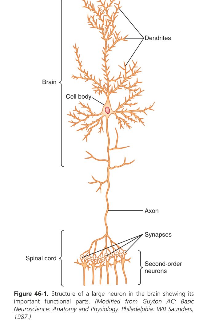
A synapse is the specialised contact between two neurons (or between a neuron and an effector). Synapses are classified in three useful ways:
| Basis | Types |
|---|---|
| Anatomical contact | Axodendritic (commonest, often excitatory) · Axosomatic (on the cell body, often powerful/inhibitory) · Axoaxonic (modulates transmitter release — pre-synaptic inhibition) |
| Function | Excitatory (depolarising) vs Inhibitory (hyperpolarising) |
| Transmission | Chemical (transmitter, >99% in CNS) vs Electrical (gap junctions; bidirectional, ~no delay; cardiac muscle, retina, embryonic CNS) |
- Pre-synaptic membrane: bears active zones with voltage-gated Ca²⁺ channels; the terminal is packed with mitochondria (ATP supply) and synaptic vesicles (~40 nm) holding transmitter quanta.
- Synaptic cleft: 20–40 nm gap containing extracellular matrix & sometimes degrading enzymes (e.g. AChE).
- Post-synaptic membrane: dense with receptor proteins (ionotropic or metabotropic) coupled to ion channels.
- Pre-synaptic AP arrives → depolarises terminal → voltage-gated Ca²⁺ channels open.
- Ca²⁺ influx triggers SNARE-mediated fusion of vesicles → quantal release of transmitter by exocytosis.
- Transmitter diffuses across the cleft → binds post-synaptic receptors.
- Receptor opening allows ion flux → EPSP or IPSP.
- Transmitter action ends by reuptake (e.g. NE, dopamine), enzymatic degradation (ACh by AChE), or diffusion.
| Potential | Ion movement | Typical transmitter | Effect |
|---|---|---|---|
| EPSP | Na⁺ influx (±Ca²⁺) | Glutamate, ACh (nicotinic) | Depolarises → toward threshold |
| IPSP | Cl⁻ influx / K⁺ efflux | GABA (brain), glycine (cord) | Hyperpolarises → away from threshold |
The axon initial segment has the lowest threshold (highest density of voltage-gated Na⁺ channels). The neuron sums all EPSPs − IPSPs reaching the hillock; if the net depolarisation reaches threshold there, an AP is fired down the axon. Inputs near the hillock (axosomatic) carry more weight than distal dendritic inputs.
- One-way conduction (Dale’s principle): transmitter is released only from the pre-synaptic side → signal flows pre → post.
- Synaptic delay (~0.5 ms): the time for Ca²⁺ entry → vesicle fusion → transmitter binding. Counting synapses in a pathway estimates delay.
- Summation: temporal (one input fires rapidly) and spatial (many inputs fire together).
- After-discharge: output continues after stimulus stops, because of reverberating circuits.
- Synaptic fatigue: sustained high-frequency input depletes transmitter → response wanes (basis of post-tetanic depression; protective against seizures).
- Sensitivity to environment: hypoxia, acidosis, alkalosis, drugs (anaesthetics, caffeine, strychnine) strongly modulate transmission; synapses are far more vulnerable than axons.
Strychnine blocks glycine receptors → loss of spinal inhibition → convulsions. Tetanus toxin blocks release of glycine/GABA from Renshaw inhibitory interneurons → spastic paralysis (“lockjaw”). Benzodiazepines potentiate GABA-A → sedation. Caffeine blocks adenosine receptors → CNS arousal.
Reflexes & the Spinal Cord
A reflex is a stereotyped involuntary response to a stimulus, mediated by a 5-element reflex arc:
- Receptor → (2) afferent (sensory) neuron → (3) centre (cord or brainstem) → (4) efferent (motor) neuron → (5) effector (muscle / gland).
Reflexes may be monosynaptic (one synapse in the centre — stretch reflex) or polysynaptic (multiple interneurons — flexor / withdrawal reflex).
| Receptor | Arrangement | Senses | Afferent | Reflex action |
|---|---|---|---|---|
| Muscle spindle (intrafusal fibres) | Parallel with extrafusal fibres | Length & rate of length change | Ia (primary, fast), II (secondary) | Stretch → excite α-motor neuron of same muscle (myotatic) & inhibit antagonist |
| Golgi tendon organ | In series at the musculotendinous junction | Tension | Ib | Excess tension → inhibit α-motor neuron → protective relaxation (inverse stretch / clasp-knife) |
The spindle is reset by γ-motor neurons that contract intrafusal fibres — keeping the spindle sensitive at any muscle length (α–γ coactivation).
| Reflex | Receptor / afferent | Synapses | Feature / clinical use |
|---|---|---|---|
| Stretch (myotatic) — knee jerk, biceps jerk, ankle jerk | Muscle spindle (Ia) | Monosynaptic | Tests integrity of one cord segment + peripheral nerve |
| Inverse stretch | Golgi tendon organ (Ib) | Disynaptic (inhibitory interneuron) | Protects against tendon rupture; basis of clasp-knife rigidity |
| Flexor (withdrawal) | Cutaneous nociceptors | Polysynaptic, multi-segmental | Limb withdrawal from painful stimulus |
| Crossed-extensor | Couples to flexor reflex | Polysynaptic, contralateral | Extends opposite limb to bear weight as the painful limb flexes |
| Babinski | Plantar cutaneous | Polysynaptic | Up-going great toe = UMN lesion (normal >2 y = down-going) |
Reciprocal inhibition: when an agonist is reflexly excited, the antagonist is simultaneously inhibited (Ia inhibitory interneuron), so movement is smooth.
Complete spinal cord transection produces three sequential phases:
- Spinal shock (days–weeks): flaccid paralysis, areflexia, atonic bladder, loss of sensation below the level — due to abrupt loss of descending facilitation.
- Recovery phase (weeks): reflexes return progressively (anal → tendon → flexor → crossed-extensor).
- Chronic phase: hyper-reflexia, spasticity, positive Babinski, mass reflexes — pure UMN syndrome.
Reflexes localise lesions: absent reflexes = LMN/sensory lesion; exaggerated reflexes + Babinski = UMN lesion. The Brown–Séquard syndrome (hemisection) loses ipsilateral motor + DCML and contralateral spinothalamic below the lesion (because spinothalamic crosses at entry, DCML crosses in medulla).
Sensory Pathways & Pain
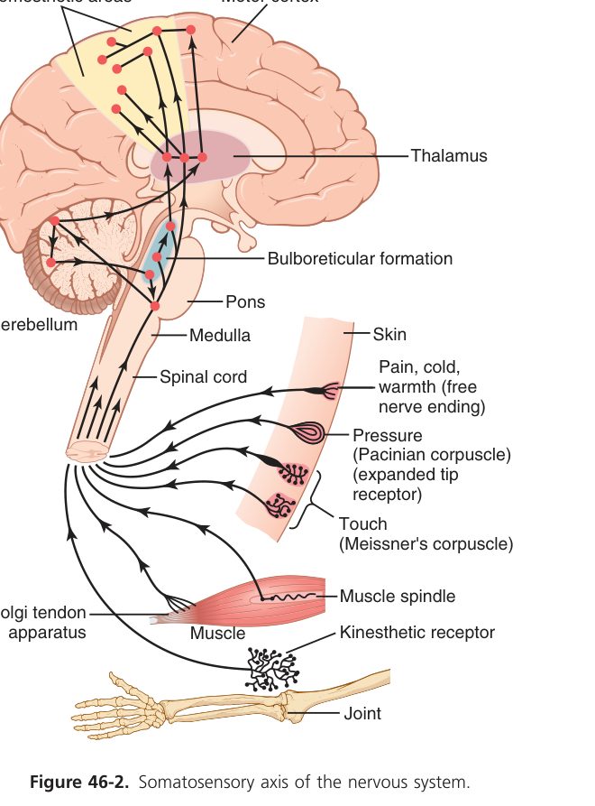
Touch a kettle, hear a violin, see the page in front of you — every sensation begins at a receptor that converts a physical stimulus into a nerve impulse. This step is called transduction, and the rest of the sensory system is just relay and decoding. Each receptor has an adequate stimulus — the type of energy it’s tuned to (light for photoreceptors, pressure for mechanoreceptors, chemical for nociceptors, etc.) — and a receptive field — the patch of body or world it samples. Two strong stimuli on adjacent fingertips feel separate because they activate different receptive fields; that’s the basis of two-point discrimination, the densest on the fingertip and lips (small receptive fields), the loosest on the back (large fields).
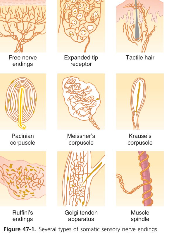
A sensory receptor converts a physical/chemical stimulus into a neural signal (transduction). Each receptor responds best to its adequate stimulus; an inadequate stimulus needs a much larger energy to fire.
| Class | Adequate stimulus | Example |
|---|---|---|
| Mechanoreceptor | Pressure / deformation | Pacinian corpuscle, muscle spindle |
| Thermoreceptor | Temperature (cold or warm) | Free nerve endings, TRPM8/TRPV1 |
| Nociceptor | Tissue damage | Free nerve endings (Aδ, C) |
| Chemoreceptor | Chemical (O₂, CO₂, taste, smell) | Carotid body, olfactory cells |
| Photoreceptor | Light (Unit 10) | Rods & cones |
Stimulus → receptor potential (graded) → if it reaches threshold in the first node of Ranvier → action potentials whose frequency encodes stimulus intensity.
| Type | Behaviour | Example |
|---|---|---|
| Rapidly adapting (phasic) | Fires at stimulus onset/offset; reports change | Pacinian (vibration), Meissner |
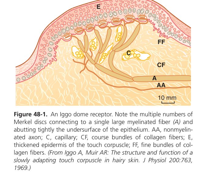
| Slowly adapting (tonic) | Continues firing throughout stimulus; reports steady state | Muscle spindle, Merkel disc, nociceptor |
Adaptation lets us ignore continuous unimportant stimuli (clothing on skin) but still detect new or changing inputs.
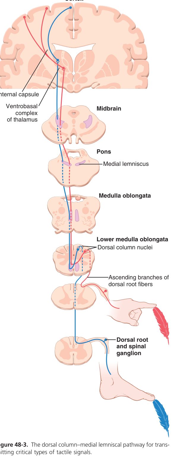
| Feature | Dorsal column–medial lemniscus (DCML) | Anterolateral / spinothalamic |
|---|---|---|
| Carries | Fine touch, vibration, conscious proprioception, 2-point discrimination | Pain, temperature, crude touch |
| 1st-order neuron | DRG → ascends ipsilaterally in dorsal column | DRG → synapses in dorsal horn at entry level |
| Where it crosses | Medulla (internal arcuate fibres → medial lemniscus) | Spinal cord (anterior white commissure within 1–2 segments) |
| Thalamic relay | VPL nucleus | VPL nucleus |
| Cortex | Postcentral gyrus (S1, somatotopic) | Postcentral gyrus (S1) |
| Fibre type | Large myelinated Aβ, fast | Small Aδ & C, slower |
DCML = “Discrimination Climbs, then Crosses” — ascends ipsilateral first, crosses in medulla. Spinothalamic = “Pain Crosses fast, then Climbs” — crosses at entry, then ascends contralaterally.
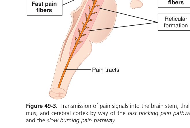
Pain is unique among sensations — it’s the one we’d most like to switch off, yet without it, life is dangerous (think leprosy, where lost pain leads to unnoticed injuries that destroy hands and feet). Pain receptors are nociceptors, bare nerve endings activated by tissue damage. They come in two functional flavours: fast Aδ fibres (thinly myelinated, sharp first pain) and slow C fibres (unmyelinated, dull burning second pain). They synapse in the dorsal horn and ascend in the contralateral spinothalamic tract to thalamus and cortex. The brain doesn’t just receive pain — it modifies it. Descending fibres from periaqueductal grey can release endogenous opioids (enkephalins) in the dorsal horn, blunting pain transmission. This is why distraction reduces pain, why stress can mask serious injury, and why opioids work where they do.
| Type | Fibre | Quality | Transmitter |
|---|---|---|---|
| Fast (first) pain | Aδ myelinated, ~10–30 m/s | Sharp, pricking, well-localised | Glutamate |
| Slow (second) pain | C unmyelinated, ~0.5–2 m/s | Dull, burning, aching, poorly localised | Substance P, glutamate |
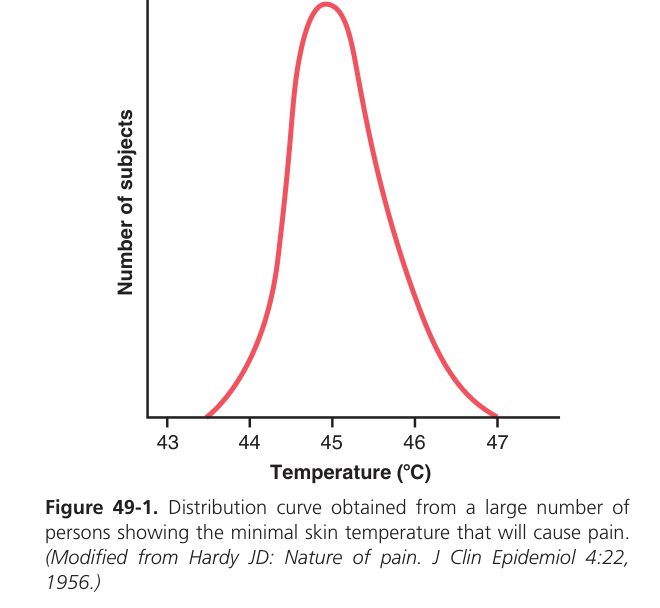
Receptors are polymodal free nerve endings activated by TRPV1 (heat / capsaicin / acid >43°C) and TRPM8 (cold / menthol). Tissue damage releases K⁺, H⁺, prostaglandins, bradykinin, histamine, serotonin — these sensitise nociceptors (hyperalgesia).
- Gate control theory (Melzack & Wall): non-painful Aβ input excites inhibitory interneurons in the dorsal horn → "closes the gate" on C-fibre pain transmission. Rubbing or TENS works by this mechanism.
- Endogenous analgesia: the periaqueductal grey & raphe magnus send serotonergic & enkephalinergic fibres that inhibit dorsal-horn pain neurons. Opioids work here.
- Referred pain: visceral pain is felt in a somatic dermatome that shares the spinal segment (cardiac → left arm/jaw via T1–T4; diaphragm → shoulder C3–C5).
Syringomyelia (central canal cyst) damages crossing spinothalamic fibres → “cape” loss of pain & temperature with preserved touch / proprioception (DCML spared). Tabes dorsalis (neurosyphilis) damages dorsal columns → sensory ataxia, positive Romberg, loss of vibration & proprioception.
Motor Pathways — UMN vs LMN
The primary motor cortex (M1) is a strip running across the back of the frontal lobe, just in front of the central sulcus. Its layout is famously somatotopic — the body is mapped onto it like a distorted little man, the motor homunculus. Body parts that need fine motor control get massive cortical real estate: hands, lips, tongue, face. Body parts moved as a whole (trunk, thigh) get tiny representations. Stimulate a spot on M1 (Penfield’s open-brain experiments showed this in conscious patients) and the corresponding muscle group twitches. A stroke that knocks out the “hand area” leaves the patient with weak hand but spared face; a stroke that knocks out the “face area” does the opposite. Knowing the map predicts the deficit.
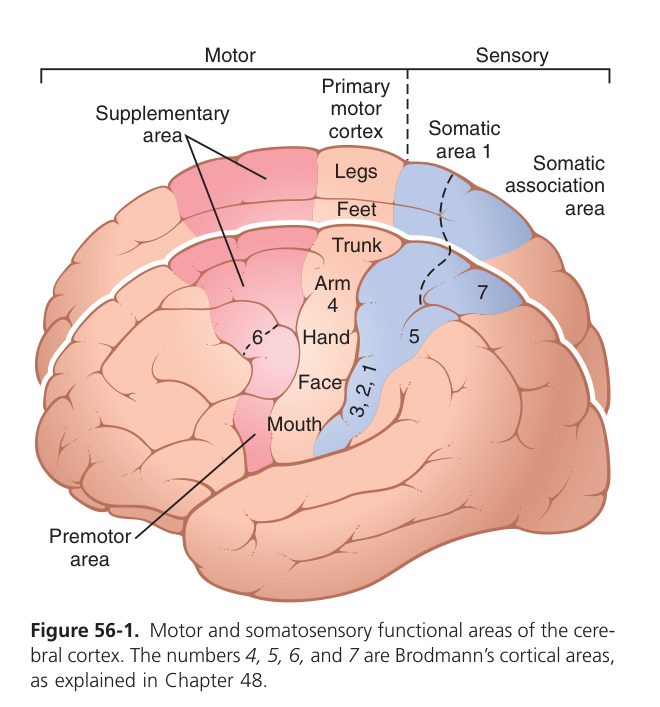
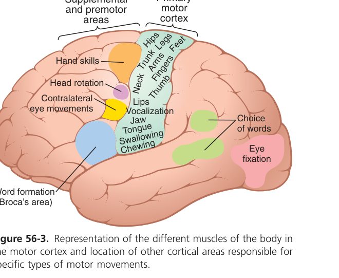
The primary motor cortex (M1, precentral gyrus, Brodmann 4) contains a somatotopic map — the motor homunculus. Body parts requiring fine control (hand, face, lips, tongue) occupy disproportionately large areas. Lateral parts of M1 control the face/hand; medial parts (paracentral lobule) control the leg.
- Premotor cortex (BA 6): plans externally-cued movement & postural set.
- Supplementary motor area (SMA, medial BA 6): plans internally-generated, sequential movement.
- Broca area (BA 44/45, dominant hemisphere): motor planning of speech.
The corticospinal tract carries voluntary motor commands directly from cortex to spinal motor neurons.
- Cell bodies in motor cortex (giant Betz cells in layer V of M1) → axons descend through the posterior limb of the internal capsule → cerebral peduncle → pons → medulla.
- In the lower medulla ~85% decussate in the medullary pyramids → form the lateral corticospinal tract (controls contralateral distal limb muscles).
- ~15% remain ipsilateral as the anterior corticospinal tract → cross at the cord segment of synapse (controls axial / proximal muscles bilaterally).
- Synapses on α-motor neurons in the ventral horn — directly or via interneurons.
The corticobulbar tract targets cranial-nerve motor nuclei. Most are bilaterally innervated; CN VII lower face & CN XII are contralateral only → UMN facial palsy spares forehead, LMN (Bell’s) affects whole half-face.
| Tract | Origin | Role |
|---|---|---|
| Rubrospinal | Red nucleus | Excites contralateral flexors (limb fine motion) |
| Reticulospinal | Pontine + medullary reticular formation | Posture, automatic gait, tone (pontine = extensor +; medullary = extensor −) |
| Vestibulospinal | Vestibular nuclei | Excites antigravity extensors for balance |
| Tectospinal | Superior colliculus | Head & eye orientation toward stimulus |
| Sign | Upper motor neuron (UMN) | Lower motor neuron (LMN) |
|---|---|---|
| Tone | ↑ (spastic) — clasp-knife | ↓ (flaccid) |
| Reflexes | Hyper-reflexia, clonus | Hypo-/areflexia |
| Babinski | Positive (up-going toe) | Negative (down-going) |
| Atrophy | Minimal (disuse only, late) | Marked, early |
| Fasciculations | Absent | Present (denervation) |
| Pattern | Whole limb / hemiplegia | Specific muscle / nerve / root |
Stroke (capsular / cortical) → contralateral UMN signs. Poliomyelitis & ALS (LMN component) → LMN signs. ALS is unique — UMN and LMN signs together (no sensory loss). Decerebrate rigidity (brainstem lesion below red nucleus): extensor posture from unopposed pontine reticulo- + vestibulospinal drive.
Cerebellum & Basal Ganglia
The cerebellum is the brain’s coach — it doesn’t initiate movement, it makes movements smooth. Every time the motor cortex sends a command down to muscle, a copy goes to the cerebellum (corollary discharge). The cerebellum simultaneously gets the sensory feedback — what the muscles actually did. It compares intended with actual and corrects the next command in real time, all without conscious awareness. Lose the cerebellum and movements still happen, but they overshoot, undershoot, wobble, and lack timing — ataxia. Importantly cerebellar signs are ipsilateral (right cerebellum → right-sided ataxia) because cerebellar output crosses to the contralateral cortex, which then crosses back via the corticospinal tract — two crosses cancel out. It also stores procedural motor memory: how to ride a bike, throw a ball, type without looking.
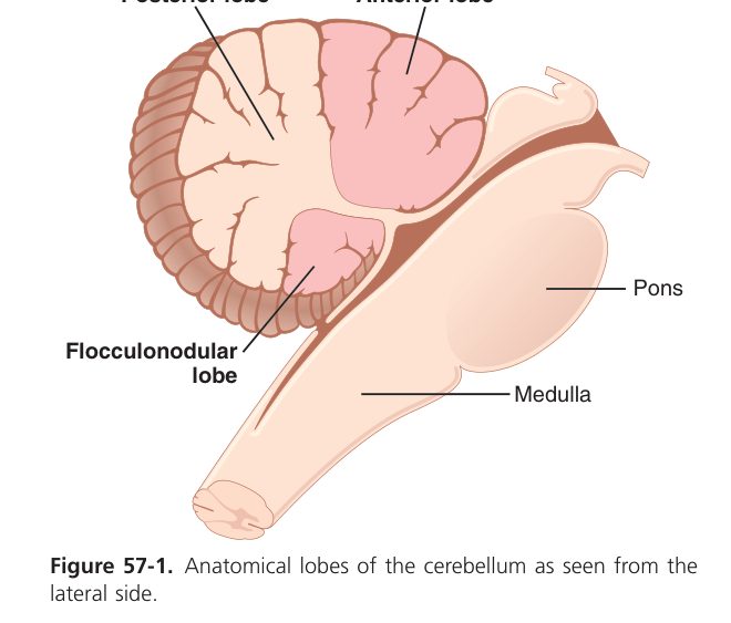
The cerebellum does not initiate movement; it compares the intended movement (cortex) with the actual movement (proprioception) and corrects timing, smoothness & force in real time.
| Lobe | Other name | Input | Function | Lesion sign |
|---|---|---|---|---|
| Vestibulocerebellum | Flocculonodular | Vestibular | Balance, eye movement | Truncal ataxia, nystagmus |
| Spinocerebellum | Vermis + paravermal | Spinal proprioception | Posture, gait, tone | Gait / truncal ataxia (vermis); limb dysmetria (intermediate) |
| Cerebrocerebellum | Lateral hemisphere | Cortex via pons | Planning, timing, motor learning | Intention tremor, dysdiadochokinesia |
Inputs arrive via mossy fibres (from pons, cord, vestibular nuclei) → granule cells → parallel fibres to Purkinje cells, and climbing fibres (from inferior olive) directly onto Purkinje cells. Purkinje cells are the sole output of the cerebellar cortex — and they are inhibitory (GABA), projecting to the deep cerebellar nuclei (dentate, interposed, fastigial). Outputs leave via the superior cerebellar peduncle → thalamus → motor cortex.
Ataxia (incoordination), intention tremor (worse as target approached), dysmetria (past-pointing), dysdiadochokinesia (poor rapid alternating movement), nystagmus, scanning dysarthria, and a broad-based ataxic gait — all on the same side as the lesion (because cerebellar output crosses to the cortex which then crosses again to act on the body).
The basal ganglia comprise the caudate, putamen, globus pallidus (internal & external), substantia nigra (pars compacta = dopamine; pars reticulata) and subthalamic nucleus. Striatum = caudate + putamen. They modulate movement via two opposing pathways:
| Pathway | Sequence | Net effect | Dopamine action |
|---|---|---|---|
| Direct (facilitatory) | Cortex → striatum → (inhibits) GPi → thalamus → cortex | ↑ movement | D₁ receptor — excites direct pathway |
| Indirect (inhibitory) | Cortex → striatum → GPe → STN → GPi → thalamus → cortex | ↓ movement | D₂ receptor — inhibits indirect pathway |
Net: dopamine from substantia nigra facilitates movement by both excitation of direct and inhibition of indirect pathways. Loss of dopamine → hypokinesia.
Parkinson disease — loss of dopaminergic neurons in the substantia nigra pars compacta → both pathways shift to inhibition of movement → resting "pill-rolling" tremor, cogwheel rigidity, bradykinesia, postural instability (TRAP). Treated with L-DOPA. Huntington disease — loss of striatal GABAergic neurons of indirect pathway → chorea. Hemiballismus — lesion of subthalamic nucleus (often lacunar stroke) → violent flinging movements of the opposite limb. Wilson disease — Cu deposition in lentiform nucleus → tremor, dystonia, dysarthria.
The Autonomic Nervous System
| Feature | Sympathetic | Parasympathetic |
|---|---|---|
| Origin | Thoracolumbar (T1–L2) | Craniosacral (CN III, VII, IX, X; S2–S4) |
| Ganglia / fibres | Near cord; short pre-, long post-ganglionic | Near organ; long pre-, short post-ganglionic |
| Post-ganglionic transmitter | Noradrenaline (except sweat glands = ACh) | Acetylcholine |
| Target receptor | Adrenergic α / β | Muscarinic |
| Overall role | "Fight or flight" (↑HR, pupils dilate, bronchodilate, ↓gut) | "Rest & digest" (↓HR, pupils constrict, ↑gut) |
All autonomic ganglia & the NMJ use ACh on nicotinic receptors. The adrenal medulla is a modified sympathetic ganglion — pre-ganglionic ACh makes it release adrenaline/noradrenaline into the blood.
Ganglia & sweat = ACh; everything else sympathetic = NE. Nicotinic = all ganglia + NMJ (fast, ionotropic); Muscarinic = parasympathetic targets (GPCR). Sympathetic = ThoracoLumbar; Parasympathetic = CranioSacral.
| Receptor | 2nd messenger | Key tissue effect |
|---|---|---|
| α₁ | Gₙ → IP₃/DAG → ↑Ca²⁺ | Vasoconstriction, pupil dilation (mydriasis), GI/urinary sphincters contract |
| α₂ | Gᵢ → ↓cAMP | Pre-synaptic auto-inhibition of NE release; ↓insulin release |
| β₁ | Gₛ → ↑cAMP | Heart — ↑HR (chronotropy), ↑contractility (inotropy), ↑conduction; renin release (kidney) |
| β₂ | Gₛ → ↑cAMP | Bronchodilation, vasodilation in skeletal muscle, uterine relaxation, ↑glycogenolysis |
| β₃ | Gₛ → ↑cAMP | Lipolysis in brown/white fat |
| M₁ | Gₙ → IP₃/DAG | CNS, gastric glands |
| M₂ | Gᵢ → ↓cAMP, K⁺ channel open | Heart — ↓HR (SA node), ↓AV conduction |
| M₃ | Gₙ → IP₃/DAG | Smooth muscle contraction (bronchi, GI, bladder detrusor), glandular secretion, pupillary constriction (miosis) |
| N (nicotinic) | Ligand-gated Na⁺ channel | All ganglia (Nn), NMJ (Nm) — fast EPSP |
"α₁ squeezes, β₁ speeds, β₂ relaxes (lungs/vessels)". M₂ in the heart is the only Gi-coupled brake. Atropine (M-blocker) → tachycardia, dry mouth, mydriasis, urinary retention — the classic "dry as a bone, blind as a bat, red as a beet, mad as a hatter".
| Organ | Sympathetic effect (receptor) | Parasympathetic effect (receptor) |
|---|---|---|
| Heart rate | ↑ (β₁) | ↓ (M₂) |
| Bronchi | Dilate (β₂) | Constrict (M₃) |
| GI motility | ↓ (α, β₂) | ↑ (M₃) |
| Bladder detrusor | Relax (β₂/β₃) | Contract (M₃) → voiding |
| Bladder sphincter | Contract (α₁) | Relax |
| Pupil | Dilate — mydriasis (α₁) | Constrict — miosis (M₃) |
| Skin vessels | Constrict (α₁) | None |
| Sweat glands | Secrete (M) — sympathetic cholinergic exception | None |
| Adrenal medulla | Releases adrenaline (Nn pre-ganglionic) | None |
Horner syndrome (sympathetic chain lesion) — ptosis, miosis, anhidrosis on the affected side. Atropine (muscarinic block) → tachycardia + dry secretions. β-blockers (propranolol, atenolol) → slow HR, lower BP. Organophosphate poisoning (AChE inhibitor) → SLUDGE-M: salivation, lacrimation, urination, defecation, GI cramps, emesis, miosis — treat with atropine + pralidoxime.
Higher Cortex, EEG & Sleep
| Area | Brodmann | Function | Lesion |
|---|---|---|---|
| Primary motor (M1) | 4 | Voluntary movement, contralateral homunculus | Contralateral spastic hemiparesis |
| Primary somatosensory (S1) | 3,1,2 | Touch / vibration / proprioception map | Contralateral cortical sensory loss |
| Primary visual (V1) | 17 | Sight | Contralateral homonymous hemianopia (macular sparing) |
| Primary auditory | 41,42 | Hearing | Subtle (bilateral input) |
| Broca (dominant frontal) | 44,45 | Motor speech | Non-fluent (expressive) aphasia |
| Wernicke (dominant temporal) | 22 | Comprehension | Fluent (receptive) aphasia |
| Prefrontal cortex | 9,10,11,12 | Executive function, judgement, personality | Disinhibition (Phineas Gage) |
| Hippocampus | — | Declarative memory consolidation | Anterograde amnesia |
| Wave | Frequency | State |
|---|---|---|
| β | >13 Hz | Alert, eyes-open, mental activity |
| α | 8–13 Hz | Awake, relaxed, eyes-closed (occipital) |
| θ | 4–7 Hz | Drowsy / light sleep, normal in children |
| δ | <4 Hz | Deep (slow-wave) sleep, abnormal in awake adult |
- Non-REM (NREM): N1 (theta) → N2 (sleep spindles, K-complexes) → N3 (slow-wave delta). Dominates first third of night; growth hormone peaks here.
- REM: low-voltage fast EEG (similar to waking), rapid eye movements, skeletal muscle atonia, vivid dreams, irregular HR/BP. Dominates last third; ~25% of sleep, every ~90 min.
- Wakefulness is maintained by the ascending reticular activating system (ARAS) using ACh, NE, 5-HT, histamine, orexin. Loss of orexin neurons → narcolepsy.
Sleepwalking, night terrors, enuresis occur in NREM N3. REM-sleep behaviour disorder (loss of REM atonia) — acting out dreams — predicts Parkinson / Lewy-body disease. Benzodiazepines & alcohol suppress REM & N3 → poor sleep quality.
Nervous system complete
Synapses, reflexes, pathways, cerebellum/basal ganglia, ANS & cortex/sleep mastered. Next: Special Senses.