Endocrine Physiology
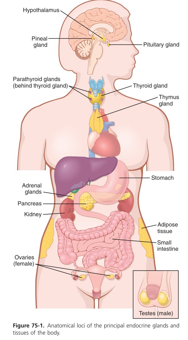
The Endocrine System & Hormones
The body has two long-distance communication systems. The nervous system is fast and precise — messages travel along wires (axons) to specific targets in milliseconds. The endocrine system is slower but vastly more diffuse — messages (hormones) are released into the blood and reach every cell in the body, but only cells with the right receptor respond. This is why endocrinology is the physiology of receptors: the same hormone causes a totally different effect in different tissues depending on which receptor is there. Insulin from one pancreatic cell tells your liver to store glucose, your muscle to take up glucose, your fat to store fat — three different effects from one signal, decoded locally.
Hormones regulate four things broadly: metabolism & energy (thyroid, insulin, glucagon, cortisol), water & electrolytes (ADH, aldosterone, PTH), growth & development (GH, thyroid, sex hormones), and reproduction (FSH, LH, oestrogen, testosterone). The same chemical (e.g. noradrenaline) can be a neurotransmitter in one place and a hormone in another — the distinction is whether it travels in blood or across a synapse.
In 1902, Bayliss & Starling showed that stimulating a dog’s intestine with HCl raised pancreatic secretion, and that injecting an extract of intestinal mucosa into a second dog reproduced the effect — the first hormone, secretin. They proposed a second regulatory system alongside the nervous system: the endocrine system.
Four functions:
- Maintains homeostasis of the internal environment.
- Facilitates growth & development.
- Regulates metabolism.
- Facilitates reproductive development & maturation; maintains reproduction.
The endocrine system is directly or indirectly controlled by the nervous system — together they form the neuro-endocrine network.
| Gland | Hormones |
|---|---|
| Pituitary (hypophysis) | 7 (GH, TSH, ACTH, FSH, LH, PRL, MSH) |
| Thyroid | 3 (T₃, T₄, calcitonin) |
| Parathyroid | 1 (PTH) |
| Adrenal glands | 4 (cortisol, aldosterone, DHEA, adrenaline/NA) |
| Islets of Langerhans | 2 (insulin, glucagon) |
| Ovary / Testis | 2 / 1 |
Plus “non-classical” sources: heart (ANP), endothelium (endothelin), thymus (thymosin), kidney (renin, EPO, calcitriol), GI tract (gastrin/CCK/secretin — Unit 6).
A hormone is a highly active chemical substance elaborated by one part of the body that controls or helps control a function elsewhere. A target cell is one that bears specific receptors for that hormone. Agonists bind receptor and trigger the effect; antagonists bind but block the agonist.
| Mode | Path | Example |
|---|---|---|
| Telecrine (endocrine) | Released into blood, acts on distant target cell | TSH, ACTH |
| Paracrine | Diffuses locally to neighbouring cells | Somatostatin in islets |
| Autocrine | Acts on the cell that secreted it | Many growth factors |
| Neurocrine | Transmitted along axon, released into blood, distant target | ADH, oxytocin (axoplasmic transport) |
| Class | Examples | Synthesis & storage |
|---|---|---|
| Peptides / proteins | Insulin, GH, ACTH, ADH | Made as prohormones in ER; cleaved in Golgi; stored in secretory vesicles. Most "nitrogenous" hormones. |
| Amines | T₃/T₄ (tyrosine), catecholamines (tyrosine), melatonin (tryptophan) | Derived from amino acids |
| Steroids | Cortisol, aldosterone, oestradiol, testosterone, progesterone | Derived from cholesterol; not stored — made on demand |
| Fatty-acid derivatives | Prostaglandins, prostacyclins (from arachidonic acid via COX) | Very rapid turnover (seconds) |
- Specificity (relative): a hormone affects only cells with its specific receptor.
- Messenger / information transmission: the hormone carries a signal from secreting cell to target cell, tissue or organ.
- Biological amplification (high efficacy): binding initiates a cascade — nanomolar hormone (T₄ 51–142 nmol/L; T₃ 1.2–3.4 nmol/L) produces large cellular effects.
- Interaction between hormones — three patterns:
- Synergistic — combined effect > sum (e.g. glucagon + adrenaline raising glucose).
- Antagonistic — one opposes the other (e.g. insulin vs glucagon).
- Permissive — hormone A has no direct effect but is required for B’s action. The TMU example: cortisol permits the vasoconstrictor action of noradrenaline.
Mechanisms of Hormone Action
Hormones change cell function in one of two ways: (1) activate a second messenger at the membrane, or (2) activate genes in the nucleus.
| Feature | Water-soluble (peptide, catecholamine) | Lipid-soluble (steroid, thyroid) |
|---|---|---|
| Receptor location | Cell membrane | Intracellular / nuclear |
| Mechanism | 2nd messenger / phosphorylation cascade | Receptor–hormone complex binds DNA → transcription → new mRNA & protein |
| Speed | Seconds–minutes | Hours (delayed but prolonged) |
| Storage in blood | Mostly free | Mostly bound to carrier proteins (TBG, albumin) |
Most water-soluble hormones (except thyroid) act on membrane receptors. Second messengers are intracellular mediators that translate the signal: cAMP, cGMP, IP₃, DAG, Ca²⁺. The receptor families:
| Family | Cascade | Example ligand |
|---|---|---|
| Gₛ-protein coupled | Receptor → Gₛ → adenylyl cyclase (AC) → cAMP → PKA → phosphorylation of target proteins | ACTH, glucagon, ADH (V₂), β-adrenergic, TSH |
| Gᵢ-protein coupled | Inhibits AC → ↓cAMP | Somatostatin, α₂-adrenergic |
| Gᶉ-protein coupled | Activates PLC → PIP₂ → IP₃ (→ Ca²⁺) + DAG (→ PKC) | TRH, GnRH, angiotensin II, α₁-adrenergic, oxytocin |
| Tyrosine-kinase receptor | Receptor dimerises & autophosphorylates → downstream cascade | Insulin, growth factors |
| Guanylyl-cyclase receptor | Generates cGMP | ANP (atrial natriuretic peptide) |
First messenger = the extracellular hormone reaching the cell. Second messenger = the intracellular molecule generated in response (cAMP, IP₃, DAG, Ca²⁺, cGMP). Effectors are protein kinases — PKA, PKC, PKG, CaMKII.
- Lipid-soluble hormone diffuses across the cell membrane.
- Binds an intracellular receptor in cytoplasm or nucleus.
- Hormone–receptor complex migrates to the nucleus and binds a hormone-response element in DNA.
- Activates transcription of specific genes → new mRNA → new proteins → cellular response.
- Down-regulation: persistently elevated hormone → receptor number falls (e.g. insulin in type 2 diabetes).
- Up-regulation: chronic hormone deficiency → receptor number rises (e.g. PRL, FSH, angiotensin II receptors).
Regulation of Hormone Secretion
- Control by a regulated variable — the controlled solute itself triggers the hormone (e.g. blood Ca²⁺ → PTH; blood glucose → insulin).
- Control by another hormone — trophic axes (CRH → ACTH → cortisol) with positive & negative feedback.
- Nervous control — via the hypothalamus, which links emotion / sensory input to endocrine output.
| Axis | Hypothalamic | Pituitary | Target gland |
|---|---|---|---|
| Thyroid axis | TRH | TSH | Thyroid → T₃ / T₄ |
| Adrenal cortex axis | CRH | ACTH | Adrenal cortex → cortisol |
| Gonadal axis | GnRH | FSH + LH | Ovary/testis → oestrogen / testosterone |
Each axis uses negative feedback: the target-gland hormone inhibits the pituitary and the hypothalamus (long loop) and the pituitary inhibits its own releasing hormone (short loop).
The Hypothalamic–Pituitary Unit
The pituitary is two glands fused in one walnut-sized lump under the brain — the anterior (adenohypophysis, true endocrine tissue) and the posterior (neurohypophysis, an extension of the brain). Their relationships with the hypothalamus are fundamentally different. The anterior pituitary receives blood from the hypothalamus via the hypophyseal portal vessels — hypothalamic releasing hormones (GnRH, TRH, CRH, GHRH, dopamine, somatostatin) reach the anterior pituitary directly without dilution in the systemic circulation, so tiny amounts can produce strong effects. The posterior pituitary receives axons — ADH and oxytocin are made by neurons in the hypothalamic supraoptic and paraventricular nuclei whose axon terminals end in the posterior pituitary; the hormones travel down the axons and are stored there until release. This is why the “posterior pituitary” isn’t really endocrine tissue at all — it’s a nerve terminal.
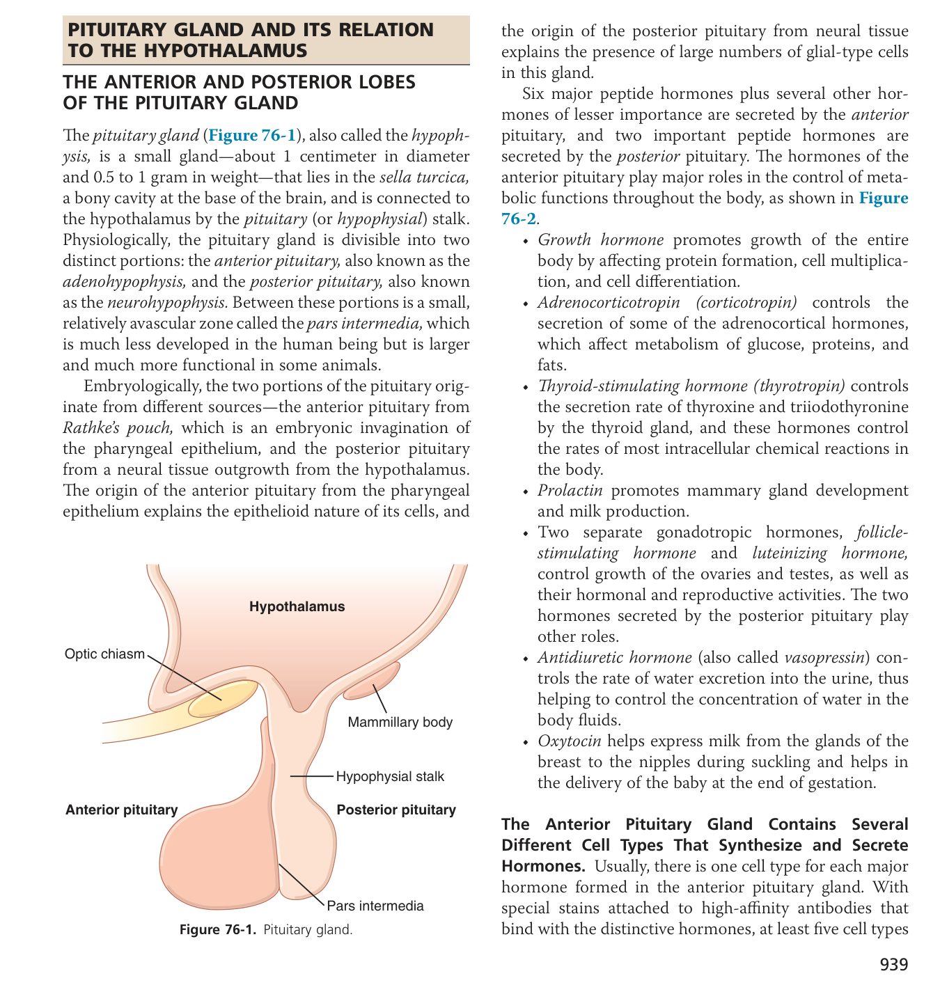
The pituitary sits in the pituitary fossa (sella turcica) beneath the brain — about the size of the tip of a little finger.
| Lobe | Tissue | Link to hypothalamus | Hormones |
|---|---|---|---|
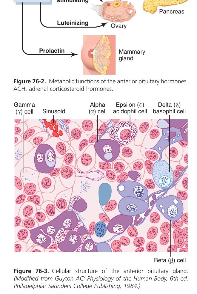
| Adenohypophysis (anterior) | Highly vascular gland tissue; 5 cell types | Hypophyseal portal system — hypothalamic peptides reach it via portal vessels | GH, TSH, ACTH, FSH, LH, PRL (+ MSH) |
| Neurohypophysis (posterior) | Glial-like cells + axon terminals | Hypothalamo-hypophyseal tract — direct axons from supraoptic & paraventricular nuclei bring hormones by axoplasmic transport | ADH (vasopressin) & oxytocin (stored, then released) |
Secreted by peptidergic neurons of the hypophysiotropic area (medial basal hypothalamus) into the portal blood. Modulated by NE, DA & 5-HT.
| Peptide (TMU list) | Length | Effect on pituitary |
|---|---|---|
| TRH — thyrotropin-releasing | 3 aa | ↑TSH, ↑PRL |
| GnRH — gonadotropin-releasing | 10 aa | ↑LH, ↑FSH |
| CRH — corticotropin-releasing | 41 aa | ↑ACTH |
| GHRH — GH releasing | 44 aa | ↑GH |
| GHRIH (somatostatin) | 14 aa | ↓GH |
| PRF / PIF (dopamine) | — | ↑ / ↓ PRL |
| MRF / MIF | ~5 / 3 aa | ↑ / ↓ MSH |
Growth Hormone, Prolactin & Posterior Pituitary Hormones
Human GH = 191 amino acids, MW ~22 000, secreted in a pulsatile pattern. Acts mostly indirectly via liver-derived IGF-1 (somatomedin C).
| Effect | Mechanism |
|---|---|
| Promotes linear growth | IGF-1 → stimulates cartilage cell proliferation & collagen synthesis → bone & muscle growth (does not act on brain) |
| ↑Protein synthesis | ↓protein oxidation throughout the body (anabolic) |
| ↑Lipolysis | Mobilises FFA from adipose tissue, ↑FFA oxidation for energy |
| ↑Blood glucose | ↓glucose utilisation by cells (diabetogenic) |
IGF-1 — secreted by the liver (95%) post-natally under GH control; mediates GH’s growth effects. IGF-2 — GH-independent; drives fetal growth.
Regulation of GH:
- Hypothalamus: GHRH stimulates, somatostatin (GHRIH) inhibits (GHRH dominates).
- Feedback: blood GH ↑ → IGF-1 ↑ → ↑GHRIH and ↓pituitary GH.
- Ghrelin (stomach brain–gut peptide) → ↑GH.
- Slow-wave sleep — first few hours of deep sleep → ↑GH (the day’s biggest peak).
- ↓Blood glucose, ↑FFA, ↑amino acids → ↑GH; testosterone, oestrogen, thyroid hormone, exercise & stress (catecholamines) all ↑GH.
GH excess: gigantism (before epiphyseal closure) or acromegaly (after — jaw, hands, feet, nose, tongue enlarge). GH deficiency in childhood → pituitary dwarfism (normal proportions, mental development normal because GH does not act on brain). Compare with cretinism in section 8.6.
Single peptide of 199 aa (similar to GH). Stimulates milk production in the breast after parturition.
- During pregnancy: high oestrogen & progesterone prepare the breast but inhibit the lactogenic effect of PRL.
- After parturition: oestrogen/progesterone fall → PRL produces milk.
- Suckling → neuroendocrine reflex → ↑PRL & ↑oxytocin (milk-ejection reflex).
- PRL release is tonically inhibited by hypothalamic dopamine (PIF); stimulated by TRH & suckling.
| Hormone | Made in | Trigger | Target / action |
|---|---|---|---|
| ADH (vasopressin) | Supraoptic nucleus | ↑Plasma osmolality, ↓blood volume/BP | Kidney collecting duct → ↑water reabsorption (Unit 7); V₁ → vasoconstriction |
| Oxytocin | Paraventricular nucleus | Suckling, cervical stretch (Ferguson reflex) | Uterine smooth-muscle contraction (parturition); myoepithelial cells of breast → milk ejection |
Thyroid Gland
The thyroid is a butterfly-shaped gland in the front of the neck, and its job is metabolic. Thyroid hormone sets the resting metabolic rate of almost every cell in your body. Too little and you slow down: cold, tired, slow heart, dry skin, sluggish bowels, weight gain even when eating little. Too much and you speed up: hot, anxious, racing heart, sweaty, hyperactive bowels, weight loss even when eating lots. Almost every clinical sign of thyroid disease follows from this one principle — a thermostat set too low or too high. The gland is unique in another way: it stores its product extracellularly, in the form of colloid (thyroglobulin) within follicles — weeks of hormone supply locked into a protein scaffold, released as needed.
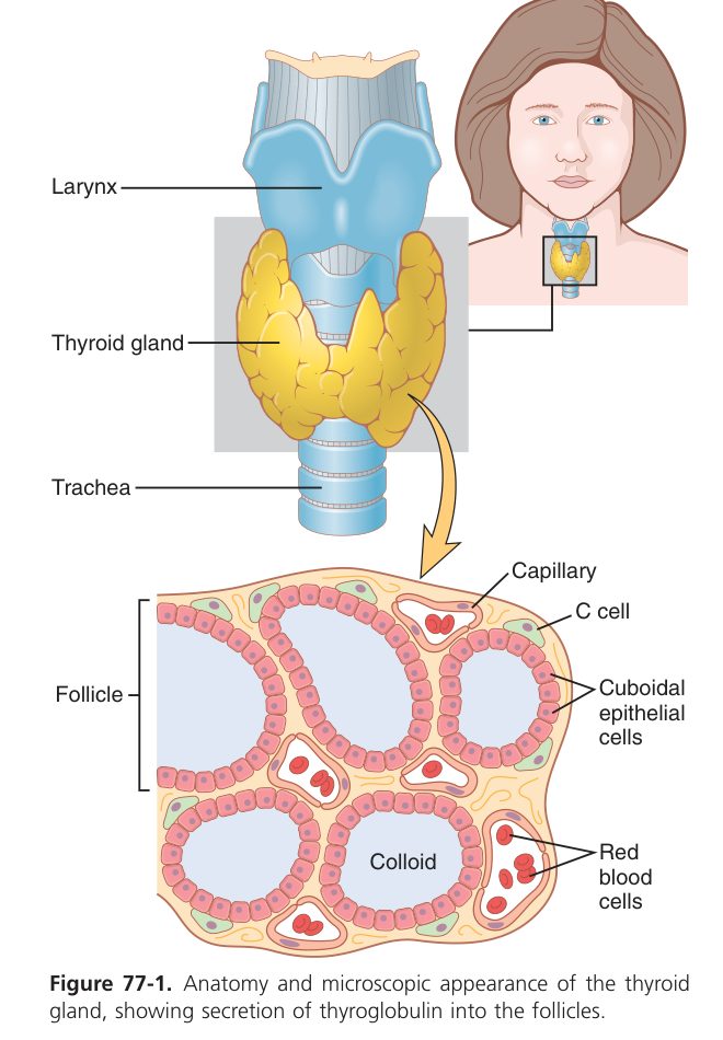
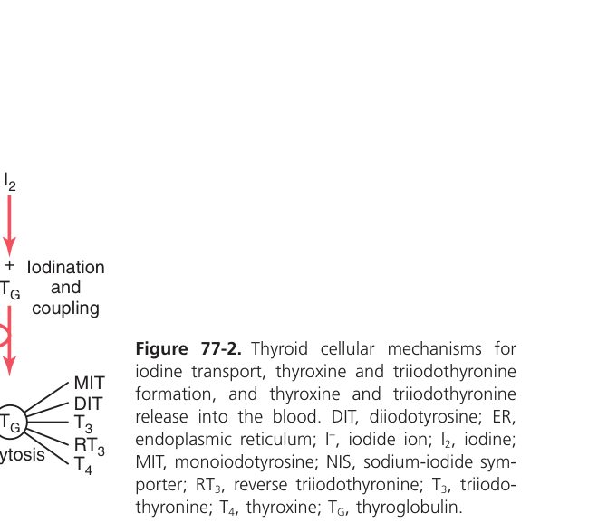
The largest endocrine gland; the only hormone stored extracellularly (in the colloid of the follicle, on thyroglobulin).
- Follicular cells → thyroid hormones (T₃, T₄).
- Parafollicular (C) cells → calcitonin.
Derivatives of tyrosine + iodine:
| Form | % secreted | Activity | Daily output |
|---|---|---|---|
| T₄ (thyroxine) | 93% | Low (a pro-hormone); converted peripherally to T₃ | ~80 µg/day |
| T₃ (triiodothyronine) | 7% | Active form — ~5× the affinity of T₄ for the receptor | ~4 µg/day |
| rT₃ (reverse T₃) | <1% | Inactive — rises in starvation/illness | ~2 µg/day |
In blood, >99% of T₃/T₄ is bound to plasma proteins, mainly thyroxine-binding globulin (TBG); only the small free fraction is biologically active.
Normal values: total T₄ 51–142 nmol/L, total T₃ 1.2–3.4 nmol/L.
- Iodide trapping — the follicular cell concentrates iodide ~30× via a Na⁺/I⁻ symporter ("iodide pump"). Daily intake required ~150 µg; gland stores ~7500 µg.
- Iodination & coupling — thyroid peroxidase (TPO) oxidises I⁻ and iodinates tyrosine residues on thyroglobulin → MIT + DIT. Coupling: DIT + DIT → T₄; DIT + MIT → T₃.
- Storage & release — thyroglobulin (carrying hormones) is endocytosed back into the follicular cell; lysosomes digest it, releasing free T₃/T₄ into the blood.
Thiouracil & propylthiouracil (PTU) inhibit TPO → block synthesis (used in hyperthyroidism). Perchlorate blocks the Na⁺/I⁻ symporter.
- Growth & development: essential for normal brain (synapses, dendrites, myelination), bone (ossification, long-bone growth) and teeth.
- Calorigenic / metabolic: ↑O₂ consumption in nearly all tissues → ↑BMR — via ↑Na⁺/K⁺-ATPase activity. The exceptions: brain, testes, uterus, lymph nodes, spleen, anterior pituitary.
- Carbohydrate: ↑intestinal glucose absorption, ↑glycogenolysis → ↑blood glucose.
- Fat: ↑cholesterol synthesis & biliary cholesterol excretion → net ↓ plasma cholesterol; ↑ FA oxidation.
- Protein: at normal levels ↑synthesis; in excess → catabolic (muscle weakness).
- Nervous system: ↑CNS excitability; a fine tremor (10–15/s) is classic in hyperthyroidism.
- Cardiovascular: ↑HR, ↑contractility, ↑CO; vasodilation in peripheral tissues.
- GI: ↑appetite, ↑motility & secretion.
Hypothalamic–pituitary–thyroid axis: TRH → TSH → T₃/T₄ — T₃/T₄ negatively feed back on TSH (↓TSH) and on TRH responsiveness.
TSH actions on the gland: ↑iodide uptake; ↑TPO & thyroglobulin synthesis; ↑T₃/T₄ release; ↑blood flow; hypertrophy/hyperplasia (chronic TSH → goitre).
- ANS: sympathetic ↑TH secretion; parasympathetic ↓.
- Cold → NE → ↑TRH → ↑TSH → ↑TH (adaptive thermogenesis).
- Autoregulation by iodine — the Wolff–Chaikoff effect: at intake > ~2 mg/day intra-glandular iodide rises high enough to suppress TPO and TH synthesis (used clinically to manage a thyroid crisis).
- Iodine deficiency → ↓TH → loss of feedback → ↑TSH → endemic (compensatory) goitre.
In adults (myxoedema): ↓BMR → weight gain without ↑intake, cold intolerance, fatigue, sleepiness, bradycardia, ptosis. Mucopolysaccharides accumulate → puffy face, large tongue, hoarseness. Constipation, hair loss, menstrual dysfunction, anaemia.
In children (cretinism): marked retardation of body stature and mental development. If present at birth and untreated for > 2–4 weeks the CNS will not mature normally — severe irreversible mental retardation. Late milestones (sitting, walking), short stature, flat broad nose, dry skin/hair, muscle weakness.
Autoantibodies bind the TSH receptor and mimic TSH → diffuse hyperplasia + autonomous hormone release. ↑BMR → weight loss despite ↑intake, heat intolerance, sweating, thirst. ↑Adrenergic drive: tachycardia, fine tremor, nervousness, labile mood, wide-eyed stare/exophthalmos. Muscle wasting.
Adrenal Cortex & Medulla
Each adrenal gland is really two glands stacked together. The cortex (outer) makes steroid hormones; the medulla (inner) makes catecholamines (adrenaline, noradrenaline). Of the two, the cortex matters more long-term — you can survive without the medulla, but adrenocortical failure is fatal in days. The cortex is organised in three concentric zones with strict division of labour, memorisable as “GFR / Salt, Sugar, Sex”: zona Glomerulosa → aldosterone (Salt); zona Fasciculata → cortisol (Sugar); zona Reticularis → androgens (Sex). Each zone is driven by a different signal: angiotensin II + plasma K⁺ for glomerulosa; ACTH for fasciculata and reticularis. Cortisol is the “stress hormone”: it raises blood glucose by gluconeogenesis, suppresses inflammation, breaks down protein, and is essential for survival of major stress (surgery, trauma, sepsis). Loss of all three (Addison disease) presents with low BP, low Na⁺, high K⁺, fatigue, weight loss — classic.
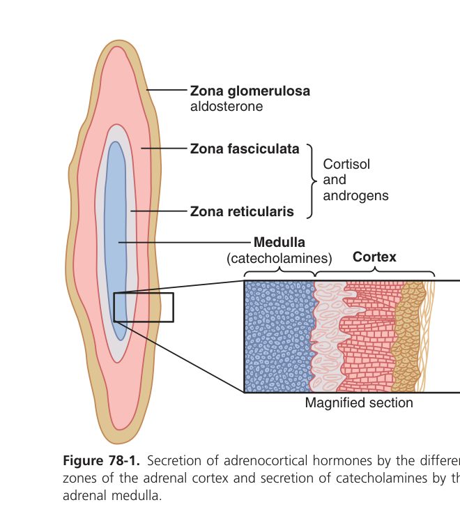
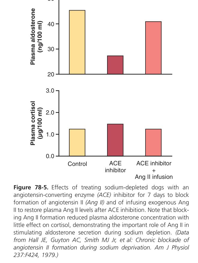
| Zone | Class | Hormone | Regulator | Main action |
|---|---|---|---|---|
| Zona Glomerulosa | Mineralocorticoid | Aldosterone | Angiotensin II + ↑plasma K⁺ | ↑renal Na⁺ reabsorption + K⁺/H⁺ secretion (DCT/CD) → ↑volume, ↑BP — Unit 7 |
| Zona Fasciculata | Glucocorticoid | Cortisol | ACTH (HPA axis) | ↑gluconeogenesis, ↓glucose uptake, protein & fat catabolism, anti-inflammatory, permissive for catecholamines, stress response |
| Zona Reticularis | Androgen | DHEA & weak androgens | ACTH | Weak androgenic effect; major source in females |
| Medulla | Catecholamine | Adrenaline (80%) + NA (20%) | Sympathetic preganglionic ACh | "Fight or flight" — ↑HR, ↑contractility, bronchodilation, glycogenolysis, lipolysis |
"GFR — Salt, Sugar, Sex" — deeper into the cortex, the further from homeostasis & closer to "selfish/sex". Medulla = modified sympathetic ganglion (chromaffin cells secrete catecholamines into blood — pre-ganglionic ACh on nicotinic receptors triggers release).
- Carbohydrate: ↑gluconeogenesis in the liver, ↓peripheral glucose uptake → ↑blood glucose ("diabetogenic").
- Protein: catabolic in muscle (→ amino acids for gluconeogenesis); anabolic in liver.
- Fat: lipolysis in extremities, lipogenesis centrally → truncal/buffalo-hump fat redistribution in chronic excess.
- Anti-inflammatory / immunosuppressive: ↓phospholipase A₂ (via lipocortin), ↓COX-2, ↓cytokines; stabilises lysosomes; lymphopenia.
- Cardiovascular: permissive for catecholamines — maintains vascular tone.
- Stress response: circulating cortisol rises with physical / emotional / surgical stress — the body cannot survive major stress without it.
- Diurnal rhythm: peak at ~6–8 a.m., trough at midnight.
- Triggered by angiotensin II (the RAAS — Unit 4 & 7) and ↑plasma K⁺; ACTH plays only a permissive role.
- Acts on principal cells of DCT & collecting duct → ↑Na⁺/water reabsorption + K⁺ & H⁺ secretion.
- Made from tyrosine: tyrosine → L-DOPA → dopamine → NA → adrenaline (last step needs cortisol — another permissive link).
- Adrenaline acts on α & β (especially β₂ bronchodilation, β₁ cardiac); NA is predominantly α (vasoconstriction).
Cushing syndrome (↑cortisol) — central obesity, moon face, buffalo hump, purple striae, hypertension, hyperglycaemia, osteoporosis, immunosuppression. Addison disease (↓cortisol + aldosterone) — fatigue, anorexia, hypotension, hyperpigmentation (ACTH ↑ → MSH), hyperkalaemia, hyponatraemia. Conn syndrome — primary hyperaldosteronism → hypertension + hypokalaemia. Phaeochromocytoma — medullary tumour → episodic hypertension, sweating, headache, palpitations.
Endocrine Pancreas — Insulin & Glucagon
The endocrine pancreas is the tiny endocrine tissue scattered inside the larger exocrine pancreas — about 1-2% of the gland by mass, clustered into ~1 million islets of Langerhans. The two main cells are simple to keep straight: β cells (~65% of islet cells, central) make insulin, the storage/anabolic hormone — lowers blood glucose by driving glucose into liver, muscle and fat, where it’s stored as glycogen and triglyceride. α cells (~25%, peripheral) make glucagon, the mobilising/catabolic hormone — raises blood glucose by gluconeogenesis and glycogenolysis in the liver. The two hormones balance each other minute by minute. Eat → insulin up, glucagon down → nutrients stored. Fast → insulin down, glucagon up → stored fuel mobilised. Diabetes is what happens when this balance breaks — Type 1 destroys β cells (no insulin); Type 2 makes target tissues resistant to insulin.
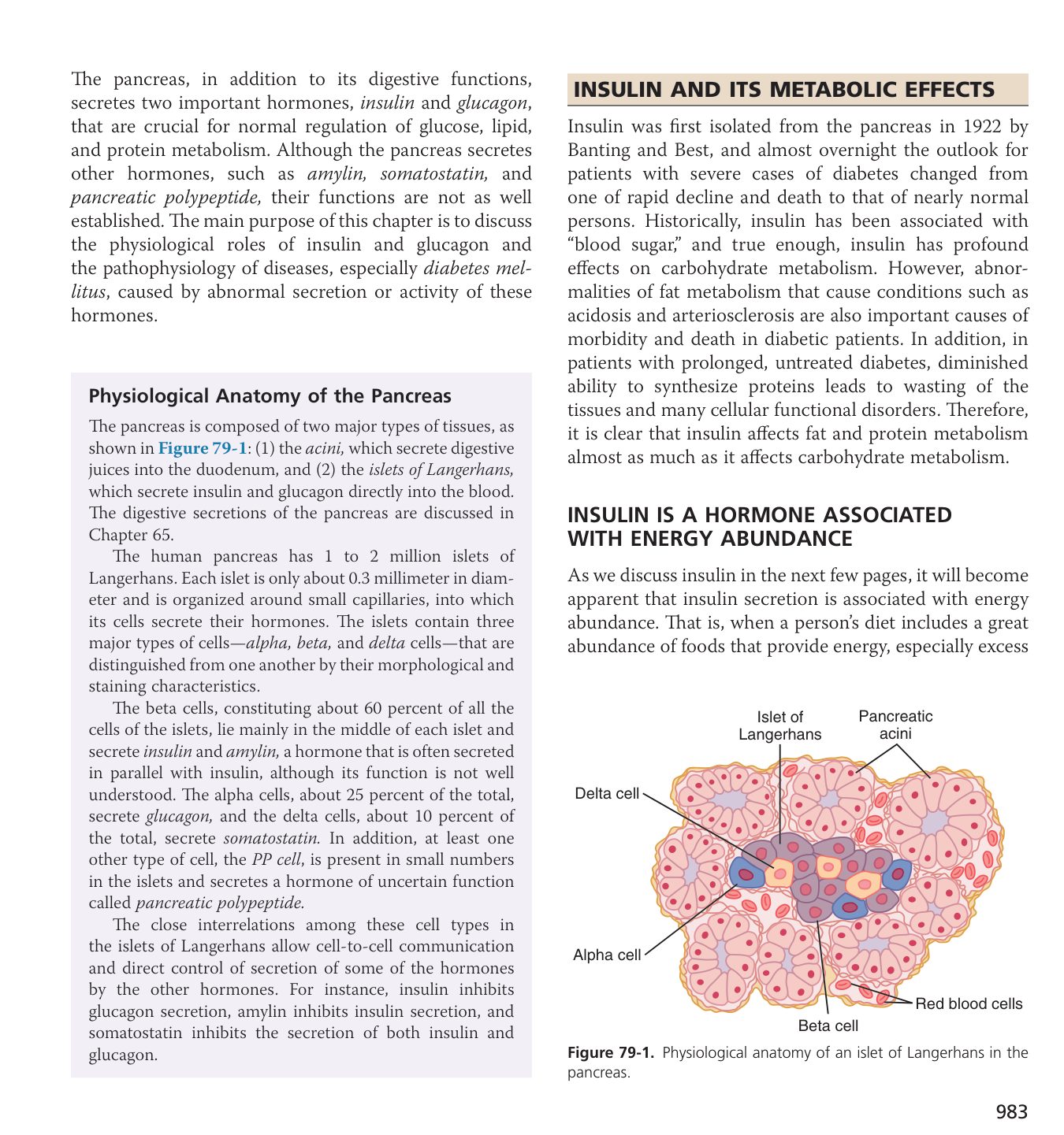
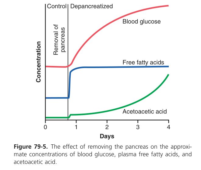
The islets of Langerhans are scattered through the exocrine pancreas. Four cell types:
| Cell | % | Hormone | Effect on glucose |
|---|---|---|---|
| β | ~70% | Insulin | Lowers — the only hypoglycaemic hormone |
| α | ~20% | Glucagon | Raises |
| δ | ~5% | Somatostatin | Inhibits both insulin & glucagon |
| PP / F | <5% | Pancreatic polypeptide | Regulates exocrine pancreas |
- Receptor: tyrosine kinase → insulin receptor substrate (IRS) → PI3K / Akt & MAPK cascades.
- Glucose uptake: inserts GLUT4 into membranes of skeletal muscle & adipose (liver uses GLUT2 — insulin-independent uptake but insulin still drives glucokinase & storage).
- Liver: ↑glycogenesis, ↑lipogenesis; ↓gluconeogenesis & ↓glycogenolysis.
- Muscle: ↑glucose uptake & glycogen storage; ↑amino acid uptake & protein synthesis.
- Adipose: ↑glucose uptake, ↑triglyceride synthesis, ↓lipolysis.
- K⁺: drives K⁺ into cells (clinical use in hyperkalaemia — insulin + glucose).
- Triggers: ↑blood glucose (main), amino acids, GI hormones (GIP, GLP-1 — "incretin effect"), parasympathetic; inhibited by sympathetic, somatostatin, adrenaline.
- Receptor: Gₛ-coupled → cAMP → PKA in liver.
- Liver: ↑glycogenolysis (fast) & ↑gluconeogenesis (sustained); ↑ketogenesis.
- Triggers: ↓blood glucose, ↑amino acids (after a protein meal — prevents reactive hypoglycaemia from concurrent insulin).
- Inhibited by hyperglycaemia, insulin, somatostatin.
Four hormones raise blood glucose — glucagon, cortisol, growth hormone, adrenaline. Only insulin lowers it.
Type 1 — autoimmune β-cell destruction → absolute insulin deficiency → hyperglycaemia, glycosuria with osmotic diuresis (Unit 7), polyuria, polydipsia, weight loss, and (in severe) ketoacidosis (uncontrolled lipolysis → FFA → ketones → metabolic acidosis — Unit 7 acid–base). Type 2 — insulin resistance + relative deficiency.
Calcium Regulation
Plasma calcium is held in a remarkably narrow window (2.2-2.6 mmol/L) because both extremes are dangerous. Too low and excitable tissues fire spontaneously — tetany, carpopedal spasm, laryngospasm, seizures. Too high and they become sluggish — constipation, polyuria, confusion, stones, fractures. The body guards Ca²⁺ with three hormones and three target organs (gut, kidney, bone). When plasma Ca²⁺ drops, the parathyroid glands sense it (via a calcium-sensing receptor) and release PTH, which raises Ca²⁺ by mobilising it from bone, increasing renal reabsorption, and stimulating renal 1α-hydroxylase to make active vitamin D (which then increases gut absorption). Plasma Ca²⁺ rises, the parathyroid notices, PTH falls — classic negative feedback. The opposite signal — high Ca²⁺ — triggers calcitonin from thyroid C cells (minor role in humans). Vitamin D is the long-term player: deficiency → impaired gut Ca²⁺ absorption → rickets (children) or osteomalacia (adults).
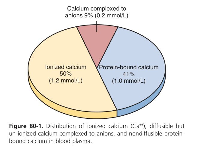
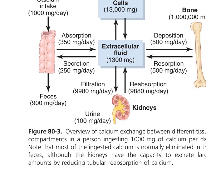
Plasma Ca²⁺ = 2.2–2.6 mmol/L (9–10.5 mg/dL). ~50% ionised (active), ~40% albumin-bound, ~10% complexed. Tightly controlled because Ca²⁺ affects:
- Neuromuscular excitability (low Ca²⁺ raises excitability → tetany).
- Skeletal & cardiac muscle contraction.
- Synaptic transmitter release (Units 1, 2).
- Clotting cascade (factor IV — Unit 3).
- Bone strength.
| Hormone | Source | Effect on Ca²⁺ | Bone | Kidney | Gut |
|---|---|---|---|---|---|
| PTH | Parathyroid chief cells | ↑ | ↑osteoclast resorption (indirect via RANKL) | ↑Ca reabsorption (distal); ↓phosphate reabsorption; activates vitamin D (↑1α-hydroxylase) | ↑Ca & PO₄ absorption (via calcitriol) |
| Calcitriol (1,25-(OH)₂-D₃) | Skin → liver → kidney | ↑ | Permits PTH’s action; ↑remodelling | ↑Ca & phosphate reabsorption | ↑↑ Ca & phosphate absorption (main effect) |
| Calcitonin | Thyroid C cells | ↓ | ↓osteoclast activity | ↑Ca & phosphate excretion | — |
Skin: 7-dehydrocholesterol ⟶ UV light → cholecalciferol (D₃). Liver: 25-hydroxylation → 25(OH)D. Kidney: 1α-hydroxylation (the regulated, PTH-driven step) → 1,25(OH)₂D (calcitriol), the active hormone.
PTH ↑Ca and ↓phosphate; calcitriol ↑both Ca and phosphate. Bone is a vast Ca/PO₄ reservoir — remodelling continuously by osteoblasts (build) and osteoclasts (resorb).
Hypocalcaemia → tetany. Loss of extracellular Ca²⁺ reduces the threshold for Na⁺-channel opening → spontaneous nerve firing → carpopedal spasm (Trousseau sign on cuff inflation), Chvostek sign (facial tap), laryngospasm in severe cases. Causes include hypoparathyroidism (post-thyroid surgery), vitamin D deficiency, severe alkalosis (↑albumin binding).
Primary hyperparathyroidism → hypercalcaemia. "Stones, bones, abdominal groans, psychic moans": kidney stones, bone resorption (subperiosteal — classic at radial side of phalanges), abdominal pain, constipation, polyuria, depression/confusion.
Vitamin D deficiency: rickets in children (soft bowed bones), osteomalacia in adults. Chronic kidney disease → ↓1α-hydroxylation → ↓calcitriol → secondary hyperparathyroidism → renal osteodystrophy.
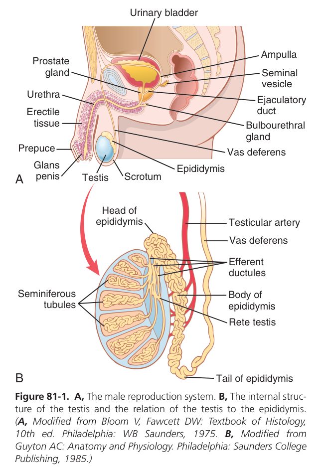
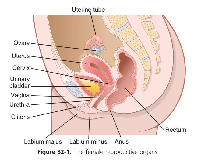
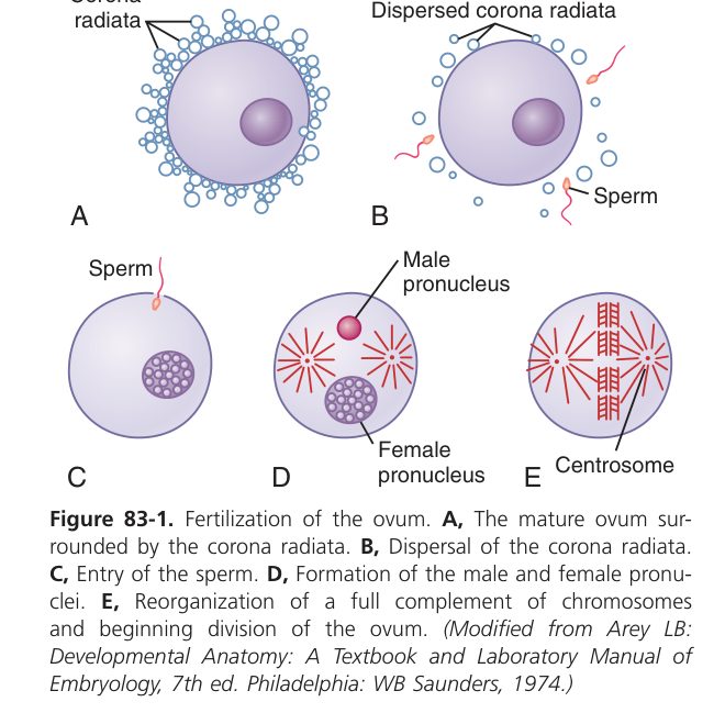
Reproductive physiology starts with one hypothalamic peptide — GnRH, released in pulses — which drives anterior pituitary FSH and LH. In males, FSH and LH act on testes: LH drives Leydig cells to make testosterone, FSH drives Sertoli cells to support spermatogenesis. In females the same FSH/LH drive the ovary in a monthly cycle: FSH grows follicles, an LH surge triggers ovulation, and the empty follicle becomes the corpus luteum that makes progesterone in the second half. Sex steroids feed back to brake the hypothalamus and pituitary — this is exactly the principle the contraceptive pill exploits.
Endocrine complete
Axes, mechanisms, thyroid, adrenal, islets & calcium mastered. Next: Nervous System.