Renal (Urinary) Physiology
Functions of the Kidney & Renal Blood Flow
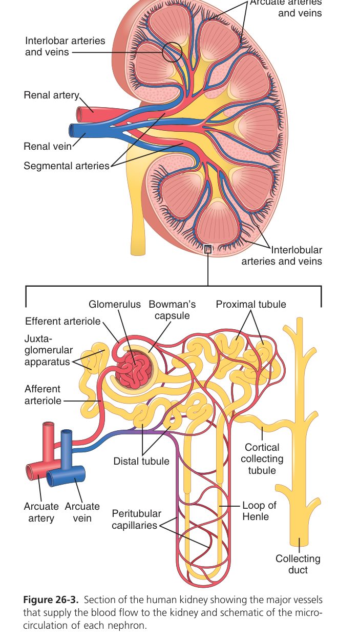
Kidneys do far more than make urine — they are the body’s main quality controller for the internal environment. Every minute, ~1.2 L of blood (a quarter of cardiac output) is delivered to organs that weigh less than 1% of body weight. The reason this disproportionate flow exists: the kidney filters plasma to clean it, then reabsorbs almost everything back, keeping only what needs removing. The output we call urine is the discarded fraction — barely 1 mL per minute. In doing this the kidney simultaneously regulates blood volume (and therefore blood pressure), plasma osmolality, pH, and the concentration of every ion that matters — Na⁺, K⁺, Ca²⁺, phosphate. It also makes hormones: renin (the trigger of the BP-controlling RAAS), erythropoietin (telling marrow to make red cells), and active vitamin D (calcitriol). Damage the kidneys and every one of these jobs starts failing — which is why renal failure presents with so many seemingly unrelated symptoms.
- Excretion of metabolic waste (urea, creatinine, uric acid) & foreign substances (drugs).
- Regulation of water & electrolyte balance, osmolality, and acid–base balance.
- Regulation of arterial pressure (volume + renin).
- Endocrine: secretes renin, erythropoietin (EPO), and activates vitamin D to calcitriol.
The kidney does not regulate body temperature, and it does not secrete ADH (that is the hypothalamus/posterior pituitary).
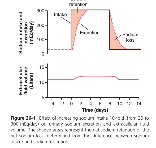
In a resting adult, renal blood flow is ~25% of cardiac output (~1200 mL/min) — very high relative to the kidney’s mass, because the kidney filters the whole plasma volume many times daily.
Glomerular Filtration
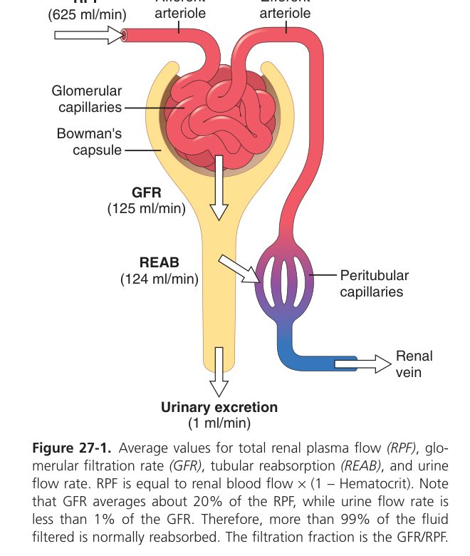
Urine formation begins with bulk filtration. The glomerulus is a high-pressure capillary tuft where ~20% of plasma volume crosses through into Bowman’s capsule every time it passes — about 180 L per day, almost 4× your entire ECF, every day. That filtrate then gets reworked downstream. The barrier is three layers: fenestrated endothelium (catches blood cells), glomerular basement membrane (size + charge selective, the main filter), podocyte slit diaphragms (fine size selectivity). Crucially, the basement membrane is negatively charged — so plasma proteins (also negative, like albumin) are repelled even though they could fit by size. Damage that negative charge (nephrotic syndrome) and albumin pours into urine; lose albumin from plasma and oedema follows.
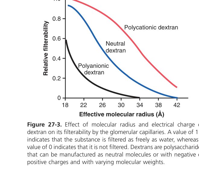
Filtrate crosses three layers: (1) fenestrated capillary endothelium, (2) basement membrane (the main size + charge barrier), (3) podocyte foot processes with filtration slits. The loop of Henle is NOT part of it. The membrane is negatively charged → cations are filtered more readily than anions (albumin, being anionic, is repelled).
Loss of the negative charge or damage to the membrane (e.g. nephrotic syndrome) → proteinuria (albumin escapes) → hypoalbuminaemia → oedema.
GFR (glomerular filtration rate) = the volume of filtrate formed by both kidneys per minute (~125 mL/min ≈ 180 L/day).
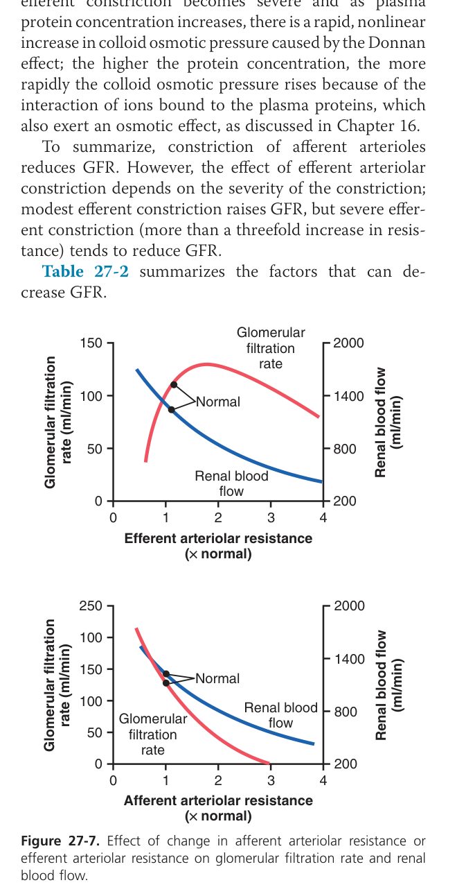
GFR is special: it has to stay constant even when your blood pressure swings between 80 and 180 mmHg. Why? Because if GFR followed BP up and down, you would either lose litres of urine when BP rose (dehydration) or filter almost nothing when BP dipped (uraemia). The kidney prevents this with autoregulation. Two mechanisms work together: the myogenic response — the afferent arteriole constricts when stretched by rising BP, opposing the rise — and tubuloglomerular feedback — if more NaCl is reaching the macula densa, the afferent constricts to drop GFR. Above ~80 mmHg the system holds GFR almost flat. Below 80 mmHg, autoregulation can’t keep up and GFR collapses, which is why severe shock causes acute kidney injury.
| Factor | Effect on GFR |
|---|---|
| Glomerular capillary pressure (BP, afferent/efferent tone) | ↑P→↑GFR (afferent dilation or efferent constriction raise it) |
| Plasma colloid osmotic pressure (πGC) | Inverse: ↑π → ↓GFR |
| Bowman’s capsule pressure (obstruction) | ↑PBS → ↓GFR |
| Filtration coefficient (membrane permeability & area) | ↑Kᶠ → ↑GFR |
| Renal blood flow | ↑RBF → ↑GFR |
Autoregulation keeps GFR nearly constant over MAP ~80–180 mmHg (myogenic + tubuloglomerular feedback). Below ~80 mmHg, GFR falls steeply.
Clearance (Cx) of a substance = the volume of plasma fully cleared of that substance per minute.
- Inulin clearance = GFR (~125 mL/min). Inulin is freely filtered, not reabsorbed, not secreted.
- Creatinine clearance ≈ GFR (slight overestimate from tubular secretion) — the clinical standard.
- PAH clearance ≈ renal plasma flow (RPF) (~625 mL/min) — PAH is filtered and almost completely secreted in PCT.
- Filtration fraction (FF) = GFR / RPF ≈ 20%. FF rises with efferent constriction (angiotensin II).
Tubular Reabsorption & Secretion
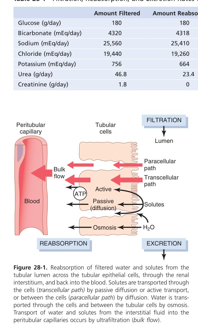
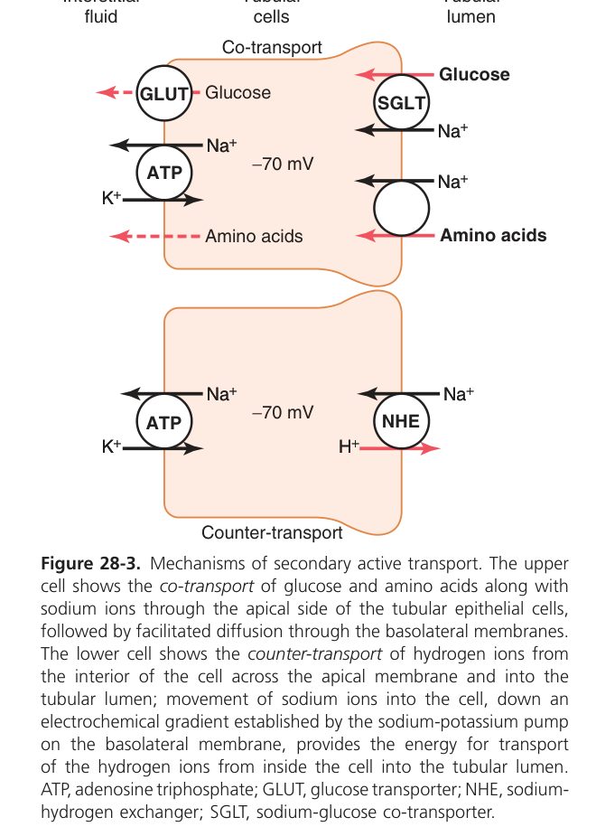
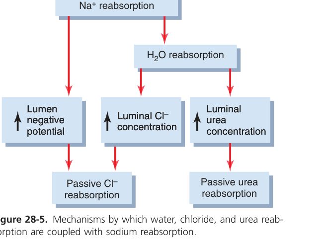
The kidney filters 180 L of plasma per day — but you produce only ~1.5 L of urine. That means tubules reabsorb >99% of what they filter. Reading nephron physiology is reading which segment grabs what. The proximal tubule is the bulk worker: ~65% of Na⁺, all glucose and amino acids, most HCO₃⁻, all leveraged off Na⁺-coupled cotransport that runs on the gradient set up by the Na⁺/K⁺-ATPase on the basolateral side. The loop of Henle generates the medullary osmotic gradient: the thick ascending limb pumps NaCl out (NKCC2 cotransporter, furosemide’s target) into the interstitium while staying water-impermeable. The distal tubule does fine-tuning (thiazides act here). The collecting duct is the last word — aldosterone tells it how much Na⁺ to grab and K⁺ to lose; ADH tells it how much water to pull out. Every diuretic in clinical use blocks one of these segments’ transporters.
| Segment | Reabsorbs | Transporter / feature |
|---|---|---|
| Proximal tubule (PCT) | ~65% of filtrate: Na⁺, all glucose & amino acids, HCO₃⁻, water | Na⁺ via Na⁺-glucose, Na⁺-amino-acid, Na⁺-H⁺ exchange; isosmotic |
| Thin descending limb (DTL) | Water only | No Na⁺ reabsorption; water-permeable |
| Thick ascending limb (TAL) | Na⁺, K⁺, Cl⁻ (water-impermeable = "diluting segment") | Na⁺-K⁺-2Cl⁻ cotransporter (NKCC2) — the furosemide target |
| Distal tubule (DCT) | Na⁺, Cl⁻ | Na⁺-Cl⁻ cotransporter (thiazide target); aldosterone-sensitive |
| Collecting duct (CD) | Na⁺ (aldosterone), water (ADH) | Principal cells; final urine concentration |
- Glucose is reabsorbed entirely in the PCT by secondary active transport (Na⁺-glucose cotransport, SGLT) at the luminal membrane — uphill, powered by the Na⁺ gradient.
- Renal threshold for glucose = the plasma glucose at which glucose first appears in the urine (the transporters saturate) — ~180 mg/dL (10 mmol/L); transport maximum (T᷀) ~375 mg/min.
Osmotic diuresis = increased urine flow caused by non-reabsorbed solute in the tubule holding water osmotically (e.g. glucose in diabetes, mannitol). Water diuresis = increased urine flow from drinking water / low ADH, producing dilute urine.
When plasma glucose exceeds the renal threshold, glucose stays in the tubule → osmotic diuresis → glycosuria, polyuria, polydipsia — the classic presentation of uncontrolled diabetes. SGLT2 inhibitors exploit this therapeutically.
Urine Concentration & Dilution
How can your kidney make urine four times more concentrated than your plasma? The trick is to build a salty inner medulla, then run the collecting duct down through it under hormone control. The loop of Henle creates the gradient via countercurrent multiplication: the ascending limb actively pumps NaCl into the interstitium (without letting water follow), so the medulla becomes increasingly hyperosmotic from cortex to papilla (up to ~1200 mOsm). The vasa recta loops alongside as a countercurrent exchanger, taking nutrients down and removing waste, without washing out the gradient. Then the collecting duct passes through this salt mountain on its way to the renal pelvis. When ADH is high (dehydration), aquaporins open in the collecting duct, water flows out into the salty interstitium, and concentrated urine emerges. When ADH is low (overhydrated), the duct stays water-tight and dilute urine pours out. One gradient, two control hormones — complete water homeostasis.
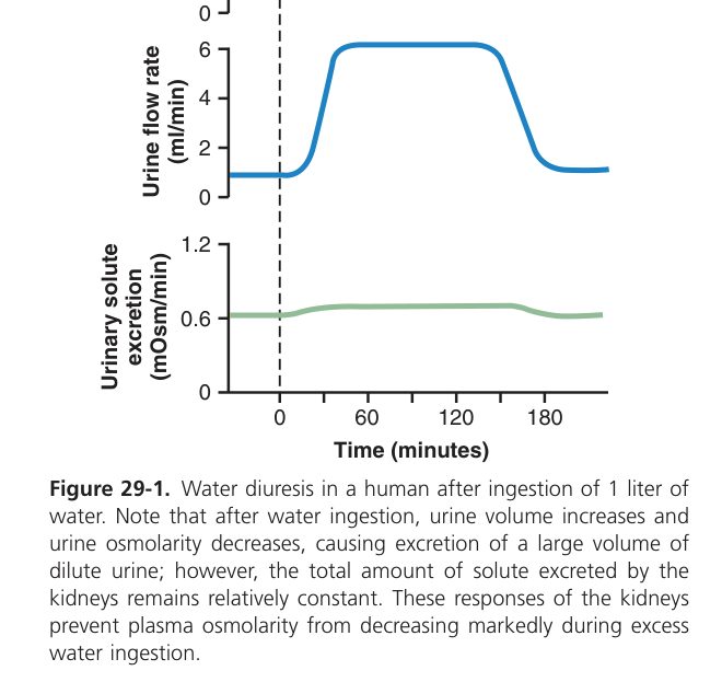
- Countercurrent multiplier (loop of Henle): the TAL pumps NaCl into the medullary interstitium (water-impermeable) while the descending limb loses water — building a hypertonic medullary gradient (up to ~1200 mOsm at the papilla).
- Countercurrent exchanger (vasa recta): the hairpin capillaries preserve that gradient.
- ADH makes the collecting duct water-permeable (inserts aquaporin-2) so water is drawn out into the hypertonic medulla → concentrated urine. Without ADH → dilute urine (water diuresis).
- Urea recycling contributes to the medullary gradient.
Hormonal Regulation
| Hormone / nerve | Trigger | Renal action |
|---|---|---|
| ADH (vasopressin) — hypothalamus | ↑Plasma osmolality or ↓volume | ↑water reabsorption in collecting duct & DCT (aquaporins) |
| Aldosterone — adrenal cortex | Angiotensin II, ↑K⁺ | ↑Na⁺ reabsorption + K⁺ secretion (DCT/CD) |
| Angiotensin II | Renin (RAAS) | Vasoconstriction (↑BP), ↑aldosterone, ↑ADH & thirst, ↑proximal Na⁺ reabsorption |
| Renal sympathetic nerve | ↓BP, stress | ↓GFR, ↑renin, ↑NaCl reabsorption (NOT ↑K⁺ reabsorption) |
Renin is released by juxtaglomerular cells in response to three signals: ↓renal perfusion pressure, ↑sympathetic activity, and ↓NaCl delivery to the macula densa. (ADH is not a renin trigger.)
Renin (kidney) → cleaves angiotensinogen (liver) → Angiotensin I → ACE (lung endothelium) → Angiotensin II. Effects of Ang II:
- Powerful systemic vasoconstriction (especially efferent arteriole → ↑filtration fraction).
- ↑Aldosterone from adrenal zona glomerulosa → ↑Na⁺ reabsorption + K⁺/H⁺ secretion (DCT/CD).
- ↑ADH & thirst via the hypothalamus.
- ↑Na⁺/H⁺ exchange in PCT → ↑Na⁺ & HCO₃⁻ reabsorption.
ACE inhibitors (-pril) and ARBs (-sartan) lower BP & are renoprotective in diabetic nephropathy (dilate efferent arteriole → ↓glomerular pressure). Spironolactone = aldosterone antagonist (K-sparing diuretic). SIADH = inappropriately high ADH → concentrated urine + hyponatraemia + euvolaemia.
| Hormone | Source / trigger | Renal action |
|---|---|---|
| ANP / BNP | Atrial / ventricular stretch (volume overload) | ↑GFR (afferent dilate, efferent constrict), ↓Na⁺ reabsorption (collecting duct), ↓renin, ↓aldosterone → natriuresis |
| PTH | ↓Ca²⁺ | ↑Ca²⁺ reabsorption (DCT), ↓phosphate reabsorption (PCT, phosphaturic), ↑1α-hydroxylase → calcitriol |
| Calcitriol (active vitamin D) | PTH, ↓Ca²⁺, ↓PO₄ | ↑Gut Ca²⁺ & phosphate absorption (kidney is the activation site, not the target) |
Potassium is dangerous in extremes — both too high and too low cause arrhythmias and death — so the kidney watches it carefully. Most filtered K⁺ is reabsorbed proximally and in the thick ascending limb; the regulated step is aldosterone-driven K⁺ secretion by principal cells of the distal nephron and collecting duct. High plasma K⁺ (after a banana or in renal failure) directly stimulates adrenal aldosterone release, which raises K⁺ secretion until balance is restored. The K⁺ exits through luminal ROMK channels driven by the Na⁺ reabsorption next door — that’s why ANY drug that increases distal Na⁺ delivery (loop diuretics, thiazides) also increases K⁺ loss and causes hypokalaemia. Conversely, drugs blocking aldosterone (spironolactone) or ENaC (amiloride) are K⁺-sparing.
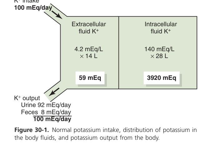
~65% reabsorbed in PCT, ~25% in TAL (NKCC2). The DCT/CD principal cells are the variable component — aldosterone drives K⁺ secretion via ROMK channels. Drivers: high plasma K⁺, alkalosis, ↑distal tubular flow.
Loop diuretics (furosemide) and thiazides → ↑distal Na⁺ delivery → ↑K⁺ secretion → hypokalaemia. Spironolactone & amiloride spare K⁺. Conn syndrome (primary aldosteronism) → hypertension + hypokalaemic alkalosis.
| Drug | Site | Mechanism |
|---|---|---|
| Mannitol | Whole tubule (mainly DTL) | Osmotic diuretic |
| Acetazolamide | PCT | Carbonic anhydrase inhibitor → HCO₃⁻ loss |
| Furosemide | TAL | NKCC2 block |
| Thiazide | DCT | NCC block |
| Spironolactone | CD | Aldosterone receptor antagonist |
| Amiloride | CD | ENaC block |
Renal Acid–Base Balance
Acid-base balance is the kidney’s slow but final answer to pH. The lungs handle CO₂ in seconds (the volatile acid). The kidney handles the fixed acids — the sulphates and phosphates produced by metabolism — over hours and days. It does this by secreting H⁺ into the tubule lumen and by reabsorbing HCO₃⁻ back into blood. The two are coupled: for every H⁺ secreted, one HCO₃⁻ is added back to plasma. In the proximal tubule, H⁺ from intracellular carbonic anhydrase exits via Na⁺/H⁺ exchange; in the collecting duct, α-intercalated cells use H⁺-ATPase to dump H⁺ against a steep gradient. Urinary H⁺ would saturate the pH limits quickly, so it’s buffered — mainly by phosphate (titratable acid) and ammonia (NH₄⁺). In acidosis the kidney makes more NH₃ from glutamine; in alkalosis it makes less and dumps more HCO₃⁻. Henderson-Hasselbalch in one line: pH ∝ HCO₃⁻/PCO₂.
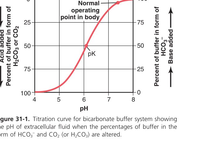
The kidney defends pH by reabsorbing filtered HCO₃⁻ and excreting H⁺ (as titratable acid + ammonium). In the PCT, for each H⁺ secreted, one HCO₃⁻ is reabsorbed (1:1). Ammonia (NH₃) buffers secreted H⁺ as NH₄⁺, aiding H⁺ elimination.
| Disorder | Primary change | Compensation |
|---|---|---|
| Respiratory acidosis | ↑PCO₂ | Kidney ↑HCO₃⁻ reabsorption (slow) |
| Respiratory alkalosis | ↓PCO₂ | Kidney ↓HCO₃⁻ reabsorption |
| Metabolic acidosis | ↓HCO₃⁻ | Lungs hyperventilate (↓PCO₂, fast) + renal H⁺ excretion |
| Metabolic alkalosis | ↑HCO₃⁻ | Lungs hypoventilate (↑PCO₂) |
Micturition
- Bladder filling stretches wall receptors → afferents to the sacral spinal cord.
- Parasympathetic (pelvic nerve, S2–S4) contracts the detrusor and relaxes the internal sphincter → voiding.
- The external (striated) sphincter is under voluntary cortical control — it allows voiding to be held or initiated.
Spinal cord injury above the sacral centre → loss of voluntary control → automatic (reflex) bladder. Damage to the sacral centre/nerves → atonic bladder with overflow incontinence.
Renal complete
Filtration, tubular handling, concentration, hormones & acid–base mastered. Next: Endocrine.