Digestive (Gastrointestinal) Physiology
GI Smooth Muscle & the Enteric Nervous System
The gastrointestinal tract is a 9-metre tube running from mouth to anus, and despite its length its job is simple to describe: it converts food into small molecules the body can absorb. Everything else — the muscle layers, the secretions, the hormones, the enteric nerves — serves four functions in sequence: motility (mixing and propelling), secretion (acids, enzymes, mucus, bile), digestion (chemical breakdown), and absorption (transfer into blood or lymph). At any given moment in a healthy gut, all four are happening at different points along the tube.
Two kinds of digestion go on side by side. Mechanical digestion — chewing, mixing, peristalsis — physically breaks food apart and mixes it with secretions; this enormously increases the surface area on which enzymes can act. Chemical digestion uses those enzymes to cleave starches to sugars, proteins to amino acids, and fats to fatty acids + glycerol. The two have to happen together: enzymes work on small particles, and small particles come from mechanical breakup. Without chewing, the same enzymes would only get to work on the outside of a swallowed lump.
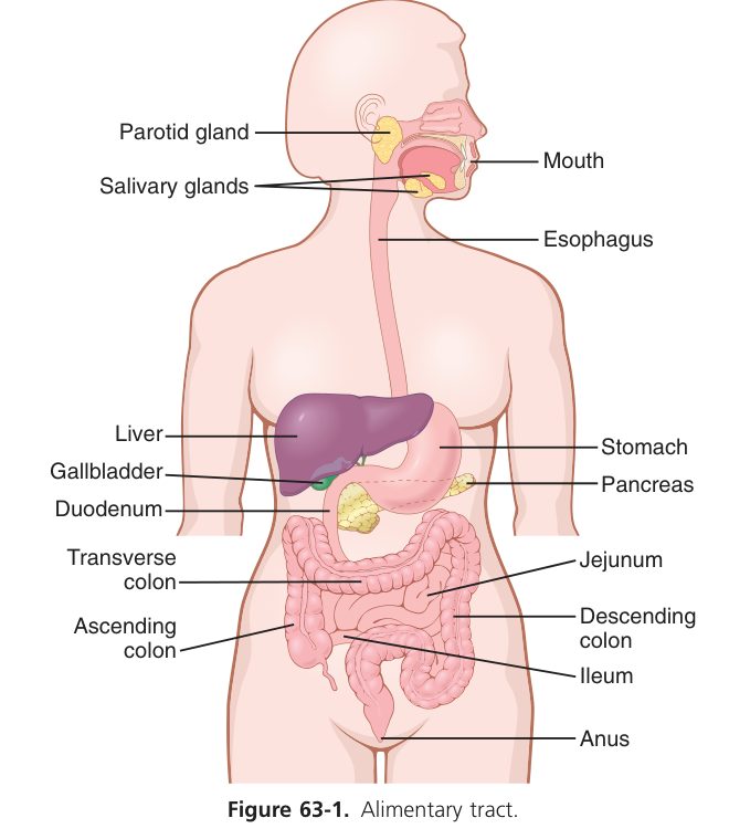
The GI tract performs motility, secretion, digestion & absorption (+ elimination). Digestion = breakdown of food into absorbable units; absorption = movement of those products across the mucosa into blood/lymph.
- Mechanical digestion — grinding, mixing with juices, propulsion.
- Chemical digestion — enzymatic breakdown into building blocks.
Gut muscle does something heart muscle doesn’t: it generates a constant background electrical rhythm called slow waves, regardless of whether you’ve eaten. These are subthreshold oscillations of membrane potential, set by specialised pacemaker cells called the interstitial cells of Cajal (ICC). Slow waves are too small to make the muscle contract by themselves — their job is to set the rhythm. When something raises the wave high enough to cross threshold (stretch, ACh, parasympathetic input), spike potentials ride on the crest and the muscle actually contracts. The frequency of slow waves determines the maximum contraction rate (3/min in stomach, 12/min in duodenum). Sympathetic noradrenaline hyperpolarises the membrane — pushing it further from threshold — which is why stress slows the gut.
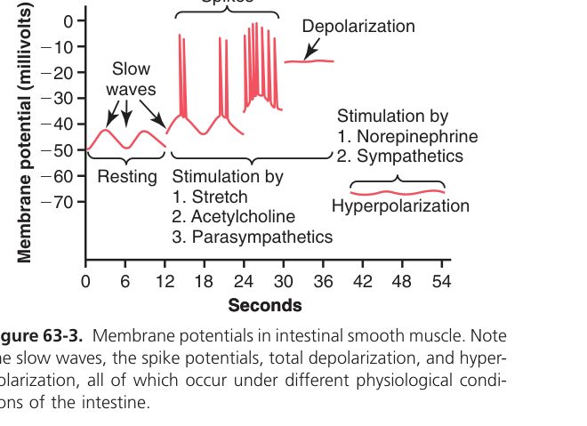
- Resting potential ~ −50 to −60 mV (set by K⁺ + electrogenic Na⁺/K⁺ pump).
- Slow waves (basic electric rhythm, BER): spontaneous, rhythmic, subthreshold depolarisations generated by the interstitial cells of Cajal (ICC = pacemaker); 3–12 cycles/min. They set the frequency of contraction but do not by themselves cause it.
- Spike potentials (APs): ride on slow-wave crests when threshold is reached — depolarisation by Ca²⁺ influx, repolarisation by K⁺ efflux; their number sets contraction amplitude.
- Stretch, ACh, parasympathetic → depolarise → more spikes/contraction; noradrenaline/sympathetic → hyperpolarise → fewer contractions.
| System | Plexus / nerve | Main role |
|---|---|---|
| Enteric (intrinsic) | Myenteric (Auerbach) — between muscle layers | Controls motility |
| Submucosal (Meissner) | Controls secretion & local blood flow | |
| Extrinsic (autonomic) | Parasympathetic (ACh, vagus) / Sympathetic (NE) | Para = excitatory; Symp = inhibitory |
• What sets the basic GI rhythm? → slow waves from interstitial cells of Cajal (ICC)
• Slow waves vs spikes? → slow waves set rhythm; spikes ride crests & cause contraction
• ACh / parasympathetic vs NE / sympathetic on gut? → ACh excites, NE inhibits
• Two enteric plexuses + roles? → myenteric (motility) · submucosal (secretion + local blood flow)
Gastrointestinal Hormones
The gut is the body’s largest endocrine organ — bigger than the pituitary and the thyroid combined. Endocrine cells scattered through the gut epithelium sample what passes by and release hormones into blood to coordinate the rest of the digestive system. Three are essential to know. Gastrin from G cells in the stomach antrum is the gas pedal — when food enters the stomach, gastrin tells the parietal cells to make acid and the stomach to start mixing. CCK from I cells in the duodenum is the “fat detector” — when fat or protein arrives in the duodenum, CCK contracts the gallbladder (to release bile), relaxes the sphincter of Oddi, and tells the pancreas to release enzymes. Secretin from S cells in the duodenum is the acid neutraliser — when H⁺ arrives from the stomach, secretin tells the pancreas and bile ducts to release water and bicarbonate, raising duodenal pH back into the safe range for enzymes. Three sensors, three timed responses, one beautifully coordinated meal.
| Hormone | Source cell | Stimulus | Main actions |
|---|---|---|---|
| Gastrin | G cells (gastric antrum) | Amino acids/peptides in stomach, vagus, distension | ↑HCl & pepsin, ↑gastric motility & emptying, trophic to mucosa |
| CCK | I cells (duodenum/jejunum) | Fat & protein in duodenum | Gallbladder contraction + Oddi relaxation, ↑pancreatic enzymes, ↑intestinal motility |
| Secretin | S cells (upper small intestine) | Acid (H⁺) entering duodenum | ↑pancreatic/biliary water & HCO₃⁻, ↓gastric acid & motility |
A brain–gut peptide is a peptide found in both the GI endocrine cells and the nervous system (brain & enteric neurons) — e.g. gastrin, CCK, VIP, somatostatin — acting as both hormone & neurotransmitter.
| Peptide | Source | Stimulus | Action |
|---|---|---|---|
| GIP (glucose-dependent insulinotropic peptide) | K cells, duodenum | Glucose, fat | ↑Insulin release (incretin effect); ↓gastric motility |
| GLP-1 | L cells, ileum | Nutrients | ↑Insulin, ↓glucagon, ↓gastric emptying, ↑satiety (target of GLP-1 agonists) |
| Motilin | M cells, duodenum | Fasting | Triggers the migrating motor complex (MMC) — "housekeeper" of the gut |
| Somatostatin | D cells (stomach, pancreas) | Acid, glucose | Broad inhibitor — ↓gastrin, ↓HCl, ↓motility, ↓all pancreatic hormones |
| VIP | Enteric neurons | Distension | Smooth-muscle relaxation, ↑intestinal water/electrolyte secretion (excess → VIPoma = WDHA syndrome) |
| Ghrelin | P/D1 cells of stomach | Fasting | ↑Appetite (the "hunger hormone") |
Mouth & Salivary Secretion
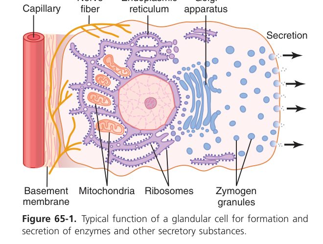
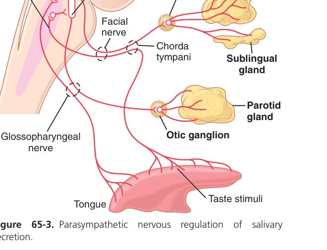
Saliva is so ordinary you barely notice it — until you stop making it (Sjögren’s, radiation) and suddenly find you can’t swallow dry bread. The salivary glands produce 1-1.5 L per day, and that fluid does five distinct jobs at once: it lubricates food for swallowing (mucin), begins starch digestion (α-amylase / ptyalin), kills bacteria (lysozyme, IgA, lactoferrin), buffers acid that flows back from the stomach (HCO₃⁻), and dilutes and washes the mouth between meals. Notice that unlike most secretions, saliva is hypotonic to plasma — it leaves the acini isotonic, then duct cells reabsorb NaCl as it travels out, but not water. Salivation is unique in another way: it’s controlled entirely by the autonomic reflex — almost no humoral input. The medulla decides.
| Property | Value |
|---|---|
| Volume | 1000–1500 mL/day |
| pH / tonicity | 6.6–7.1; hypotonic to plasma; ~99% water |
| Key components | Salivary α-amylase (ptyalin), lysozyme, mucus, lingual lipase, IgA, lactoferrin |
Functions: (1) lubrication (mucus) for swallowing; (2) starch digestion (amylase: starch → maltose); (3) protection/excretion of noxious substances; (4) antiseptic (lysozyme); (5) neutralisation & dilution. Control is reflex (centre in the medulla), dominated by the parasympathetic supply.
Gastric Secretion (Gastric Juice)
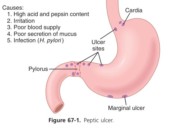
Your stomach makes about 2 L of gastric juice every day, and one component is so corrosive that the mucosa wraps itself in a thick mucus blanket to survive its own secretion: hydrochloric acid at pH 1. The acid does three useful things at once — activates pepsinogen to pepsin, denatures protein so enzymes can attack, and kills most bacteria entering with food. Parietal cells also make intrinsic factor, the chaperone that lets you absorb vitamin B₁₂ in the terminal ileum — lose parietal cells (autoimmune gastritis, gastrectomy) and you get pernicious anaemia. Chief cells make pepsinogen, the inactive zymogen of pepsin, activated by stomach acid the moment it hits the lumen. Surface mucous cells + neck cells secrete a mucus-bicarbonate gel that lines the stomach wall: bicarbonate trapped in mucus neutralises the H⁺ that would otherwise reach the epithelium. Damage that barrier — with NSAIDs (block prostaglandin protection) or H. pylori (a barrier-burrowing bacterium) — and you get peptic ulcer.
Gastric juice: pH 0.9–1.5, ~1.5–2.5 L/day.
| Component | Cell of origin | Function |
|---|---|---|
| HCl | Parietal (oxyntic) cell (H⁺/K⁺-ATPase; basal 0–5, max 20–25 mmol/h) | Activates pepsinogen, denatures protein, kills bacteria, aids Ca²⁺/Fe²⁺ absorption, stimulates pancreatic/bile secretion |
| Pepsinogen | Chief (peptic) cell | HCl converts it to pepsin (active pH ~2, inactive pH >5) → digests protein |
| Mucus + HCO₃⁻ | Surface mucous & neck cells | Forms the mucus–bicarbonate barrier protecting the mucosa from acid & pepsin |
| Intrinsic factor | Parietal cell | Binds vitamin B₁₂ for absorption in the ileum |
A weakened mucus–bicarbonate barrier (NSAIDs, H. pylori) lets acid/pepsin injure the mucosa → peptic ulcer. Loss of parietal cells (autoimmune gastritis) → no intrinsic factor → pernicious (B₁₂) anaemia — links to Unit 3.
Acid is generated by the H⁺/K⁺-ATPase ("proton pump") on the apical (canalicular) membrane, exchanging cytosolic H⁺ for luminal K⁺. The H⁺ comes from intracellular carbonic anhydrase (CO₂ + H₂O → H₂CO₃). HCO₃⁻ leaves the basolateral side in exchange for Cl⁻ (the "alkaline tide" in venous blood after a meal); Cl⁻ then follows H⁺ into the lumen as HCl. Three secretagogues converge on parietal cells:
- ACh (M₃) and gastrin (CCKB) → Gₙ → IP₃/Ca²⁺.
- Histamine (H₂) → Gₛ → cAMP. This is the dominant final pathway, which is why H₂-blockers (ranitidine) and PPIs (omeprazole, irreversible H⁺/K⁺-ATPase inhibitors) work so well.
- Somatostatin (D cells) → Gᵢ → ↓cAMP.
Zollinger–Ellison syndrome — a gastrinoma (often pancreatic) drives massive acid secretion → recurrent peptic ulcers, diarrhoea (acid inactivates pancreatic enzymes → steatorrhoea).
- Stimulators of acid: ACh (vagus), histamine (ECL cells → H₂ receptor), gastrin.
- Inhibitors: somatostatin, secretin, and acid itself (low duodenal/antral pH → negative feedback).
- Three phases: cephalic (sight/taste → vagus → ~30%), gastric (distension + peptides → gastrin → ~60%), intestinal (small, then inhibitory).
Gastric Motility & Emptying
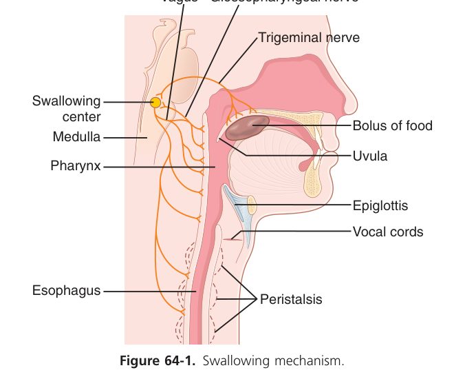
Swallowing alone is a brilliant feat of coordination — about 25 muscles must fire in the right order over 1 second, or food goes down the wrong tube. It has three phases: voluntary (tongue pushes bolus to oropharynx); pharyngeal reflex (soft palate seals nasopharynx, epiglottis covers larynx, UES opens, peristalsis sweeps bolus down); oesophageal (peristalsis carries bolus to stomach, LES relaxes). Once in the stomach, food doesn’t just sit — the antrum mixes and grinds while the fundus relaxes (receptive relaxation) to accommodate without pressure rise. Gastric emptying rate is tightly controlled: fluids > solids; small > large particles; carbohydrate > protein > fat. Fat triggers maximum CCK release, which slows emptying to give the small intestine time to digest it — the enterogastric reflex.
- Receptive relaxation — fundus/body relax on swallowing to accommodate food (vagal).
- Peristalsis — waves from body to antrum; the antrum mixes & grinds; only a little chyme passes the pyloric sphincter each wave (retropulsion).
- Tonic contraction — sustained background tone.
Gastric emptying = expulsion of chyme from stomach into duodenum. Rate: fluids > solids; small > large particles; iso-osmotic > hyper-/hypo-osmotic; carbohydrate > protein > fat. A regular meal empties in ~4–6 h.
- Promoted by: gastric distension (→ vago-vagal & intrinsic reflex) and gastrin.
- Inhibited by the enterogastric reflex (duodenal fat, acid, hyperosmolarity, stretch) and hormones secretin & GIP.
Pancreatic Juice & Bile
pH 7.8–8.4, ~1500 mL/day, isotonic. Two parts:
- HCO₃⁻ (from duct cells) — neutralises gastric acid (protects duodenal mucosa) & gives the alkaline pH pancreatic enzymes need. Driven by secretin.
- Enzymes (from acinar cells) — three groups: proteases, lipase, amylase. Driven by CCK & vagus.
| Class | Enzyme(s) | Note |
|---|---|---|
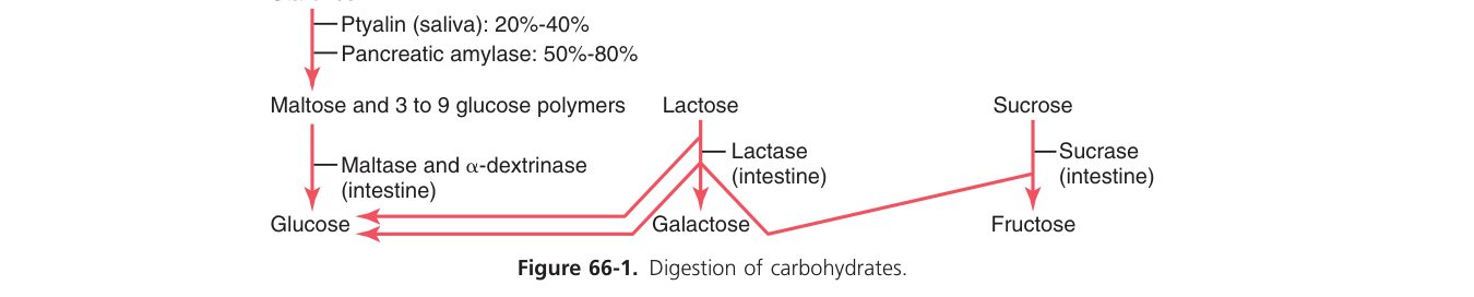
| Carbohydrate | Pancreatic amylase | Starch → maltose/oligosaccharides |
| Fat | Lipase (+ colipase), cholesterol esterase, phospholipase | TG → monoglyceride + fatty acids |
| Protein | Trypsinogen, chymotrypsinogen, procarboxypeptidase | Secreted as inactive zymogens |
Proteases are stored/secreted as inactive zymogens, plus a trypsin inhibitor is present. Enterokinase (intestinal brush border) activates trypsinogen → trypsin in the lumen; trypsin then activates the rest. Premature activation → acute pancreatitis.
- Made by the liver, stored/concentrated in the gallbladder; contains NO digestive enzymes.
- Bile salts are amphipathic: they emulsify fat (↑surface area for lipase) and form micelles that carry monoglycerides, fatty acids & fat-soluble vitamins (A, D, E, K) to the mucosa for absorption.
- Enterohepatic circulation: ~95% of bile salts are reabsorbed in the terminal ileum and reused.
Bile-salt/bile-flow problems (obstruction, ileal disease) → fat malabsorption & steatorrhoea + deficiency of vitamins A, D, E, K.
Small-Intestinal Digestion & Absorption
If the small intestine were a smooth tube, it would have ~0.5 m² of absorbing surface — nowhere near enough. Instead, three nested folds blow that surface to ~200 m², roughly the area of a tennis court. The mucosa is gathered into circular folds (plicae); the surface of each fold carries finger-like villi; and every villus epithelial cell is itself capped by hundreds of microvilli (the brush border). Each villus has its own capillary network and a central lymphatic lacteal — sugars and amino acids leave through capillaries to the portal vein, fats leave as chylomicrons via the lacteal to lymph (and so bypass the liver first-pass). Combined with the long transit time (~3-8 h), this is why 90% of absorption happens here.
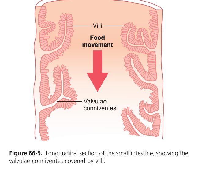
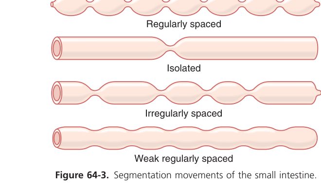
Huge surface area (plicae × villi × microvilli ≈ 200–250 m²), rich blood/lymph supply, brush-border enzymes, and a long transit time (chyme stays 3–8 h). Intestinal juice (pH ~7.6, 1–3 L/day) supplies enterokinase + brush-border maltase/lactase/peptidase. Motility: tonic contraction, segmentation (mixing), peristalsis (propulsion).
| Nutrient | Absorbed as | Mechanism | Route |
|---|---|---|---|
| Carbohydrate | Monosaccharides | Glucose/galactose = secondary active (Na⁺ cotransport, SGLT); fructose = facilitated diffusion (GLUT5) | Blood (portal) |
| Protein | Amino acids, di-/tri-peptides | Na⁺-coupled secondary active transport | Blood (portal) |
| Fat | Monoglycerides + fatty acids (via micelles) | Diffuse in → re-formed into TG → chylomicrons | Lymph (lacteal); short/medium-chain FA → blood |
Fat is the odd one out — long-chain fats leave the gut as chylomicrons via lymph (lacteals), while sugars & amino acids go straight into portal blood. Glucose follows Na⁺ ("where sodium goes, glucose follows").
Large Intestine, Defecation & Vomiting
- Water & electrolyte absorption — ~1.5 L of ileal effluent per day is reduced to ~100 mL of stool (Na⁺ absorption is driven by ENaC; H₂O follows osmotically).
- Storage of faeces (proximal colon mixes; distal colon stores).
- Bacterial fermentation of undigested carbohydrate → short-chain fatty acids (acetate, propionate, butyrate — energy for colonocytes), vitamin K, gas.
- Mucus secretion for lubrication.
- Haustration — segmental sac-like contractions that knead the contents.
- Mass movement — 1–3 powerful peristaltic waves per day, often after a meal (gastrocolic reflex), propelling contents toward the rectum.
- Defecation reflex: rectal distension → parasympathetic (S2–S4) → colonic contraction + internal anal sphincter relaxation (involuntary); voluntary relaxation of the external sphincter (pudendal, somatic) allows evacuation; the Valsalva manoeuvre assists.
Coordinated by the vomiting centre in the medulla oblongata. Triggers reach it via: chemoreceptor trigger zone (CTZ, area postrema — sensitive to toxins, opioids, dopamine, 5-HT3), vestibular system (motion), GI vagal afferents (irritation), and higher centres (sights/smells/emotion).
Sequence: deep inspiration → glottis closes → soft palate elevates → abdominal/diaphragmatic contraction raises intragastric pressure → lower oesophageal sphincter relaxes → contents expelled. Consequences: loss of H⁺ & Cl⁻ → hypochloraemic metabolic alkalosis, dehydration, hypokalaemia.
Antiemetic targets follow the CTZ pharmacology: ondansetron (5-HT3 antagonist), metoclopramide (D2 antagonist + prokinetic), cyclizine (H₁ antagonist for motion sickness), aprepitant (NK1 antagonist for chemotherapy).
Digestive system complete
Smooth muscle, hormones, secretions, motility, absorption, colon & defecation mastered. Next: Renal/Urinary.