Respiratory Physiology
Lung Volumes, Capacities & Ventilation
Take a normal breath in, then breathe out normally. You just moved about 500 mL of air — the tidal volume. But your lungs can hold far more than that. You can inhale further (~3000 mL more, the inspiratory reserve), or exhale further (~1100 mL, the expiratory reserve). And even after the deepest possible exhale, your lungs still hold ~1200 mL that simply cannot be blown out — the residual volume — because the chest wall’s outward pull keeps the lungs partly expanded. These four primary volumes add up to total lung capacity (~5800 mL in an adult man).
Why does this matter? Because almost every lung disease shifts one of these numbers. Asthma traps air → RV rises. Pulmonary fibrosis stiffens lungs → all volumes shrink. A spirometer (a sealed bell over water) records every breath as a tracing — but it cannot measure RV (because RV never leaves the lungs), so capacities containing RV (FRC, TLC) require helium dilution or body plethysmography. Knowing what spirometry can and cannot show is half the clinical use.
| Term | Definition | Typical (adult ♂) |
|---|---|---|
| Tidal volume (TV) | Air in/out per quiet breath | ~500 mL |
| Inspiratory reserve volume (IRV) | Extra air inhaled beyond TV | ~3000 mL |
| Expiratory reserve volume (ERV) | Extra air exhaled beyond TV | ~1100 mL |
| Residual volume (RV) | Air left after maximal expiration (cannot be exhaled) | ~1200 mL |
| Vital capacity (VC) | IRV + TV + ERV = max air expelled after max inspiration | ~4600 mL |
| Functional residual capacity (FRC) | ERV + RV (air left after quiet expiration) | ~2300 mL |
| Total lung capacity (TLC) | VC + RV | ~5800 mL |
RV, FRC & TLC contain RV, so they cannot be measured by spirometry (need helium dilution/body plethysmography).
Dead space = the volume that is ventilated but takes no part in gas exchange (anatomical: conducting airways ~150 mL). Only the air reaching alveoli (TV − dead space) is useful.
Mechanics of Breathing & Surfactant
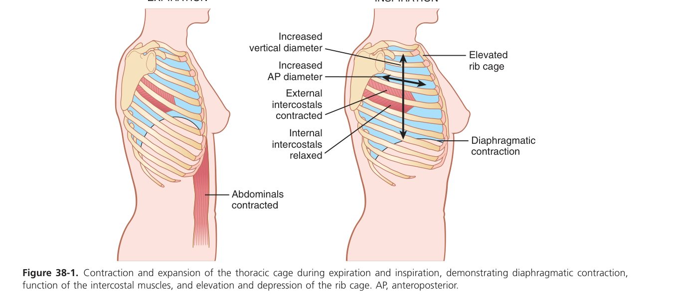
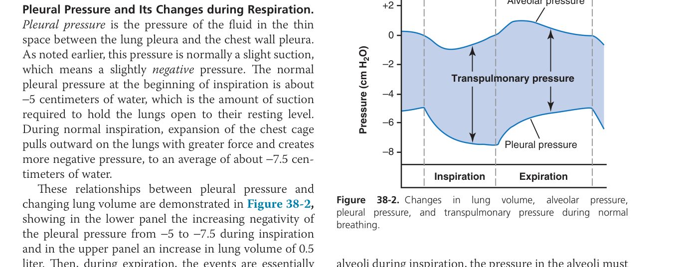
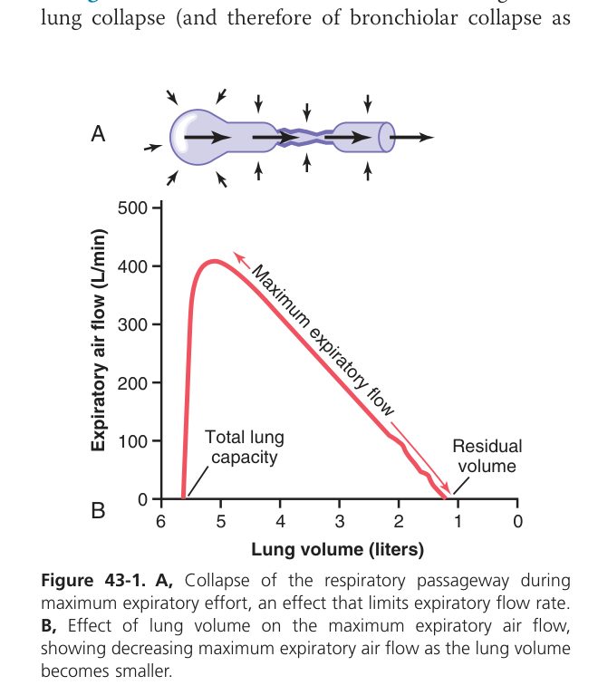
Air doesn’t flow into your lungs by magic. It flows because you create a pressure gradient. When the diaphragm contracts and the chest wall expands, the intrapleural space becomes more negative (it’s already slightly negative at rest, ~−5 cmH₂O); the lungs follow the chest wall outward; alveolar pressure drops below atmospheric; air rushes in. Expiration is the reverse, and at rest it’s passive — elastic recoil of the lung tissue. The whole system runs on two opposing forces: chest wall wants to spring outward, lungs want to collapse inward. They tug against each other across the pleural space, leaving the lungs gently stretched at rest.
The lung’s collapse-tendency has two sources. Tissue elasticity (elastin fibres) accounts for ~1/3. The other 2/3 is something more subtle: surface tension at the alveolar air-water interface. Wherever water meets air, the water molecules pull on each other, trying to minimise surface area — and in an alveolus that means pulling inward. Without something to counter this, alveoli would collapse like soap bubbles, especially the small ones (Laplace: P = 2T/r). That something is surfactant, made by Type II pneumocytes — you’ll meet it next.
- Inspiration is active (diaphragm + external intercostals contract → thorax expands → intra-alveolar pressure falls below atmospheric → air in). Quiet expiration is passive (elastic recoil).
- Intrapleural pressure is negative (~−5 cmH₂O) — keeps the lung expanded against its recoil. If air enters the pleural space (pneumothorax), the lung collapses.
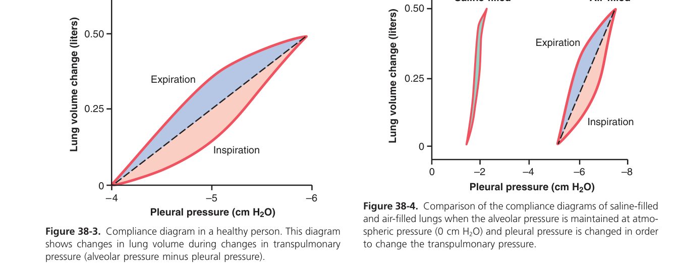
- Compliance = ΔV/ΔP — the distensibility of the lung. Surface tension at the air–liquid interface reduces compliance and (by Laplace, P = 2T/r) rises as an alveolus shrinks, tending to collapse small alveoli into large ones.
Here is the elegance of surfactant in one line: it solves the small-alveolus problem. Without it, the smallest alveoli would collapse and empty into bigger ones (Laplace), and the lung would degenerate into a few huge sacs with terrible exchange. Surfactant is a phospholipid (dipalmitoyl-phosphatidylcholine) that lines the alveolar air-water interface and dramatically lowers surface tension — and its effect is greater where it’s more concentrated, which is exactly the small alveoli where the molecules pack tighter. So small alveoli get the biggest tension reduction, stop collapsing, and stay open. That is also why a premature baby (born before Type II pneumocytes can make enough surfactant, <~34 weeks) develops neonatal respiratory distress syndrome — stiff lungs, atelectasis, severe hypoxia — and why we give the mother steroids before premature delivery to accelerate surfactant production.
Surfactant is a phospholipid (mainly dipalmitoyl-phosphatidylcholine) secreted by type II alveolar (pneumo)cytes; it adsorbs to the alveolar air–liquid interface and lowers surface tension.
Physiological significance:
- ↓ Surface tension → ↑ lung compliance → easier inflation (less work of breathing).
- Stabilises alveoli of different sizes — its effect is greater in small alveoli, so small ones don’t empty into large ones (prevents collapse, anti-Laplace).
- Prevents atelectasis (alveolar collapse) at end-expiration.
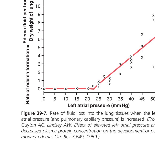
- Keeps alveoli "dry" — reduced surface tension lowers the pull that would draw fluid into the alveoli (anti-oedema).
Neonatal respiratory distress syndrome (NRDS / hyaline membrane disease) — premature babies (<~34 weeks) lack surfactant → high surface tension → stiff lungs, atelectasis, hypoxia. Managed with antenatal steroids (mature type II cells) & exogenous surfactant.
Restrictive vs Obstructive Disease
FVC = forced vital capacity (max air forcibly exhaled after max inspiration). FEV₁ = volume exhaled in the first second. The FEV₁/FVC ratio distinguishes the two patterns:
| Feature | Obstructive (asthma, COPD) | Restrictive (fibrosis, NRDS) |
|---|---|---|
| Problem | ↑ airway resistance (can’t get air out) | ↓ compliance / expansion (can’t get air in) |
| FEV₁ | ↓↓ (markedly reduced) | ↓ (proportionate) |
| FVC | ↓ (slightly) | ↓↓ |
| FEV₁/FVC | ↓ (<70%) | Normal or ↑ |
| Lung volumes (RV/TLC) | ↑ (air trapping) | ↓ (small stiff lungs) |
Gas Exchange & V/Q Matching
Gas exchange happens in the deepest 0.5 µm of your lung. An O₂ molecule has to cross five layers to reach the haemoglobin in a red cell: surfactant, type I pneumocyte, fused basement membrane, capillary endothelium, plasma. Each layer is barely there, so under healthy conditions diffusion is staggeringly fast — an RBC equilibrates with alveolar air in ~0.25 seconds, only a third of its transit through the capillary. That huge margin is why even with one healthy lung you can do most daily activities. But thicken the membrane (oedema, fibrosis, pneumonia) and equilibration becomes the slow step — especially during exercise when transit time is shortest — and the patient becomes hypoxic.
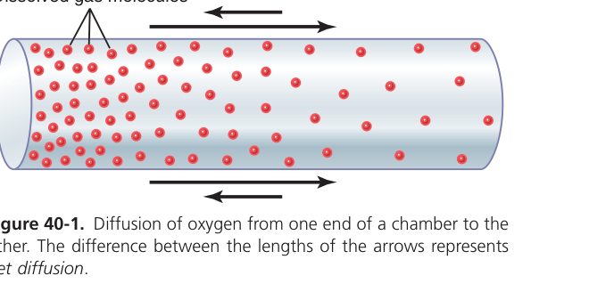
Gases diffuse down partial-pressure gradients across the respiratory (blood–air) membrane. Oxygen crosses: surfactant layer → type I pneumocyte (alveolar epithelium) → fused basement membrane → capillary endothelium → plasma → RBC membrane → Hb — no smooth muscle is in the path.
Airflow order (inhalation): nasal cavity → naso-pharynx → larynx → trachea → bronchi → bronchioles → alveoli.
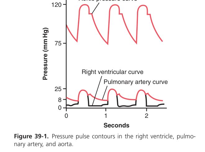
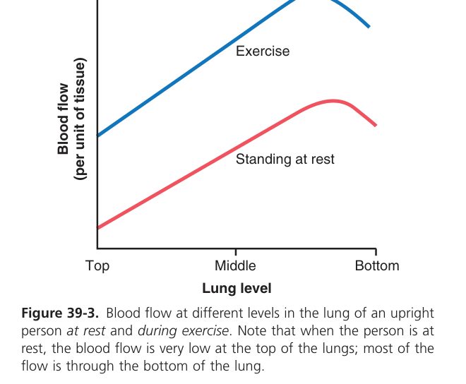
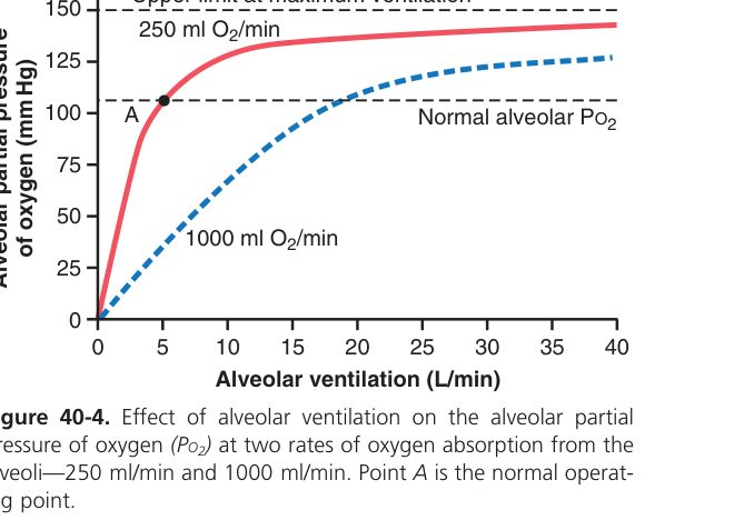
Two things must reach every alveolus for gas exchange to work: air (ventilation, V) and blood (perfusion, Q). If only one arrives, no gas exchange happens. A region with air but no blood is wasted ventilation (high V/Q, dead-space-like — think pulmonary embolism). A region with blood but no air is wasted perfusion (low V/Q, shunt-like — think pneumonia, atelectasis). The average V/Q across normal lungs is ~0.8, but it varies regionally. In an upright person, both V and Q are higher at the base than the apex (gravity), but Q grows faster — so the apex is relatively over-ventilated (V/Q ~3) and the base relatively under-ventilated (V/Q ~0.6). The body has one elegant fix: hypoxic pulmonary vasoconstriction — alveoli that are poorly ventilated get their blood diverted away (the opposite of every other vascular bed, where hypoxia causes dilatation). This redirects blood toward better-ventilated regions and protects oxygenation.
V/Q ratio = alveolar ventilation ÷ pulmonary blood flow. Overall ~0.8. Ideal exchange needs ventilation matched to perfusion.
- High V/Q (ventilation > perfusion) → wasted ventilation (dead-space-like); seen at the lung apex & in pulmonary embolism.
- Low V/Q (perfusion > ventilation) → wasted perfusion (shunt-like); seen at the base & in airway obstruction/pneumonia.
- Hypoxic pulmonary vasoconstriction diverts blood away from poorly ventilated alveoli to improve matching.
Therefore diffusion is worsened by ↓area (emphysema), ↑thickness (oedema/fibrosis), and ↓gradient (altitude). DLCO is the diffusion capacity for CO and is the clinical test — reduced in fibrosis, emphysema, anaemia.
| Type | Mechanism | PaO₂ | SaO₂ | CaO₂ |
|---|---|---|---|---|
| Hypoxic hypoxia | Low alveolar PO₂ (altitude, hypoventilation, V/Q mismatch, diffusion defect, shunt) | ↓ | ↓ | ↓ |
| Anaemic hypoxia | ↓Hb or dysfunctional Hb (anaemia, CO poisoning, methaemoglobinaemia) | Normal | Normal* | ↓ |
| Stagnant (ischaemic) | Reduced blood flow (shock, heart failure, local thrombosis) | Normal | Normal | Normal (arterial), ↓ tissue |
| Histotoxic | Tissue cannot use O₂ (cyanide blocks cytochrome oxidase) | Normal | Normal | Normal — ↑venous PO₂ |
*Pulse oximetry over-reads in CO poisoning because it cannot distinguish HbCO from HbO₂.
• V/Q at rest, apex vs base (upright)? → apex high (~3), base low (~0.6), average ~0.8
• Unique pulmonary vascular response to hypoxia? → vasoconstriction (everywhere else it’s vasodilation) — diverts blood to better-ventilated alveoli
• Four types of hypoxia? → hypoxic / anaemic / stagnant / histotoxic
• CO poisoning gives which hypoxia + curve shift? → anaemic; left shift → tissue hypoxia despite normal PaO₂
Oxygen Transport & the O₂–Hb Dissociation Curve
If oxygen had to dissolve in water to travel in your blood, you would last about 4 seconds — plasma can dissolve barely 0.3 mL of O₂ per dL of blood. Haemoglobin solves this. Each Hb molecule carries up to four O₂ (one per haem-iron), giving ~20 mL O₂ per dL of blood — nearly 70 times the plasma alone. That’s the entire reason red cells exist. The other ~1.5% of O₂ transport stays dissolved in plasma, and although tiny, it’s the part that actually equilibrates with the alveolus and with tissue — it sets PaO₂, which then determines how saturated Hb becomes.
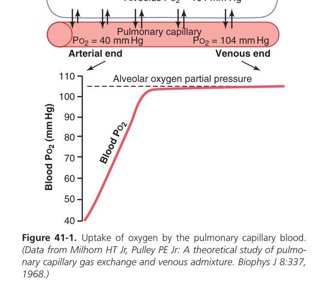
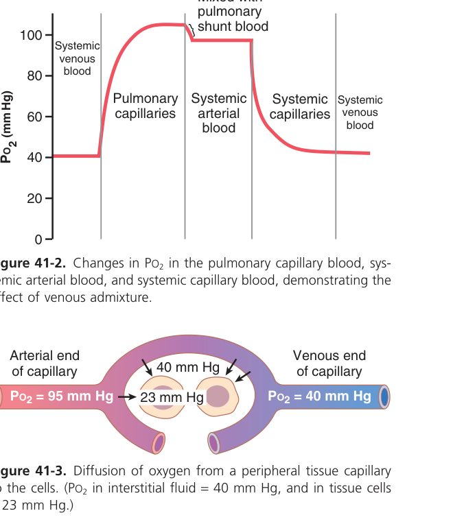
O₂ travels ~98.5% bound to haemoglobin (as oxyhaemoglobin; 1 g Hb carries ~1.34 mL O₂) and ~1.5% dissolved in plasma. O₂ saturation of Hb = the % of available Hb O₂-binding sites that are occupied.
The O₂-Hb dissociation curve is sigmoid because each O₂ that binds makes the next one bind more easily — cooperative binding. The shape is what makes Hb so good at its job: the flat top (PaO₂ > ~60 mmHg) means even modest oxygen drops don’t affect saturation much, so Hb fully loads in the lungs even when alveolar PO₂ isn’t perfect. The steep middle (PaO₂ 20-40 mmHg) is where most unloading happens at the tissue — a small fall in PO₂ releases a large amount of O₂, exactly when tissues need it. P₅₀ — the PO₂ at 50% saturation — is the curve’s landmark, ~27 mmHg normally. Drag the curve right (Bohr effect: more CO₂, more H⁺, warmer, more 2,3-DPG) and unloading is easier; drag it left (CO, fetal Hb) and Hb holds onto its O₂ tighter.
The O₂–Hb curve plots O₂ saturation against PO₂. It is sigmoid (S-shaped) (cooperative binding): a flat upper plateau (loading in lungs is safe despite PO₂ falls) and a steep lower part (large O₂ unloading for small PO₂ falls in tissues). P₅₀ ≈ 26–27 mmHg (PO₂ at 50% saturation).
| Shift | Hb–O₂ affinity | Effect | Causes |
|---|---|---|---|
| Right shift | Decreased | Facilitates O₂ unloading to tissues | ↑CO₂, ↑H⁺ (↓pH), ↑temperature, ↑2,3-DPG, exercise (the Bohr effect) |
| Left shift | Increased | Facilitates O₂ loading in lungs (holds O₂ tighter) | ↓CO₂, ↓H⁺ (↑pH), ↓temperature, ↓2,3-DPG, CO, fetal Hb |
The Bohr effect = an increase in CO₂/H⁺ (as in metabolising tissue) lowers Hb’s O₂ affinity (right shift), promoting O₂ release exactly where it is needed.
Carbon monoxide (CO) binds Hb ~240× tighter than O₂, forming carboxyhaemoglobin and left-shifting the curve → severe tissue hypoxia despite "normal" PO₂. Fetal Hb (HbF) has higher O₂ affinity (left-shifted) to draw O₂ across the placenta.
Carbon Dioxide Transport
| Form | ~% | Note |
|---|---|---|
| Bicarbonate (HCO₃⁻) | ~70% | CO₂ + H₂O ↔ H₂CO₃ ↔ H⁺ + HCO₃⁻ (carbonic anhydrase in RBC); the chloride shift moves Cl⁻ in as HCO₃⁻ leaves |
| Carbamino-Hb | ~23% | CO₂ bound to Hb amino groups |
| Dissolved | ~7% | In plasma |
The Haldane effect = oxygenation of Hb in the lungs reduces its ability to carry CO₂, promoting CO₂ release. (The counterpart of the Bohr effect.)
Control of Breathing
Breathing has to be automatic (you can’t consciously choose to breathe every second), yet also responsive (it must speed up when you exercise or fall asleep). The brainstem solves this with a respiratory centre in the medulla, modulated by the pons, that fires the diaphragm and intercostals in rhythm without conscious input. But the rhythm is constantly adjusted by the body’s chemistry. The central chemoreceptors (medulla) are the most important: they don’t sense O₂ or CO₂ directly — they sense the H⁺ in CSF, which rises when CO₂ crosses the blood-brain barrier and forms H₂CO₃. So when arterial CO₂ rises (e.g. during exercise, or because you held your breath), CSF H⁺ rises within seconds, ventilation accelerates, CO₂ falls. The peripheral chemoreceptors (carotid + aortic bodies) are the backup: they sense PaO₂ directly, but they only fire strongly when PaO₂ drops below ~60 mmHg. That’s why severe hypoxia drives breathing only in extreme cases — CO₂ is the primary stimulus, hypoxia is the safety net.
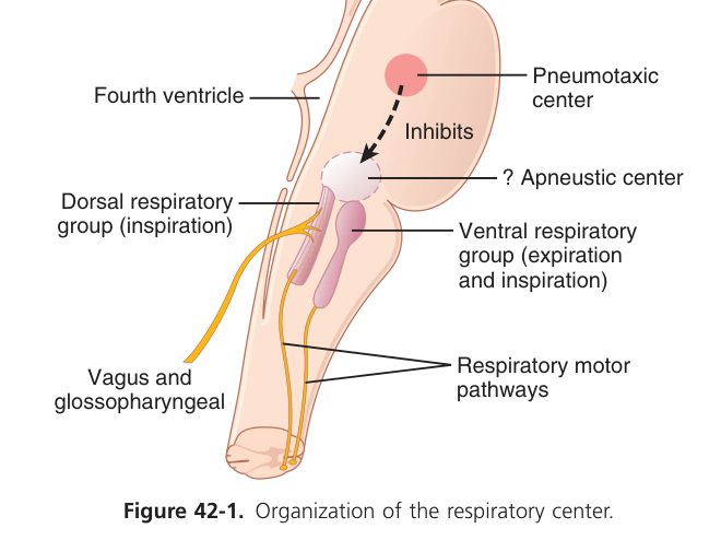
- Respiratory centre in the medulla oblongata (dorsal & ventral respiratory groups) sets the basic rhythm; the pons (pneumotaxic/apneustic) modulates it. The medulla is also the vital centre for cardiovascular & GI control.
- Central chemoreceptors (in the medulla) are the most important drive — they respond to arterial PCO₂ (via H⁺ produced in the CSF). A rise in PCO₂ powerfully increases ventilation.
- Peripheral chemoreceptors — the carotid & aortic bodies — respond mainly to ↓arterial PO₂ (hypoxia), and also to ↑CO₂ / ↓pH.
In chronic CO₂ retainers (severe COPD), central chemoreceptors become desensitised and the hypoxic drive (peripheral) dominates — giving high-flow O₂ can paradoxically suppress breathing.
- Hering–Breuer inflation reflex: pulmonary stretch receptors fire when the lung is over-inflated → inhibit inspiration via vagal afferents — protective at high tidal volumes; minor at rest in adults but important in infants.
- J receptors (juxta-capillary): respond to pulmonary congestion / oedema → rapid shallow breathing, dyspnoea.
- Irritant receptors: cough, bronchoconstriction.
The lungs control CO₂, the volatile acid; the kidneys control HCO₃⁻. The Henderson–Hasselbalch frame: pH ∝ HCO₃⁻ / PCO₂.
| Disturbance | Primary change | Compensation | Cause |
|---|---|---|---|
| Respiratory acidosis | ↑PCO₂, ↓pH | Renal ↑HCO₃⁻ (slow, days) | Hypoventilation, COPD, opioid OD |
| Respiratory alkalosis | ↓PCO₂, ↑pH | Renal ↓HCO₃⁻ | Hyperventilation, anxiety, altitude |
| Metabolic acidosis | ↓HCO₃⁻, ↓pH | Hyperventilation (Kussmaul) → ↓PCO₂ | DKA, lactic acidosis, renal failure |
| Metabolic alkalosis | ↑HCO₃⁻, ↑pH | Hypoventilation → ↑PCO₂ | Vomiting, diuretics |
Climb a high mountain and atmospheric pressure falls, so PaO₂ falls — hypoxic hypoxia. The body adapts on three timescales. Within seconds, peripheral chemoreceptors drive hyperventilation (which paradoxically causes respiratory alkalosis — the kidneys take a day or two to compensate by dumping bicarbonate). Within days, 2,3-DPG rises in red cells, right-shifting the O₂-Hb curve to favour unloading at tissue. Within weeks to months, hypoxia drives renal EPO release → polycythaemia → more Hb to carry the available O₂. Capillaries also proliferate in muscle. Acclimatisation is real and impressive: Sherpas live and work at altitudes where unacclimatised climbers struggle to think. When acclimatisation fails — usually because the climb is too fast — you get acute mountain sickness, and if untreated, life-threatening HAPE (pulmonary oedema) or HACE (cerebral oedema).
Atmospheric pressure falls with altitude → lower PO₂ → hypoxic hypoxia. Acclimatisation over days–weeks:
- Acute: peripheral chemoreceptors drive hyperventilation → respiratory alkalosis (renal HCO₃⁻ loss compensates over days).
- ↑2,3-DPG in RBCs → right shift → eases O₂ unloading.
- Hypoxia → EPO release from kidney → polycythaemia (weeks).
- ↑Capillary density, ↑mitochondria, hypoxic pulmonary vasoconstriction (can cause pulmonary hypertension).
- Failure to acclimatise → acute mountain sickness, HAPE, HACE.
Respiratory complete
Volumes, surfactant, O₂/CO₂ transport & control mastered. Next: Digestive (GI).