Blood
Composition & Physicochemical Properties
Blood is the only tissue in the body you can hold in a tube. That alone makes it the most-tested system in the lab, and it is also the way every other organ gets fed, heated, defended, and kept at the right pH. Think of blood as five jobs done at once by one fluid: it transports O₂, CO₂, nutrients, hormones; it buffers pH; it moves heat around the body; it seals leaks through clotting; and it defends through white cells and antibodies. Read every fact below knowing it serves one of those five jobs.
In adults blood makes up about 8% of body weight — roughly five litres — and splits cleanly into two parts. Spin it in a tube and you see plasma (the straw-coloured liquid, ~55%) sitting on top of the formed elements (~45%, almost all of it red cells). The percentage of red cells in that tube is the haematocrit, and it shifts the moment anything goes wrong — falling in anaemia, rising in dehydration or polycythaemia. That single number gives you a quick read of the whole system.
Functions of blood (5): transport · buffering (pH) · temperature regulation · haemostasis · defence (immunity). Blood = formed elements (cells/fragments) suspended in plasma (the liquid internal environment).
| Parameter | TMU value |
|---|---|
| Total blood volume | ~8% of body weight; 70–80 mL/kg; 5000–6000 mL in a 70-kg man |
| Plasma fraction | ~55% of whole blood |
| Formed elements | ~45% (RBC ~45%, buffy coat WBC+platelets <1%) |
| Haematocrit | ♂ 40–50%; ♀ 37–48%; neonate 55% (= % volume occupied by RBC) |
- Water 91–92%.
- Plasma proteins 5–8% (60–80 g/L): albumin 40–50 g/L (osmotic, transport), globulin 15–30 g/L (immune, transport), fibrinogen (clotting). Made in the liver (except γ-globulins from plasma cells).
- Electrolytes: plasma — Na⁺ & Cl⁻ are major; ICF — K⁺ & HPO₄²⁻ are major.
- Organic compounds (amino acids, hormones, enzymes, nitrogenous waste, nutrients) & gases (O₂, CO₂, N₂).
Transport · nourishment · immune function · colloid osmotic pressure · blood coagulation · buffering.
| Property | Blood | Plasma | Determined by |
|---|---|---|---|
| Specific gravity | 1.050–1.060 | 1.025–1.030 (RBC 1.090–1.092) | RBC number / plasma protein / Hb |
| Viscosity (water = 1) | 4–5 | 1.6–2.4 | RBC number / plasma protein |
Two completely different osmotic pressures live in your plasma, and they protect two completely different things. The big one (~99% of the total) comes from electrolytes — tiny ions like Na⁺ and Cl⁻, which are too numerous and small to ignore. This crystalloid osmotic pressure decides whether water flows in or out of cells; if it drops, cells swell, if it rises, cells shrivel. The much smaller one (~25 mmHg) comes from plasma proteins, mainly albumin. These are too big to cross capillary walls, so this colloid osmotic pressure decides whether water stays in your capillaries or leaks out into the tissues. Lose albumin (liver failure, nephrotic syndrome, kwashiorkor) and you lose that pull — fluid escapes and the patient swells. That is the entire mechanism of one of the most common signs in medicine, oedema.
Total plasma osmotic pressure ≈ 313 mOsm. Osmotic pressure depends on the NUMBER of particles, NOT their chemical nature or size. Two components:
| Component | Generated by | Magnitude | Physiological role |
|---|---|---|---|
| Crystalloid OP | Electrolytes (mainly NaCl) — small, numerous | Large (~99%) | Maintains intra-/extra-cellular water balance (cells cross the cell membrane) |
| Colloid OP | Plasma proteins (mainly albumin) — large, few | Small (~1.3 mOsm, ~25 mmHg) | Maintains intra-/extra-capillary (plasma vs interstitial) water balance |
Low albumin (liver failure, nephrotic syndrome, malnutrition) → ↓ colloid OP → fluid leaves capillaries into the interstitium → oedema. Crystalloid OP guards cells (cell swelling/shrinkage); colloid OP guards capillary fluid balance.
0.9% NaCl & 5% glucose are both iso-osmotic and isotonic. 1.9% urea is iso-osmotic but NOT isotonic — urea crosses the RBC membrane, so the cell still takes up water and lyses (tonicity counts only the non-penetrating particles).
Plasma pH = 7.35–7.45 (narrow). <7.35 = acidosis, >7.45 = alkalosis; the range compatible with life is 6.8–7.8.
- Buffers: bicarbonate (NaHCO₃/H₂CO₃ — main), plasma proteins, phosphate, and haemoglobin (the major RBC buffer).
- Organ regulation: the respiratory system (blows off CO₂ → ↓H⁺) and the kidneys (excrete fixed acids: sulfuric, phosphoric).
• What does the haematocrit measure? → % volume occupied by RBCs (♂ 40-50%, ♀ 37-48%)
• What determines osmotic pressure? → number of particles — not size or composition
• Which OP protects cells vs capillaries? → crystalloid OP → cells; colloid OP (albumin) → capillaries
• Plasma pH range? → 7.35–7.45 (life-compatible 6.8–7.8)
Erythrocytes (Red Blood Cells)
A red blood cell is a beautifully specialised vehicle — and almost every weird thing about it is in service of one job: carrying as much oxygen as possible without getting stuck in tiny capillaries. It is shaped like a biconcave disc (a doughnut without the hole), which gives it far more surface area per volume than a sphere — so O₂ loads and unloads fast across the whole surface. It has thrown out its nucleus and organelles, freeing the entire inside for one molecule: haemoglobin. And it is endlessly flexible — an 8 µm disc has to squeeze through 3 µm splenic slits without rupturing, billions of times in its 120-day life.
A haemoglobin molecule is a four-piece team: four globin chains (2 α + 2 β), each cradling one haem ring with an iron atom. Each iron can grip one O₂, so one Hb carries four O₂. That iron must stay Fe²⁺; oxidise it to Fe³⁺ and you get methaemoglobin, which cannot carry oxygen at all. This is why so much of physiology obsesses over Hb — it is both your gas-transport molecule and a major plasma buffer.
| Feature | Value |
|---|---|
| Shape | Biconcave disc, non-nucleated, no organelles |
| Diameter / volume | 7–8 µm; thickness 2.5 µm (edge) & ~1 µm (centre); MCV 90–95 fL |
| RBC count | ♂ 4.5–5.5 × 10¹²/L; ♀ 3.8–4.6 × 10¹²/L |
| Haemoglobin | ♂ 120–160 g/L (~15 g/dL); ♀ 110–150 g/L |
| Lifespan | ~120 days |
The biconcave shape gives a large surface-area:volume ratio (~140 µm² : 90 µm³) → fast gas diffusion, flexibility to squeeze through capillaries (<1 µm pores), and tolerance of volume change. Hb = globin (4 chains: 2α+2β) + 4 haem (Fe²⁺); functions: O₂ & CO₂ transport and acid–base buffering.
- Plastic deformation: RBCs deform to pass capillaries/sinusoid pores <1 µm.
- Osmotic fragility: sensitivity to osmotic change. In hypotonic solution the RBC swells & bursts (haemolysis); in hypertonic it shrinks (crenation). Normal haemolysis begins at 0.42–0.45% NaCl and is complete at 0.30–0.33% NaCl. Increased fragility → haemolysis at higher salt (e.g. hereditary spherocytosis).
- Suspension stability & ESR: RBCs stay evenly suspended; the erythrocyte sedimentation rate (ESR) measures how far they settle in 1 h. Normal ♂ 0–15 mm/h, ♀ 0–20 mm/h.
ESR depends mainly on plasma factors (not the RBC). Rouleaux formation (stacking of RBCs) makes them settle faster → ↑ ESR. ESR ↑ with fibrinogen, globulins, cholesterol (inflammation, pregnancy, TB); ESR ↓ with albumin, lecithin.
• RBC lifespan? → ~120 days
• Three RBC characteristics? → plastic deformation · osmotic fragility · suspension stability (ESR)
• What raises ESR? → rouleaux from ↑ fibrinogen/globulins (inflammation, pregnancy)
• Hb structure? → 4 globin chains (2α+2β) + 4 haem with Fe²⁺
Erythropoiesis & Its Regulation
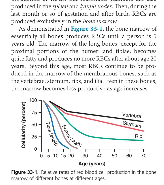
If a red cell only lives 120 days, your body has to replace them at an astonishing rate — about 2–3 million per second, every second of your life. That production line lives in your red bone marrow, and it starts from one extraordinary cell: a pluripotent haematopoietic stem cell (PHSC) that can become any blood cell. The PHSC divides into committed lineages, each producing one cell type. In adults the marrow narrows to the flat bones (sternum, ribs, pelvis, vertebrae) and the ends of long bones; in babies, every bone is busy making blood.
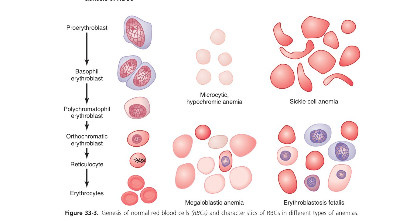
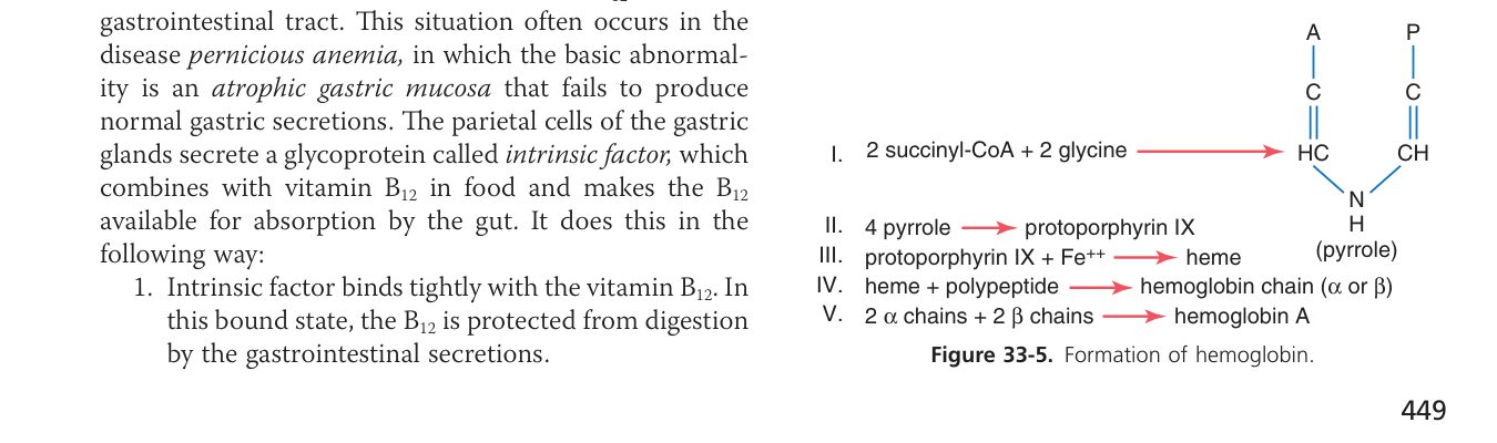
Building a red cell needs two distinct ingredient pools, and missing one gives a different disease. Iron and protein are the bricks of haemoglobin itself — lack them and the cells come off the line small and pale (microcytic, hypochromic anaemia). Vitamin B₁₂ and folate are needed to make DNA so the nucleus can mature in step with the cytoplasm — lack them and you get oversized, immature-nucleated cells (megaloblastic anaemia). And to absorb B₁₂ you also need intrinsic factor from the stomach’s parietal cells — lose those (autoimmune gastritis) and even a perfect diet cannot save you (pernicious anaemia).
| Requirement | Role | Deficiency → |
|---|---|---|
| Iron + protein | Build haemoglobin (Fe in haem) | Microcytic hypochromic (iron-deficiency) anaemia — small cell, low Hb |
| Vitamin B₁₂ + folic acid | DNA synthesis → nuclear maturation/division | Megaloblastic (macrocytic) anaemia — large cells |
| Intrinsic factor (parietal cells of stomach) | Needed to absorb B₁₂ in the ileum | Pernicious anaemia (no IF → only ~1/50 B₁₂ absorbed) |
How does the marrow know when to crank production up or back down? The kidney watches. Specialised cells in the renal cortex (peritubular interstitial cells) sense local oxygen, and when tissue O₂ falls — for any reason: blood loss, lung disease, high altitude — they release the hormone erythropoietin (EPO). EPO travels through blood to the marrow and tells erythroid precursors to multiply and finish maturation. Within a few days the haematocrit rises, more O₂ reaches the kidney, and EPO release falls back down. A textbook negative-feedback loop.
At the other end of life, after ~120 days, the cell membrane starts to fail. The spleen (the body’s red-cell graveyard) traps stiff old cells in its narrow sinuses, and macrophages there phagocytose them. The iron is salvaged and reused (we are remarkably stingy with iron); globin is broken down to amino acids; and the haem ring becomes bilirubin, which travels to the liver, gets conjugated, and is excreted in bile. When this breakdown runs faster than the liver can handle, bilirubin spills into the blood and stains the eyes and skin — jaundice.
- EPO is a hormone from the kidney (O₂ sensor); hypoxia / low blood O₂ → ↑EPO → ↑RBC production (a negative-feedback loop that raises O₂ carriage). Also stimulated by androgen, thyroid hormone, growth hormone.
- Destruction (after ~120 days): aged RBCs lose deformability → trapped & phagocytosed mainly in the spleen & liver (extravascular ~90%; intravascular ~10%). Hb → globin (→amino acids) + haem → iron (recycled) + bilirubin (→ liver → bile).
Chronic kidney disease → ↓EPO → anaemia (treated with recombinant EPO). High altitude / chronic hypoxia → ↑EPO → secondary polycythaemia. Excess RBC breakdown → ↑bilirubin → jaundice.
• Iron deficiency → what anaemia? → microcytic hypochromic
• B₁₂ / folate deficiency → what anaemia? → megaloblastic (macrocytic)
• Where does B₁₂ absorb + needs what? → terminal ileum; intrinsic factor (parietal cells)
• Two fates of haem? → iron recycled; bilirubin → liver → bile
Leukocytes (White Blood Cells)
White cells are the body’s mobile defence force. They make up <1% of blood volume, yet the moment a barrier breaks — a cut, an inhaled bacterium, a swallowed virus — they pour out of the bloodstream into the tissue to deal with it. The five types are not redundant: each is a specialist. Neutrophils are the front-line phagocytes, the first to arrive at any bacterial infection. Monocytes follow and become tissue macrophages, doing the longer cleanup and presenting bits of the enemy to the immune system. Lymphocytes are the immune memory — B cells make antibodies, T cells kill infected cells. Eosinophils target parasites and modulate allergy. Basophils (and their tissue cousin, the mast cell) release histamine in allergic reactions.
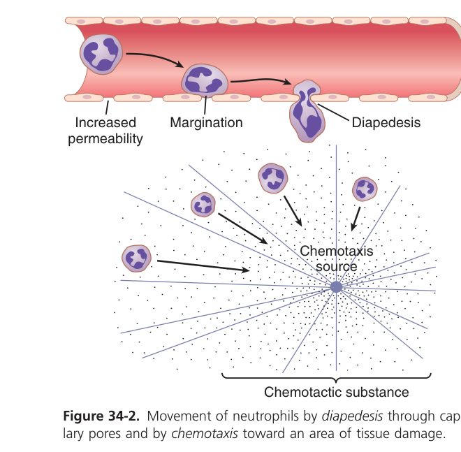
An important physiological move you must remember: WBCs don’t do their job in blood. They travel in blood; they work in tissue. To get there they squeeze between endothelial cells — a process called diapedesis. So a high blood neutrophil count doesn’t mean the neutrophils are killing bacteria in the bloodstream; it means the marrow is releasing them in droves to deploy into infected tissue.
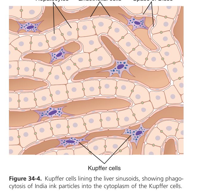
Total WBC = 4–10 × 10⁹/L. Two groups by granules: granulocytes (neutrophil, eosinophil, basophil) and agranulocytes (lymphocyte, monocyte).
| Cell | % (TMU) | Main function |
|---|---|---|
| Neutrophil | 50–70% | Phagocytosis & killing of bacteria (acute infection) |
| Lymphocyte | 20–40% | Immunity (B = antibody, T = cell-mediated) |
| Monocyte | 2–8% | → macrophage; phagocytosis, antigen presentation |
| Eosinophil | 1–4% | Anti-parasite; modulates allergy |
| Basophil | 0–1% | Histamine + heparin (allergy) |
Genesis: granulocytes & monocytes form only in bone marrow; lymphocytes & plasma cells mainly in lymphoid tissue (nodes, spleen, thymus, tonsils). Granulocytes/monocytes defend by phagocytosis; lymphocytes/plasma cells serve the immune system.
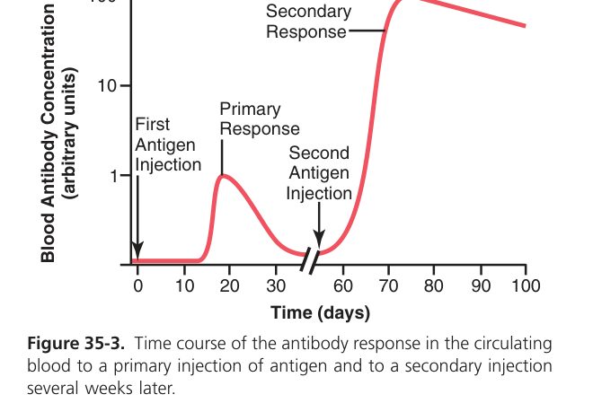
• Most abundant WBC + role? → neutrophil (50-70%) — phagocytosis of bacteria
• Immunity is the job of which cells? → lymphocytes (B = antibody, T = cell-mediated) + plasma cells
• What is diapedesis? → WBCs squeezing between endothelial cells to leave the bloodstream
• Allergy-related cells? → basophils (blood) + mast cells (tissue) release histamine
Platelets (Thrombocytes)
Platelets are unique in that they are not whole cells — they are pinched-off pieces of cytoplasm from giant marrow cells called megakaryocytes. Despite having no nucleus and a short life (about a week), they are the first responders to any vessel injury. They carry a tiny but vicious chemical arsenal in their granules: ADP, serotonin, thromboxane A₂, platelet-derived growth factor — everything needed to recruit more platelets and stop bleeding within seconds. Their production is driven by thrombopoietin (TPO), made mainly by the liver.
Platelets are anucleate cytoplasmic fragments (2–3 µm) shed from megakaryocytes in marrow; production is driven by thrombopoietin (TPO) from the liver. Count = 100–300 × 10⁹/L; lifespan 7–14 days (functional first ~2 days); removed in spleen/liver/lung.
| Property (the "5") | Detail |
|---|---|
| Adhesion | Bind exposed collagen via vWF & GPIb (after endothelial injury) |
| Aggregation | Clump via fibrinogen + GPIIb/IIIa; promoted by ADP, TXA₂, thrombin (↑Ca²⁺, ↓cAMP); inhibited by PGI₂ & NO |
| Release/secretion | Dense bodies (ADP, 5-HT, Ca²⁺); α-granules (fibrinogen, vWF); synthesise TXA₂ |
| Contraction | Actin/myosin contract → clot retraction |
| Adsorption | Concentrate clotting factors on their surface |
Functions: (1) maintain endothelial integrity; (2) haemostasis — form the platelet plug & provide phospholipid surface for coagulation.
Thrombocytopenia (↓count, <50×10⁹/L → purpura, easy bruising/bleeding); thrombocytosis (↑count → thrombosis risk). Aspirin blocks COX → ↓TXA₂ → ↓aggregation (antiplatelet).
Haemostasis & Blood Coagulation
You nick yourself shaving, and somehow in seconds the bleeding stops. The body solves the “stop the leak” problem with a beautiful three-act sequence, each act buying time for the next. First, the injured vessel constricts (vascular spasm), instantly cutting the flow into the wound — a temporary squeeze. Second, platelets rush to the exposed collagen, stick to it via von Willebrand factor, then activate and recruit more platelets to clump into a soft plug — primary haemostasis. This is enough for tiny wounds; thousands of these silent plugs form daily in your microvessels. But for anything bigger, the plug needs reinforcement, and act three is the coagulation cascade — a chemical chain reaction that builds a tough mesh of fibrin threads through and around the platelet plug, turning the soft mass into a stable scab (secondary haemostasis).
- Vascular spasm (vasoconstriction) — injured vessel constricts → ↓blood loss.
- Platelet plug — platelets adhere (collagen/vWF) → activate → aggregate (fibrinogen) → loose plug (primary haemostasis).
- Blood coagulation — the cascade lays down fibrin to reinforce the plug into a stable clot (secondary haemostasis).
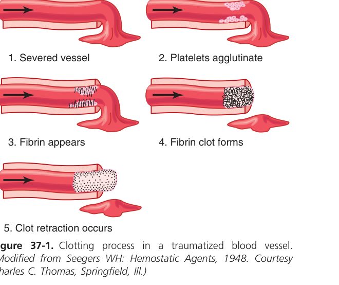
Physiological haemostasis = the body’s process to stop bleeding from a damaged vessel (spasm → plug → coagulation). Blood coagulation = conversion of liquid blood into a solid gel; its fundamental reaction is the conversion of soluble fibrinogen → insoluble fibrin.
The cascade is engineering by design: each factor activates the next, and each step amplifies. A tiny amount of trigger at the top can produce a vast amount of fibrin at the bottom — which is exactly what you need to seal a wound fast. Two routes get the cascade started. The extrinsic pathway begins outside the vessel: damaged tissue releases tissue factor (factor III) which immediately partners with factor VII. This is the fast route — bleeding into tissue is the most common emergency. The intrinsic pathway begins inside the vessel: contact with damaged collagen activates factor XII, then XI, IX, VIII. Both pathways meet at the same gateway, factor X, and from there it’s a single common path: Xa converts prothrombin (II) to thrombin (IIa); thrombin converts fibrinogen (I) to fibrin; factor XIII cross-links the fibrin strands into a mesh.
Four of these factors — II, VII, IX, X — need vitamin K to function (remember the mnemonic “1972”). That is why warfarin, a vitamin-K antagonist, thins the blood; why neonates get a vitamin K shot to prevent haemorrhagic disease of the newborn; and why severe liver disease causes coagulopathy — the liver makes the factors and stores vitamin K. The cascade is regulated by anticoagulants like antithrombin III and the protein C/S system; deficient anticoagulant function → thrombophilia (clots forming when they shouldn’t).
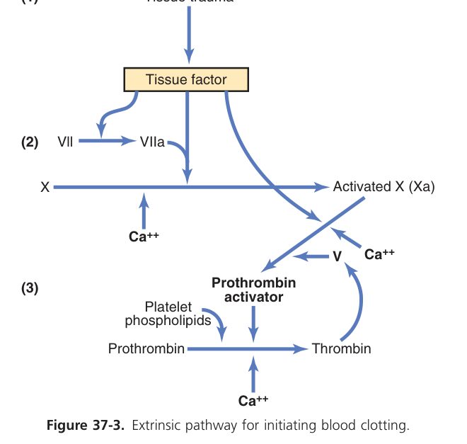
A cascade of plasma clotting factors (mostly liver-made, several vitamin-K-dependent: II, VII, IX, X); most steps need Ca²⁺ (factor IV) and a platelet phospholipid surface. Two initiating pathways converge on a common pathway:
| Pathway | Trigger | Key factors |
|---|---|---|
| Intrinsic | Contact with damaged surface (collagen) | XII → XI → IX → VIII |
| Extrinsic | Tissue factor (III) from injured tissue | TF + VII (faster) |
| Common | Both converge at factor X | X → Xa; prothrombin (II) → thrombin (IIa); fibrinogen (I) → fibrin; XIII cross-links it |
Factor X is the meeting point of intrinsic & extrinsic pathways (the start of the common pathway). Thrombin (IIa) is the enzyme that converts fibrinogen → fibrin. Vitamin-K factors = “1972” → II, VII, IX, X.
Haemophilia A = factor VIII deficiency; Haemophilia B = factor IX. Warfarin blocks vitamin-K recycling → ↓II,VII,IX,X (monitored by PT/INR). Heparin potentiates antithrombin (monitored by APTT). Liver disease & vitamin-K deficiency → bleeding. Citrate/EDTA chelate Ca²⁺ → prevent clotting in blood-bank tubes.
• Two pathways meet at? → factor X (start of common pathway)
• Final enzymatic step? → thrombin (IIa) converts fibrinogen → fibrin
• Vitamin-K-dependent factors? → II, VII, IX, X (the "1972" mnemonic)
• Why does warfarin work? → antagonises vitamin K → blocks II, VII, IX, X
• What does Ca²⁺ do in clotting? → factor IV; essential cofactor at most cascade steps
Blood Groups & Transfusion
Why can’t you give any blood to any patient? Because RBC membranes carry sugar-protein labels (antigens / agglutinogens) that the recipient’s immune system reads. If the donor cells wear a label the recipient’s plasma has antibodies against, the donor cells clump and burst — a transfusion reaction that can kill within minutes. The strange fact about ABO is that you make these antibodies without ever being exposed: by about six months of age, gut bacteria carrying similar sugars have trained your immune system to make whatever antibody does NOT match your own type. So a type A person has anti-B antibodies floating in plasma at all times; a type B person has anti-A; type O has both; type AB has neither.
Two practical consequences. Type O is the universal donor — its cells wear no A or B label, so they can be given to anyone (in an emergency). Type AB is the universal recipient — their plasma has no anti-A or anti-B, so they can accept any RBCs. In practice every transfusion is still cross-matched, because surface antigens beyond ABO (the Rh system below, and many minor systems) can also cause reactions.
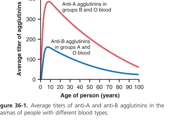
Blood groups are classified by the antigens (agglutinogens) on the RBC membrane; the plasma carries the opposite antibodies (agglutinins).
| Group | RBC antigen (agglutinogen) | Plasma antibody (agglutinin) | Note |
|---|---|---|---|
| A | A | anti-B | |
| B | B | anti-A | |
| AB | A and B | none | Universal recipient |
| O | none | anti-A and anti-B | Universal donor |
Mixing a recipient’s antibody with the matching donor antigen → agglutination + haemolysis (a transfusion reaction). Hence cross-matching before transfusion; ideally give same-ABO, same-Rh blood.
- Rh(D) antigen: present = Rh-positive, absent = Rh-negative. Unlike ABO, anti-D is not natural — it forms only after prior exposure (sensitisation).
- Haemolytic disease of the newborn: an Rh− mother carrying an Rh+ fetus is sensitised at the first delivery; in a later Rh+ pregnancy her anti-D crosses the placenta → fetal RBC haemolysis. Prevented by anti-D immunoglobulin given to the mother.
• Universal donor / recipient? → donor = O (no A/B antigen); recipient = AB (no anti-A/anti-B)
• Why are anti-A / anti-B antibodies present without prior transfusion? → cross-reactive antigens on gut bacteria
• Is anti-D natural? → no — only forms after exposure to Rh+ blood (sensitisation)
• HDN prevention? → anti-D immunoglobulin to Rh− mother after delivery / sensitising event
Blood complete
Plasma, RBC, coagulation cascade & blood groups mastered. Next: Cardiovascular.