Nerve & Muscle
Motor Unit & Muscle Types
Start with a question you already know the answer to from your own body: why can your eyes flick to land precisely on this exact word, while your thigh muscle can launch your whole body up off a chair? Both are muscle. The difference is how the nervous system wires them — and that wiring unit is the motor unit.
A motor unit is one motor neuron plus every muscle fibre it personally connects to. When that neuron fires, all of its fibres contract together, in one go — there is no half-firing. So the size of a motor unit decides how finely you can control that muscle. In the muscles that move your eyes, a single neuron commands only a handful of fibres, so the brain can add force in tiny, delicate steps. In the quadriceps, one neuron drives hundreds of fibres at once: far less precision, but enormous power per command.
That raises the obvious problem: if every motor unit is all-or-none, how does a whole muscle produce a smooth range of force, from threading a needle to lifting a suitcase? The body has two dials. First, recruitment — switching on more motor units. Second, rate coding — making each active neuron fire faster. Together they turn a collection of all-or-none units into beautifully graded whole-muscle force.
Before going further, it helps to place this muscle in its family. Under the microscope, muscle that shows stripes (cross-striations) is striated muscle — that means skeletal muscle and cardiac muscle. Muscle without stripes is smooth muscle, the muscle of your gut, blood vessels and bladder. This unit is mostly about skeletal muscle, with a comparison to smooth muscle at the end.
A motor unit = one motor neuron + all the muscle fibres it innervates. All fibres of a motor unit contract simultaneously when the neuron fires. Small motor units (few fibres, e.g. extra-ocular) give fine control; large units (hundreds of fibres, e.g. quadriceps) give power. Graded whole-muscle force is produced by recruiting more units and by raising firing frequency.
There is a clever order to recruitment, and it is not random. When you begin to lift something light, your nervous system switches on the smallest motor units first, and only calls in the big ones as the load grows. This is Henneman’s size principle. The reason it matters: small neurons run small, fatigue-resistant fibres, perfect for fine and sustained effort; the large, powerful, easily-tiring units are held in reserve for when you truly need maximal force. That ordering is exactly why you can pick up a feather and a dumbbell with the same hand and never feel a jarring jump in force — the force is added in graded steps, smallest contributions first.
Think of recruitment like a dimmer switch, not an on/off switch. You bring in little lights first for a soft glow (fine control), and only flip on the big floodlights when you need full brightness (maximal force). Save the floodlights for last because they burn out fastest.
Not all skeletal fibres are built the same, and the difference is a trade-off between endurance and speed/power — you cannot maximise both in one fibre. Slow fibres are packed with mitochondria, myoglobin and capillaries, so they make ATP aerobically and keep going for hours; that red colour you see in chicken “dark meat” is the myoglobin. Fast glycolytic fibres skip all that machinery for raw speed, burn fuel anaerobically, and tire quickly — the pale “white meat”. The table below is the comparison you will be examined on; read it knowing why each row falls the way it does (more mitochondria → more aerobic → more fatigue-resistant → redder).
| Feature | Type I (slow oxidative) | Type IIa (fast oxidative-glycolytic) | Type IIx/IIb (fast glycolytic) |
|---|---|---|---|
| Speed | Slow | Fast | Fastest |
| Fatigue | Resistant | Moderate | Rapid |
| Mitochondria / capillaries | Many | Many | Few |
| Myoglobin / colour | High — red | High — red/pink | Low — white |
| Metabolism | Oxidative | Oxidative + glycolytic | Glycolytic |
| Role | Posture, endurance (soleus) | Sustained running | Sprint, jumping (gastrocnemius white) |
Endurance training shifts fibres toward more Type I/IIa profile; sprint training favours IIx hypertrophy — which is why a marathoner and a sprinter end up with visibly different muscle.
• Two ways to grade whole-muscle force? → recruitment + rate coding
• Recruitment order? → smallest → largest (Henneman size principle)
• Posture / endurance fibre? → Type I (slow, red, fatigue-resistant)
• Sprint / power fibre? → Type IIx (fast, white, fatigable)
The Neuromuscular Junction (Motor End-plate)
Here is a real scene to anchor everything that follows. A woman notices that by evening her eyelids droop and she sees double, yet after a night’s rest she is fine again. Her muscles are healthy and her nerves are healthy — the fault is in the tiny gap between them. That gap is the neuromuscular junction, and her disease, myasthenia gravis, is the reason examiners love this topic: it only makes sense once you understand each step of transmission. So let’s build the junction first, then walk a signal across it.
The nerve cannot simply “touch” the muscle and pass on its electrical signal — there is a physical cleft in the way. So the message has to change form: electrical → chemical → electrical again. The chemical messenger is acetylcholine (ACh). Picture three parts: the nerve ending stocked with ACh in vesicles, the narrow cleft (patrolled by an enzyme that destroys ACh), and the muscle’s end-plate studded with receptors waiting to catch ACh.
The neuromuscular junction (NMJ) is the synapse between a motor-neuron terminal and a skeletal muscle fibre. Components: pre-synaptic terminal (ACh-filled vesicles + voltage-gated Ca²⁺ channels), the synaptic cleft (contains acetylcholinesterase, AChE), and the post-synaptic membrane / motor end-plate (deeply folded, packed with nicotinic (Nₘ) ACh receptors = ligand-gated Na⁺ channels).
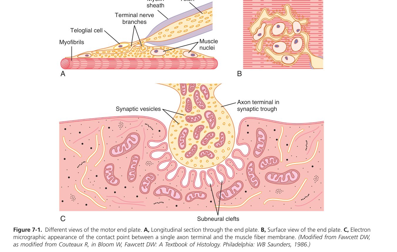
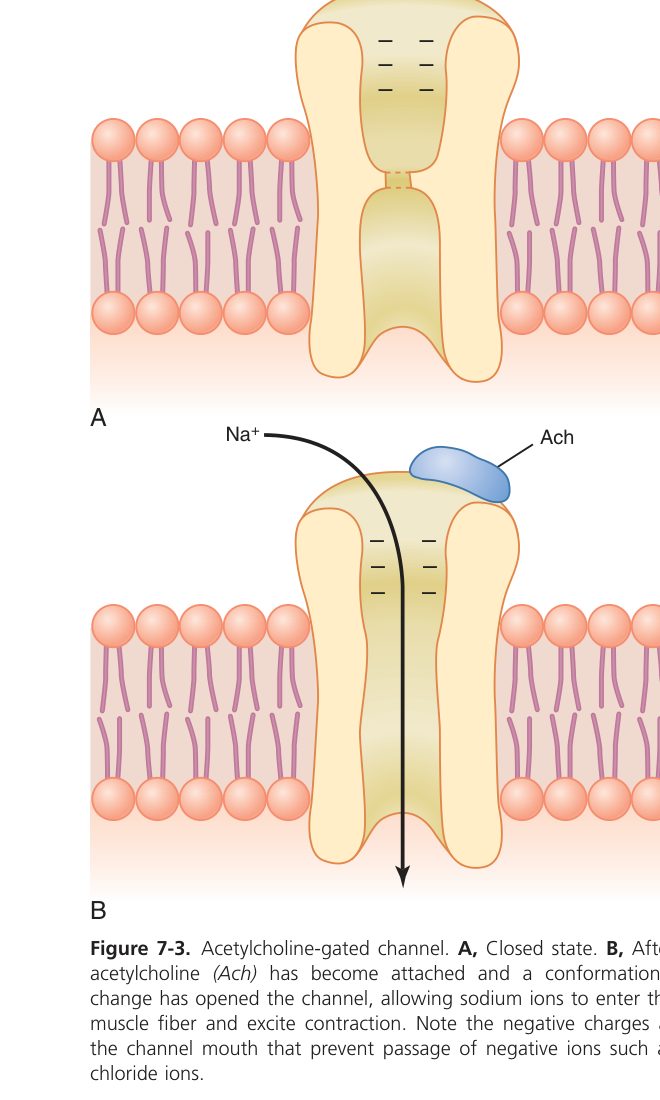
Now follow one nerve impulse across the gap. The electrical spike races down the axon and reaches the terminal. Its arrival flings open voltage-gated calcium channels, and calcium rushing in is the trigger — remember this single fact above all others: no calcium, no transmitter release. Calcium makes the ACh vesicles fuse with the membrane and dump their contents into the cleft. ACh drifts across, lands on nicotinic receptors, and pops them open. Because these receptors are sodium channels, sodium pours in and the end-plate depolarises — this local voltage is the end-plate potential (EPP). The EPP is normally far bigger than it needs to be (a built-in “safety factor”), so it comfortably drags the membrane to threshold and ignites a full muscle action potential that sweeps across the fibre. Finally, to stop the signal jamming “on”, the enzyme acetylcholinesterase (AChE) chops up the ACh within milliseconds, the choline is recycled, and the junction resets for the next impulse.
- AP arrives at the axon terminal → depolarisation opens voltage-gated Ca²⁺ channels.
- Ca²⁺ influx into the terminal triggers exocytosis of ACh-containing vesicles into the cleft (Ca²⁺ is essential — no Ca²⁺, no release).
- ACh diffuses across the cleft and binds nicotinic receptors on the end-plate.
- Receptor channels open → Na⁺ influx (> K⁺ efflux) → local depolarisation = end-plate potential (EPP) — a graded, non-propagated potential.
- The large EPP (high “safety factor”) easily reaches threshold → fires a muscle action potential that sweeps along the sarcolemma.
- AChE in the cleft rapidly hydrolyses ACh → acetate + choline (choline is recycled) → the end-plate repolarises, ready for the next impulse.
The EPP is a graded local potential (like an EPSP): it summates, it decrements with distance, it has no refractory period. Its only job is to trigger the all-or-none muscle AP. One nerve impulse normally produces exactly one muscle AP (1:1, thanks to the high safety factor).
Each step you just learned is a place where disease or a drug can strike — this is why the NMJ is so “clinical”. Myasthenia gravis (our drooping-eyelid patient): autoantibodies block/destroy the post-synaptic nicotinic receptors, so EPPs shrink and start failing to reach threshold → fatigable weakness that worsens through the day; AChE inhibitors (neostigmine) help by letting ACh linger.
Lambert–Eaton syndrome: antibodies against the pre-synaptic Ca²⁺ channels → too little ACh released. Organophosphates / nerve agents: block AChE → ACh piles up → persistent depolarisation and spastic paralysis. Curare: competitively blocks nicotinic receptors → flaccid paralysis (the classic arrow poison). Botulinum toxin: stops ACh release by cleaving SNARE proteins (and is exactly how Botox relaxes a muscle).
• Receptor on the end-plate? → nicotinic ACh receptor (a Na⁺ channel)
• What ends the signal? → AChE breaks down ACh
• EPP vs muscle AP? → EPP = graded local trigger; muscle AP = all-or-none
• Myasthenia gravis vs Lambert–Eaton? → MG = receptor antibodies; L–E = pre-synaptic Ca²⁺-channel antibodies
The Sarcomere & Contractile Filaments
Now that the signal has reached the muscle, what does it actually move? Zoom into a muscle fibre and you find the same little machine repeated end to end, thousands of times: the sarcomere, running from one Z-disc to the next. It contains two sets of overlapping rods — thin actin filaments and thick myosin filaments — and the whole job of contraction is to make these two sets slide past each other.
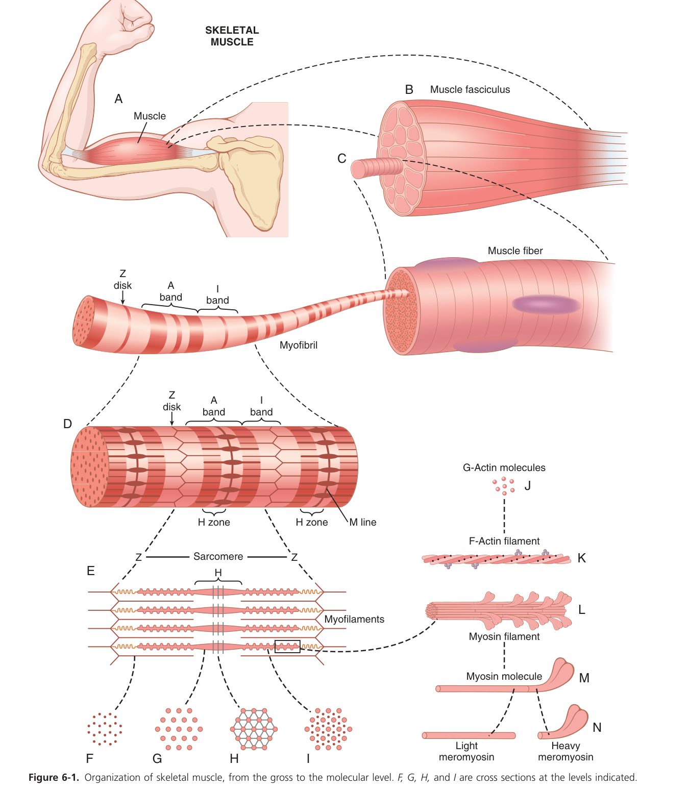
The famous “bands” you see under the microscope are simply where these filaments do or don’t overlap. The key insight for exams: when the muscle shortens, the filaments themselves never get shorter. They just slide. So the bands that contain only the thin filament shrink, while the band measured by the thick filament stays exactly the same length. Hold that idea while you read the table.
| Band / line | Contains | During contraction |
|---|---|---|
| A band (dark, anisotropic) | Whole thick filament (+ overlap) | Length unchanged (= thick filament length) |
| I band (light, isotropic) | Thin filament only (no overlap) | Shortens (may disappear) |
| H zone | Thick filament only (no overlap) | Shortens / disappears |
| Z disc | Anchors thin filaments | Z discs move closer |
| M line | Anchors thick filaments (centre) | Unchanged |
The letters tell you the truth: A band = Always the same length. I band & H zone shrink. (And remembering the colours: A = dArk, the “eye” in lIght for the I band.)
To understand contraction and its on/off switch, you need to know the parts. The thin filament is not just actin: lying along it is a long thread, tropomyosin, which physically covers the spots where myosin wants to grab. Sitting on top at intervals is the troponin complex — think of it as the gatekeeper with three jobs split among three subunits: TnC catches calcium, TnI holds the brakes on (inhibits), and TnT ties the complex to tropomyosin. The thick filament is a bundle of myosin molecules, each with a long tail and two golf-club heads that stick out as cross-bridges; every head is both a motor (it splits ATP for energy) and a hand (it binds actin).
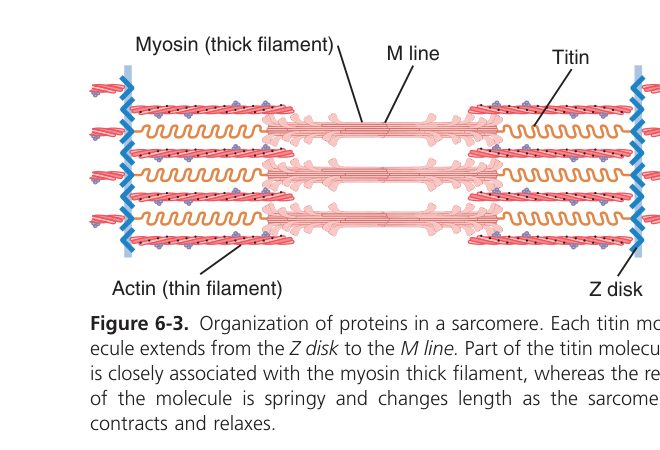
- Thin filament: two F-actin strands twisted together; in the groove lies tropomyosin; troponin attaches at intervals — TnC (binds Ca²⁺), TnI (inhibits actin–myosin), TnT (binds tropomyosin).
- Thick filament: bundled myosin molecules — a long tail (backbone) + two globular heads that protrude as cross-bridges. The head has ATPase activity (splits ATP for energy) and an actin-binding site.
A sarcomere would fall apart under its own forces without a support crew, and one of these proteins is the reason a whole disease exists. Titin is the giant molecular spring that runs from Z-disc to M-line, keeping the thick filament centred and giving relaxed muscle its springy “passive” recoil. Nebulin acts like a ruler that sets thin-filament length; α-actinin bolts the thin filaments onto the Z-disc. The one to remember for the wards is dystrophin: it tethers the whole contractile machine to the cell membrane. Lose it, and every contraction tears the membrane a little — which is exactly what happens in Duchenne muscular dystrophy.
- Titin — the largest protein known; spans Z-disc to M-line, acts as a molecular spring; gives the muscle passive elasticity and centres the thick filament in the sarcomere.
- Nebulin — aligns thin filaments and sets their length.
- α-actinin — anchors thin filaments at the Z-disc.
- Dystrophin — links the sarcomere to the sarcolemma and extracellular matrix; absent in Duchenne muscular dystrophy.
Duchenne muscular dystrophy (X-linked): dystrophin gene deletion → sarcolemmal fragility → muscle fibre necrosis, calf pseudo-hypertrophy, Gowers sign, raised CK, death from cardiopulmonary failure in adolescence. Becker MD = milder allelic variant (some functional dystrophin survives).
Put it together and you have the central idea of muscle physiology. Contraction is not the filaments scrunching up — it is the thin filaments being dragged inward over the thick filaments, deeper into overlap, pulling the Z-discs toward each other. The sarcomere shortens, the I band and H zone narrow, but the A band (the length of the thick filament) never changes. Once you can recite which bands change and which don’t, you have the whole theory.
During contraction the filaments themselves do not shorten; the thin filaments slide over the thick filaments toward the M line, increasing overlap. Therefore the sarcomere & I band shorten, the H zone narrows, but the A band stays constant; the lengths of the thick and thin filaments are unchanged.
• Which band stays constant in contraction? → A band (I band & H zone shrink)
• Do the filaments shorten? → no — they slide (sliding-filament theory)
• Which troponin subunit binds Ca²⁺? → TnC
• Protein lost in Duchenne? → dystrophin
Excitation–Contraction Coupling
We now have two separate stories: an electrical action potential travelling on the membrane (section 2.2) and a mechanical sliding of filaments (section 2.3). Something must connect them — and that something is a single ion, calcium. The whole process of turning the electrical signal into a mechanical pull is called excitation–contraction (E-C) coupling, and calcium is its messenger.
The problem the muscle has to solve is depth. A muscle fibre is fat, and the action potential only travels on the surface — so how do the filaments deep in the centre get the message at the same instant as those at the surface? The answer is plumbing: the surface membrane dives inward as narrow tunnels called T-tubules that carry the electrical signal into the core of the fibre. Running alongside them is the cell’s calcium warehouse, the sarcoplasmic reticulum (SR). When the AP travels down a T-tubule, a voltage sensor there (the DHP receptor) nudges open the SR’s calcium gates (the ryanodine receptors, RyR), and stored calcium floods out around the filaments. That calcium lands on troponin-C, the gatekeeper shifts, tropomyosin slides off the actin binding sites — and contraction can begin.
- Muscle AP propagates over the sarcolemma and down the T-tubules (deep into the fibre).
- The T-tubule voltage sensor (DHP receptor) signals the SR terminal cisternae.
- The ryanodine receptor (RyR) opens → Ca²⁺ floods from the sarcoplasmic reticulum into the sarcoplasm.
- Ca²⁺ binds troponin-C → troponin–tropomyosin shifts → actin–myosin binding sites uncovered → contraction begins.
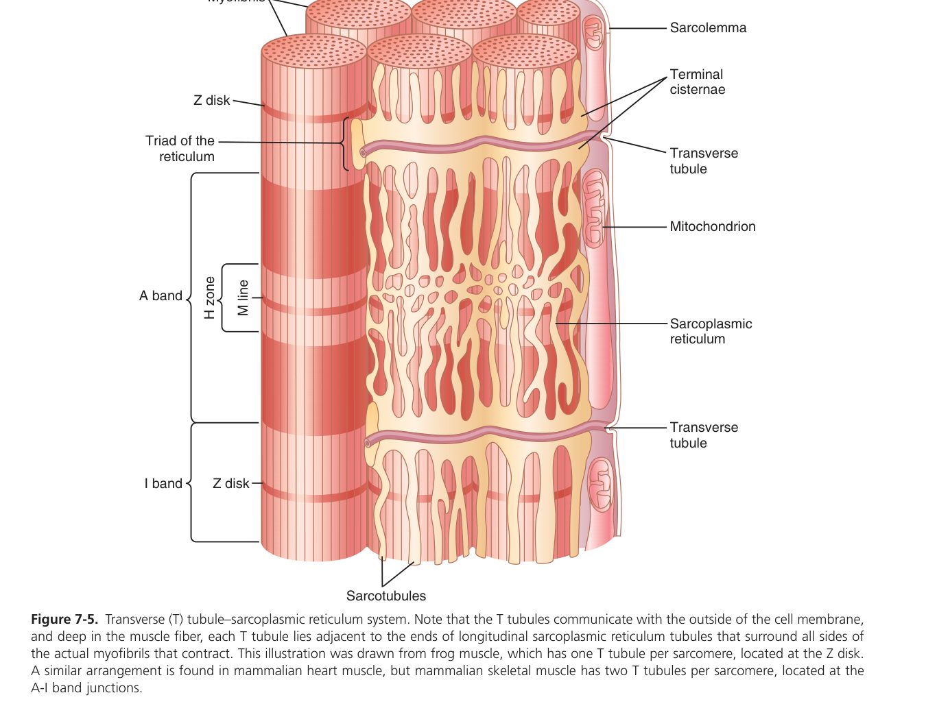
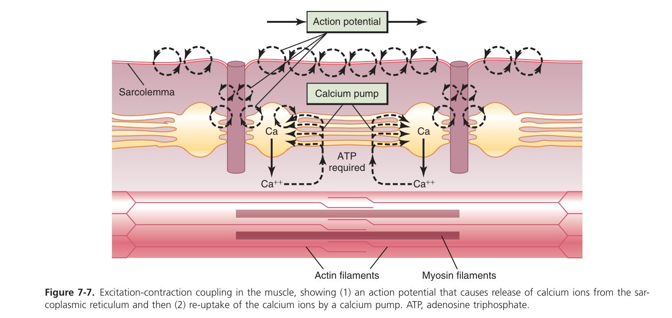
“AP → T-tubule → SR → Ca²⁺ → troponin-C → tropomyosin moves → cross-bridge.” Ca²⁺ is the coupling messenger; the SR is the Ca²⁺ store; the T-tubule carries the signal inward so the whole fibre fires together.
• Path of the signal inward? → AP → T-tubule → DHP sensor → RyR → Ca²⁺ from SR
• What does the Ca²⁺ bind? → troponin-C → tropomyosin uncovers actin
• Where is the Ca²⁺ stored? → sarcoplasmic reticulum (SR)
Cross-bridge Cycle & Relaxation
With the binding sites finally uncovered, the myosin heads get to work — and they do it like a crew rowing a boat. Each head grabs the thin filament, pulls (the “power stroke”), lets go, resets, and grabs again, hauling the actin a little further inward each time. Because thousands of heads do this slightly out of step, the pull is smooth and continuous, like many oars dipping at different moments to keep a boat gliding. Follow one cycle and notice where the ATP is spent — this is heavily examined.
- Attachment: the energised myosin head (carrying ADP + Pᵢ) binds exposed actin.
- Power stroke: Pᵢ then ADP are released; the head pivots, pulling the thin filament toward the M line → force / shortening.
- Detachment: a new ATP binds myosin → head releases actin.
- Re-cocking: myosin ATPase hydrolyses ATP → ADP + Pᵢ re-energise and re-cock the head, ready to re-attach. Cycles repeat while Ca²⁺ is present.
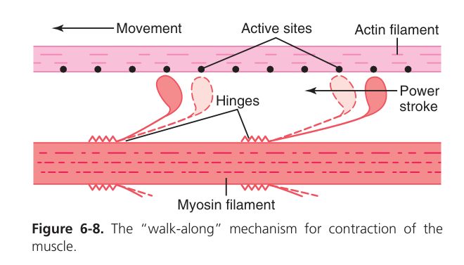
Here is the detail students miss: ATP is needed not to pull, but to let go. The power stroke runs on energy already stored in the cocked head; fresh ATP is what unsticks the head from actin so the cycle can repeat. That one fact explains a body you may have seen.
Rigor mortis: after death ATP production stops, so myosin heads have nothing to unstick them and cannot detach from actin → muscles lock rigid — a direct, visible proof that ATP is the “release” fuel. Malignant hyperthermia: a mutant RyR dumps excess Ca²⁺ when triggered by volatile anaesthetics or succinylcholine → uncontrolled contraction, massive heat and hypermetabolism — an anaesthetic emergency treated with dantrolene.
Contraction continues only as long as calcium is around the filaments, so relaxation is simply the job of removing that calcium. A pump called SERCA actively hauls calcium back into the SR store. With cytosolic calcium falling, troponin lets go, tropomyosin slides back over the actin sites, the heads can no longer attach, tension fades, and the muscle returns to rest. Note that this pump also costs ATP — so relaxation, like detachment, is an active, energy-requiring process, not just “letting go”.
- Ca²⁺ is actively pumped back into the SR by the SERCA Ca²⁺-ATPase.
- Ca²⁺ leaves troponin-C → tropomyosin re-covers the actin sites.
- Cross-bridges detach → tension falls → the muscle returns to resting length.
• Why does rigor mortis happen? → no ATP → heads can’t detach from actin
• How does the muscle relax? → SERCA pumps Ca²⁺ back into the SR
• Cause of malignant hyperthermia? → mutant RyR dumps Ca²⁺ (anaesthetic trigger)
Muscle Mechanics
A contracting muscle can do one of two things, and your own body shows both right now. Lift a cup and the muscle shortens while the tension stays roughly constant — that is isotonic (“same tension”) contraction, and it moves a load. Push against a wall that won’t budge and the muscle’s tension climbs hard while its length doesn’t change at all — that is isometric (“same length”) contraction, the kind that holds your posture all day. The distinction matters because force and movement are not the same thing.
| Type | Tension | Length | Example |
|---|---|---|---|
| Isotonic | Constant | Shortens (moves a load) | Lifting a weight, walking |
| Isometric | Rises | Unchanged (no movement) | Pushing a wall, posture |
One nerve impulse gives one brief twitch. But here is the quirk that makes sustained movement possible: the electrical action potential is over in 1–2 milliseconds, while the mechanical twitch it produces lasts about 100 milliseconds — roughly fifty times longer. Because the membrane has long since recovered while the muscle is still mid-contraction, a second impulse can land before the first twitch has relaxed, and the two pulls stack on top of each other. This stacking is summation. Fire fast enough and the twitches fuse into one smooth, sustained, maximal contraction — tetanus. Almost every real movement you make is tetanic, not single twitches.
| Response | Stimulus frequency | Mechanics |
|---|---|---|
| Single twitch | Single / very low | Full relaxation between stimuli |
| Incomplete (unfused) tetanus | Moderate | Partial relaxation between summated peaks (wavy) |
| Complete (fused) tetanus | High | No relaxation; smooth sustained maximal contraction |
Tetanic tension can reach about 4× the single-twitch tension, because rapid firing keeps cytosolic Ca²⁺ high so far more cross-bridges are engaged at once.
Your heart deliberately cannot do this. Cardiac muscle has a refractory period almost as long as its whole contraction, so impulses can’t summate — which is the safety feature that stops the heart locking up in tetanus and lets it fill between beats (revisited in Unit 4).
How hard a muscle can pull depends on how stretched it was to begin with — and the reason is purely geometric. Force comes from cross-bridges, and cross-bridges only form where thick and thin filaments overlap. Stretch a muscle too far and the filaments barely overlap, so few bridges form and force is weak; let it go too slack and the thin filaments collide and get in each other’s way. Somewhere in between is an optimal length (L₀) with ideal overlap and maximal active force. This single idea, applied to the heart, becomes the Frank–Starling law: a heart filled more before it beats stretches toward its optimum and ejects more blood.
- Preload = the stretch on a muscle before it contracts (resting length / initial filament overlap).
- Afterload = the resistance the muscle works against during contraction.
- Length–tension relation: active tension is maximal at the optimal length (L₀) where thin/thick filament overlap is ideal; too short or too stretched → fewer cross-bridges → less force.
- Total tension = active tension (cross-bridges) + passive tension (titin elastic stretch). Above L₀, passive tension rises steeply.
- This is the structural basis of the cardiac Frank–Starling law — greater end-diastolic stretch → greater stroke volume (Unit 4).
There is an everyday truth hidden here: the heavier the load, the slower you can move it — and with a heavy enough load, you cannot move it at all. Plotted out, shortening velocity against load gives the classic Hill curve. With no load, the muscle shortens at its top speed (Vmax, set by how fast the myosin ATPase works). Pile on load and velocity drops, until at the muscle’s maximum force (P0) it stops shortening entirely — that is an isometric contraction. Load it beyond that and the muscle is actually stretched while contracting (an eccentric contraction), which generates the highest force of all and, notably, causes the most next-day soreness.
- Zero load → maximum velocity (Vmax) — depends on myosin ATPase isoform (fast vs slow).
- As load rises → velocity falls.
- Load = maximum (P0) → zero velocity — an isometric contraction; no shortening, only force.
- Above P0 the muscle is forcibly lengthened (eccentric contraction) at the highest force (~1.8×P0) — explains why eccentric work causes the most soreness.
Muscle is greedy for ATP, and it spends it in three places you have already met: the power stroke, the SERCA pump that ends contraction, and the Na⁺/K⁺ pump that resets the membrane after every action potential. The catch is that a fibre stores only a few seconds’ worth of ready ATP — so the body keeps a tiered set of refills, each trading speed for staying-power. First a quick chemical battery (creatine phosphate) tops ATP back up for a sprint; then anaerobic glycolysis takes over for about a minute (paying in lactate); finally, for anything sustained, oxidative phosphorylation burns glucose and fat with oxygen. This tiering is why a 100 m sprint and a marathon feel completely different to run.
- ATP needs: (1) myosin power stroke, (2) SERCA Ca²⁺ pump, (3) Na⁺/K⁺-ATPase restoring the sarcolemmal gradient.
- Energy sources in order of use: (a) free ATP (~6 s), (b) creatine phosphate (rephosphorylates ADP via creatine kinase, ~15 s of maximal work), (c) anaerobic glycolysis → lactate (~1 min), (d) oxidative phosphorylation from glucose/fatty acids (sustained).
- Oxygen debt = the extra O₂ consumed after exercise to replenish CP, oxidise lactate & restock muscle/blood O₂ — the reason you keep panting after you stop running.
- About 75% of muscle energy is dissipated as heat (only ~25% does mechanical work) — the basis of shivering thermogenesis, how your body warms itself.
McArdle disease (myophosphorylase deficiency) blocks muscle glycogenolysis → early fatigue, painful cramps and myoglobinuria after exercise, with a classic “second wind” once fat metabolism kicks in — a living demonstration of how much peak effort leans on glycolysis. Mitochondrial myopathies cripple oxidative phosphorylation → exercise intolerance despite normal resting strength.
• How do twitches become smooth force? → summation → tetanus (~4× twitch)
• Why is force greatest at optimal length? → best filament overlap (→ Frank–Starling)
• Velocity at maximum load (P₀)? → zero (isometric)
• Fuel order? → ATP → creatine phosphate → glycolysis → oxidative; ~75% lost as heat
Smooth Muscle (Comparison)
Skeletal muscle is built for fast, voluntary, powerful bursts. But the muscle in your gut wall, your bladder and your blood vessels has the opposite job: slow, involuntary, and able to hold tension for hours without tiring or you even noticing. That is smooth muscle, and almost every difference from skeletal muscle flows from this change of job description. It has no sarcomeres (so no stripes), it answers to the autonomic nervous system, hormones and stretch rather than your will, and crucially it switches on through a completely different calcium pathway.
| Feature | Skeletal | Smooth |
|---|---|---|
| Striations / sarcomeres | Yes, organised | No (filaments oblique) |
| Ca²⁺ sensor | Troponin-C | Calmodulin → MLCK |
| Ca²⁺ source | SR (T-tubules) | SR + extracellular Ca²⁺ |
| Control | Voluntary (somatic) | Involuntary (autonomic, hormones, stretch) |
| Speed / fatigue | Fast, fatigues | Slow, sustained (“latch”), economical |
| Gap junctions | No | Yes (single-unit = syncytium) |
The key switch to remember: skeletal muscle reads calcium through troponin, but smooth muscle has no troponin at all. Instead, calcium teams up with calmodulin, which switches on an enzyme (MLCK) that phosphorylates the myosin head so it can start cycling. Relaxation needs the opposite enzyme (MLCP) to take that phosphate off again. This “phosphorylate to switch on” design is slower than skeletal muscle’s, but it is exactly what lets smooth muscle be controlled by the drugs you will prescribe most.
Ca²⁺ + calmodulin activate myosin light-chain kinase (MLCK), which phosphorylates the regulatory myosin light chain (MLC20) so the head can cycle on actin. There is no troponin. Relaxation requires myosin-light-chain phosphatase (MLCP); cytosolic Ca²⁺ is lowered by SERCA and the plasma-membrane Ca²⁺-ATPase + Na⁺/Ca²⁺ exchange.
Smooth muscle comes in two wiring styles. In most hollow organs the cells are joined by gap junctions and behave as one connected sheet — a single-unit (visceral) muscle that can set its own rhythm and squeeze as a wave, which is exactly what your gut and ureter need. A few places (the iris, the vas deferens) instead use multi-unit smooth muscle, where each cell is controlled separately like a tiny motor unit, for precise independent adjustment.
| Property | Single-unit (visceral / unitary) | Multi-unit |
|---|---|---|
| Coupling | Cells linked by gap junctions → act as a syncytium | Cells act independently — like motor units |
| Pacemaker activity | Spontaneous slow waves + APs | Quiet; needs neural drive |
| Examples | GI tract, ureter, uterus, small vessels | Iris, ciliary, vas deferens, large airways |
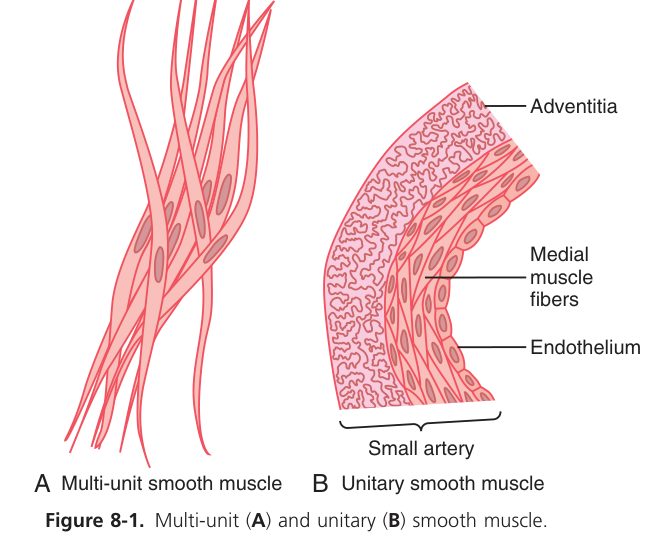
Now the trick that makes smooth muscle special. Your arteries hold a steady squeeze on your blood every second of your life, and your sphincters stay shut for hours — if they used energy the way skeletal muscle does, they would exhaust themselves in minutes. Smooth muscle avoids this with the latch state: after the initial calcium-driven activation, calcium falls and MLCK quietens, but a fraction of cross-bridges stay stuck on, cycling extremely slowly. The result is sustained force held at a tiny fraction of the ATP cost — tension almost for free. This is the physiology behind blood-pressure tone, and it is exactly where blood-pressure drugs act.
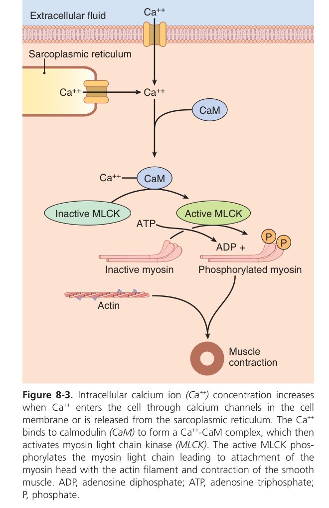
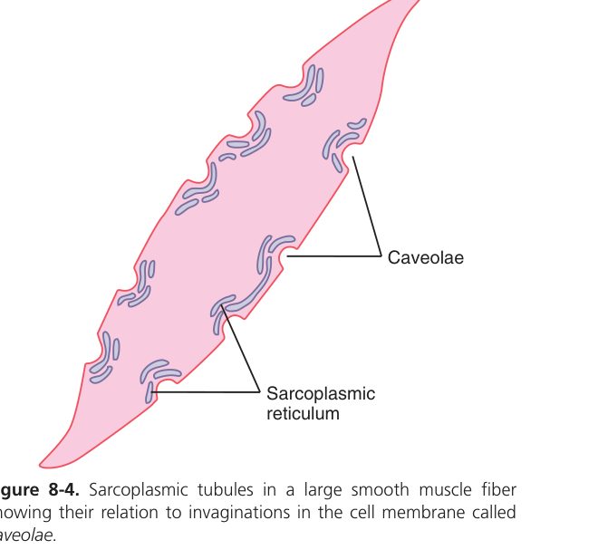
Smooth muscle uniquely maintains tension cheaply: after initial activation Ca²⁺ falls, MLCK activity drops, but a fraction of cross-bridges remain attached in a slow-cycling "latch" state — producing sustained force at a fraction of the ATP cost of skeletal muscle. This is essential for tonic vasoconstriction, sphincter tone and sustained organ contraction without fatigue.
Almost every antihypertensive works by relaxing vascular smooth muscle along the pathway you just learned: Ca²⁺-channel blockers (amlodipine, nifedipine) cut Ca²⁺ entry → less MLCK activation → vasodilation; nitrates / NO donors → cGMP → activate MLCP (the “off” enzyme) → relaxation; α₁-blockers (prazosin) stop sympathetic Gₙ signalling to vascular smooth muscle. Knowing the pathway tells you the drug.
• How is force switched off? → MLCP removes the phosphate
• Single-unit vs multi-unit? → gap-junction syncytium (gut, ureter) vs independent cells (iris, vas deferens)
• What is the latch state? → cheap, sustained tone at low ATP/Ca²⁺
• Where do antihypertensives act? → vascular smooth muscle (CCBs, nitrates, α₁-blockers)
Nerve & Muscle complete
NMJ, sliding-filament, E-C coupling and mechanics mastered. Next: Blood.