Cell Physiology
Homeostasis & the Internal Environment
Anatomy tells you what the parts of the body look like. Physiology tells you what those parts do, and how they keep doing it through fevers, runs, fasts, blood loss, hot afternoons and cold mornings. Every disease you will learn about later is, at its core, a physiological process gone wrong — so the better you understand normal function, the easier pathology becomes.
It helps to study the same body at two zoom levels. Zoom in to the cell / molecular level and you see ion channels, pumps, receptors, second messengers. Zoom out to the organ / system level and you see the heart adjusting cardiac output, the kidney holding blood pressure, the lungs balancing CO₂. Both layers describe the same patient — one in the language of molecules, the other in the language of organs talking to each other.
Physiology is the study of the normal functions of cells, organs and systems of the living body, and the mechanisms that regulate them. It is studied at two main levels: the cell (molecular) level and the organ / system level.
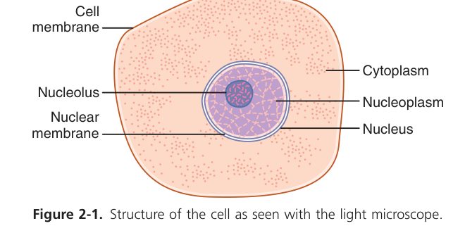
Here is the question that opens physiology: a human cell sitting in your liver looks remarkably like the single-celled amoeba in pond water — same membranes, same ATP, same K⁺ gradient. So how come the amoeba is at the mercy of every change in its puddle, while your liver cell carries on unbothered while you sit in air-conditioning, then a sauna, then a swimming pool? Because your liver cell never sees the outside world at all. It sees only the extracellular fluid bathing it — and your body works obsessively to keep that fluid’s composition steady, no matter what is happening outside.
The 19th-century French physiologist Claude Bernard called this fluid the milieu intérieur — the internal environment. His insight: the freedom of complex organisms to live in changing surroundings exists because their cells live in an unchanging one. Notice that the internal environment is not “blood”. Blood in a vessel never touches most cells; cells touch interstitial fluid, the watery space between them, which is in continuous exchange with plasma. ECF = interstitial fluid + plasma. That is the real address of your cells.
The internal environment is the extracellular fluid (ECF) that directly bathes the cells — not blood inside vessels alone. Cells live in and exchange with the ECF, so the ECF (not the outside world) is the true environment of the cell. Term coined by Claude Bernard.
Internal environment = the extracellular fluid (interstitial fluid + plasma).
Homeostasis (Walter Cannon) = the maintenance of nearly constant conditions (volume, osmolarity, pH, temperature, ion & nutrient concentrations) in the internal environment by coordinated physiological mechanisms. It is dynamic, not fixed.
Body fluids in a 70-kg adult (total body water ≈ 60% of body weight ≈ 42 L):
| Compartment | % body weight | Volume (70 kg) | Note |
|---|---|---|---|
| Intracellular fluid (ICF) | ~40% | ~28 L | High K⁺, organic phosphate, protein |
| Extracellular fluid (ECF) | ~20% | ~14 L | The internal environment |
| · Interstitial fluid | ~15% | ~11 L | High Na⁺, Cl⁻; little protein |
| · Plasma | ~5% | ~3 L | Like interstitial fluid + plasma proteins |
Think of homeostasis like a thermostat in a house. The temperature is never exactly 22 °C — it dips a little, the heater clicks on, it overshoots, the heater clicks off. The number you live at is the average around a set point. Your blood glucose, pH, sodium, blood pressure all behave the same way: forever wobbling within narrow limits, never frozen still.
• Who coined “milieu intérieur”? → Claude Bernard
• Who introduced the term homeostasis? → Walter Cannon
• Is homeostasis static or dynamic? → dynamic steady state around a set point
• Body water as % weight? → ~60% (ICF 40, ECF 20: 15 interstitial + 5 plasma)
Control Systems & the Reflex Arc
If the body wants to keep ECF steady, it needs ways to send messages to adjusting organs — fast for emergencies, slow for the long haul. Evolution settled on three channels. The fastest is nervous regulation — an action potential racing along a single wire (the axon) to a single target, like dialling one phone number. The slowest but most widespread is humoral regulation — pour a hormone into blood and it reaches every cell in the body, but the message takes minutes to hours, like a broadcast newsletter. The third is auto-regulation: a tissue defending itself without nerve or hormone — think of a kidney keeping its own blood flow constant even when your blood pressure rises. The same body uses all three on different time scales.
| Mode | Mediator | Speed / range | Example |
|---|---|---|---|
| Nervous regulation | Nerve impulses / neurotransmitters | Rapid, brief, precise (point-to-point) | Baroreceptor reflex |
| Humoral regulation | Hormones / local chemicals in blood | Slow, prolonged, diffuse | Insulin & glucose |
| Auto-regulation | Intrinsic property of the tissue itself | Local, independent of nerve/hormone | Renal & cerebral blood flow constancy |
All those control systems share one strategy — feedback. The clever default is negative feedback: when something rises above the set point, the response is to push it back down; when something falls, the response is to push it back up. Every classic regulatory loop you will learn — baroreceptor reflex, thermoregulation, insulin/glucose — runs on this principle. It is stabilising precisely because it always opposes the disturbance.
But there are moments when stabilising is the wrong answer — you actually want an event to go to completion as fast as possible. Childbirth is one: oxytocin makes the uterus contract, which stretches the cervix, which makes more oxytocin, which makes stronger contractions — an explosive runaway until delivery. That is positive feedback: rare in the body, used only where a powerful one-shot output is needed (the upstroke of an action potential, blood clotting, ovulation). It is self-limiting because the trigger eventually ends. Finally, feed-forward is the body anticipating: you start salivating at the sight of food, before any food has reached your mouth, because your nervous system is preparing the gut for the meal it predicts is coming.
- Negative feedback (the dominant control): the response opposes / reverses the initiating change → restores the set point → stabilising. e.g. ↑BP → baroreceptors → ↓BP; thermoregulation; glucose control.
- Positive feedback: the response reinforces / amplifies the change → explosive, self-limiting events. e.g. the upstroke of the action potential (Na⁺ entry → more Na⁺ channels open), blood clotting cascade, oxytocin in parturition, LH surge before ovulation.
- Feed-forward: anticipatory adjustment before the disturbance is sensed (e.g. cephalic-phase gastric secretion at the sight of food).
Negative feedback is the thermostat — correcting back to centre. Positive feedback is rolling a snowball downhill — once it starts, it amplifies itself. Feed-forward is grabbing an umbrella when you see the dark cloud, before a single drop has fallen.
When the nervous system controls something, it almost always does it through a reflex — a hard-wired, automatic response that happens faster than you can think about it. Tap the patellar tendon and the leg kicks before you are even aware of the tap. That speed comes from the fact that the loop is short and pre-built: stimulus → receptor → sensory nerve → spinal cord → motor nerve → effector. Five links, no conscious decision. Notice the stimulus itself is not part of the arc — it is what triggers the receptor. Every reflex you will meet in this course (deep tendon jerks, baroreceptor, cough, micturition) is built from this same five-element template.
A reflex is a stereotyped, involuntary response to a stimulus, mediated by the nervous system over a reflex arc with 5 components:
Receptor → Afferent (sensory) nerve → Centre (CNS) → Efferent (motor) nerve → Effector. “RACEE”. The stimulus is NOT a component of the arc — it acts on the receptor.
A stimulus is characterised by three parameters: intensity, duration, and rate of change (slope).
• Dominant feedback type? → negative — opposes the change, stabilises
• Two named examples of positive feedback? → AP upstroke; oxytocin in labour (also clotting, LH surge)
• Five elements of reflex arc? → receptor → afferent → centre → efferent → effector (RACEE)
• Is the stimulus part of the arc? → no — it acts on the receptor
Cell Membrane & Membrane Transport
The cell membrane has a tough problem to solve. It must keep the cell’s insides in, the outside world out, yet let in the food, ions and signals the cell needs — selectively, on demand. The body’s elegant answer is a lipid bilayer with proteins floating in it. Fatty tails facing each other, polar heads facing the water on both sides — like a soap film. Water-soluble things bounce off the oily middle and cannot cross; only fat-soluble things (O₂, CO₂, steroids, alcohol) slip through.
Singer and Nicolson called this the fluid-mosaic model: “mosaic” because membrane proteins are scattered through the lipid sea like tiles in a mosaic, and “fluid” because the lipids and proteins drift sideways instead of being locked in place. Cholesterol tucks between phospholipids and controls how fluid the membrane is — too much fluid and the membrane would tear, too little and it could not change shape during things like endocytosis. The proteins are not decoration: they are the working machinery — channels, pumps, receptors, enzymes. Everything interesting that crosses the membrane crosses it through a protein.
A lipid bilayer (phospholipid + cholesterol) with proteins floating in/through it (Singer & Nicolson fluid-mosaic model). The bilayer is a barrier to water-soluble substances; proteins do the selective transport and signalling. TMU lecture composition (by mass):
| Component | ~ % (TMU slide) | Role |
|---|---|---|
| Protein | 55% | Channels, carriers, pumps, receptors, enzymes |
| Phospholipid | 25% | Bilayer; amphipathic (polar head out, fatty tails in) |
| Cholesterol | 13% | Controls fluidity & stability |
| Other lipids | 4% | e.g. glycolipids |
| Carbohydrate | 3% | Glycocalyx (cell recognition, on outer surface only) |
Membrane protein functions: ion channels · carriers (facilitated diffusion) · pumps (active transport) · receptors (bind hormones/neurotransmitters) · enzymes · structural anchors · cell-identity markers.
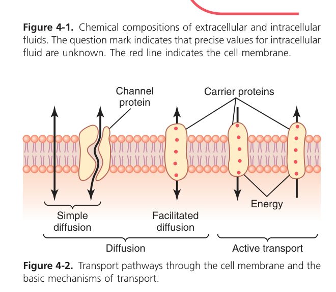
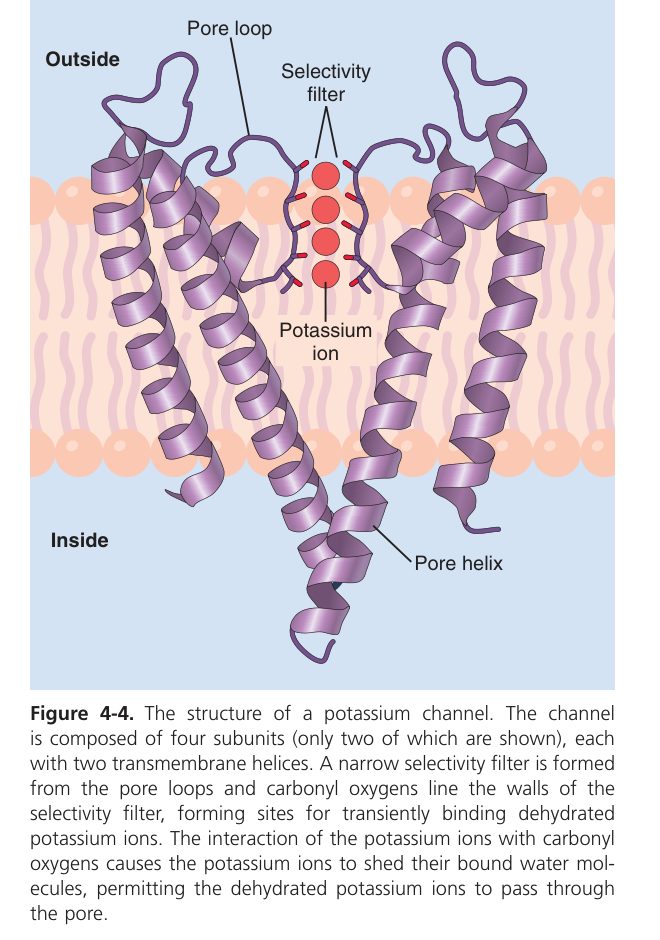
Every transport mechanism the body uses boils down to one question: are you moving the molecule down its concentration gradient (downhill, free) or up against it (uphill, costs energy)? Downhill movement is passive — no ATP needed. Lipid-soluble gases like O₂ and CO₂ just dissolve through the bilayer (simple diffusion). Water-soluble things like glucose or ions cannot — they need a protein gateway (facilitated diffusion: still passive, still downhill, but now selective and saturable).
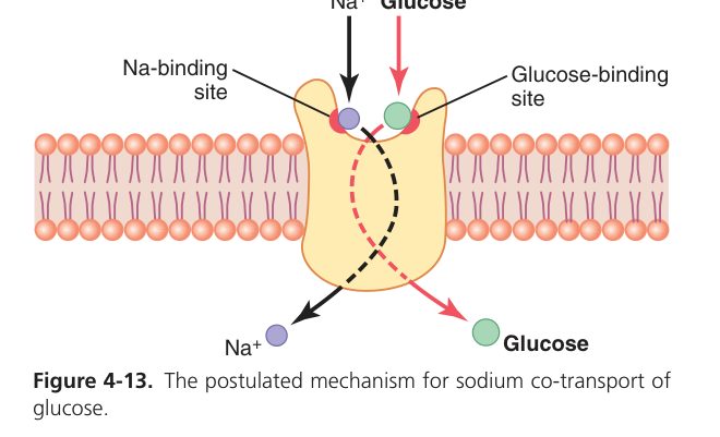
Uphill movement is active — it requires ATP, either directly (primary, like the Na⁺/K⁺ pump burning ATP itself) or indirectly (secondary, riding the Na⁺ gradient that an ATP-powered pump already created). Secondary active transport is a beautiful efficiency trick — one upstream ATP buys downstream uphill movement of many other solutes for hours. Finally, when something is too big to fit through any protein (a bacterium, a hormone-filled vesicle), the cell does endo/exocytosis — it wraps a piece of its own membrane around the cargo.
| Type | Energy? | Direction | Mechanism / carrier | Examples |
|---|---|---|---|---|
| Simple diffusion | No (passive) | Down gradient | Directly through lipid (lipid-soluble) or through channels | O₂, CO₂, alcohol, steroids, fatty acids, N₂ |
| Facilitated diffusion | No (passive) | Down gradient | Carrier-mediated (saturable) or channel-mediated | Glucose (GLUT), ions, amino acids, water (aquaporins) |
| Primary active transport | Yes — ATP directly | Against gradient | Pump hydrolyses ATP | Na⁺/K⁺-ATPase, Ca²⁺-ATPase, H⁺/K⁺-ATPase |
| Secondary active transport | Yes — ATP indirectly (uses Na⁺ gradient) | Solute against gradient | Symport (co-transport) / antiport (counter-transport) | Na⁺-glucose (SGLT, symport); Na⁺-Ca²⁺ & Na⁺-H⁺ (antiport) |
| Endo- / exocytosis | Yes (vesicular) | Bulk in / out | Membrane-bound vesicles | Phagocytosis, pinocytosis, receptor-mediated; secretion |
Both are passive (no ATP, move down the electrochemical gradient, no net energy). The difference: facilitated diffusion needs a membrane protein (carrier/channel), is faster for its substrate, and is saturable (has a Tmax) and can be competitively inhibited; simple diffusion is non-saturable and rises linearly with the gradient.
Every cell in your body is running this pump right now, and it accounts for roughly a third of the resting energy you burn at rest. That is how important the K⁺ gradient is — the cell is willing to spend a huge fraction of its ATP just to maintain it. The pump does one job per ATP: it shoves 3 Na⁺ out and 2 K⁺ in, both uphill. Notice the asymmetry — one more positive charge leaves than enters — which makes the pump slightly electrogenic, contributing a few millivolts of inside-negativity directly. But its bigger contribution is the gradient itself: high K⁺ inside, high Na⁺ outside. Every resting potential, every action potential, every glucose absorption in your gut is paid for, ultimately, by this pump.
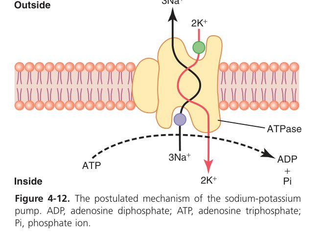
- Per ATP hydrolysed: pumps 3 Na⁺ OUT and 2 K⁺ IN — both against their gradients.
- Electrogenic (3 out / 2 in → net +1 charge leaves → contributes a few mV of negativity to the resting potential).
- Maintains the high-K⁺-inside / high-Na⁺-outside gradients that power the resting potential, the action potential, secondary active transport and cell-volume control.
Cardiac glycosides (digoxin) inhibit the Na⁺/K⁺-ATPase → intracellular Na⁺ rises → the Na⁺-Ca²⁺ antiporter slows → intracellular Ca²⁺ rises → stronger cardiac contraction (positive inotropy). Toxicity causes hyperkalaemia and arrhythmias.
Cystic fibrosis = mutation of the CFTR Cl⁻ channel → thick secretions in lung & pancreas (a channel-transport disease).
• Difference between simple and facilitated diffusion? → facilitated uses a protein, is saturable, faster
• Na⁺/K⁺ pump ratio per ATP? → 3 Na⁺ out, 2 K⁺ in → electrogenic
• What powers secondary active transport? → the Na⁺ gradient (set up by the primary pump)
• Why does digoxin strengthen the heart? → blocks Na⁺/K⁺-ATPase → ↑intracellular Na⁺ → ↑intracellular Ca²⁺ → ↑contractility
Intercellular Communication & Signal Transduction
A trillion cells cannot work together unless they can talk to each other — and they have two completely different ways to do it. The fastest is to physically share cytoplasm: gap junctions are tiny channels that link the insides of neighbouring cells, so ions and small molecules flow straight from one to the next. This is how every cardiac muscle cell knows when its neighbours fire, and why your heart contracts as a single unit. The slower (but far more flexible) route is chemical: one cell releases a signal molecule, another cell picks it up at a receptor. Depending on how far that signal travels, we call it endocrine (through the blood, like insulin reaching the liver), paracrine (next door, like a histamine release in tissue), autocrine (back on itself), or neurocrine (across a synapse).
- Gap junctions (connexons): direct intercellular channels permitting free passage of ions and small molecules (<1 kDa) — an electrical/electrotonic synapse. Allow synchronised activity, e.g. the simultaneous contraction of cardiac muscle and visceral smooth muscle (functional syncytium).
- Chemical messengers: endocrine (hormone via blood), paracrine (local diffusion to neighbours), autocrine (acts on the secreting cell), neurocrine (neurotransmitter across a synapse).
Once a chemical signal arrives at the cell surface, it needs to be translated into something the inside of the cell can act on — opening channels, changing enzyme activity, switching genes on or off. There is no single way to do this; evolution kept everything that worked, and the body uses them all. Some receptors are ion channels themselves: ACh binds the nicotinic receptor and the channel literally pops open in microseconds. Others are G-protein-coupled: the receptor activates a G-protein, which activates an enzyme that makes a second messenger (cAMP, IP₃, DAG), which then turns on intracellular kinases. A third type are tyrosine kinases like the insulin receptor — binding the hormone turns the receptor itself into an enzyme. And then steroid and thyroid hormones, being fat-soluble, just diffuse straight into the cell and bind nuclear receptors that act on DNA directly.
The big idea: one extracellular signal can trigger many intracellular responses, because second messengers amplify and branch. One adrenaline molecule binding a β-receptor can cause millions of cAMP molecules to be made — which is how you go from "I saw a tiger" to a full sympathetic surge in seconds. This is also why so many drugs target receptors: changing the start of the signal changes everything downstream.
The first messenger is the extracellular signal (hormone / neurotransmitter) binding the surface receptor. A second messenger is an intracellular molecule generated in response that mediates the intracellular responses to many different hormones and neurotransmitters (e.g. cAMP, cGMP, IP₃, DAG, Ca²⁺).
| Receptor class | Transduction | Second messenger | Example ligand |
|---|---|---|---|
| Ligand-gated ion channel (ionotropic) | Channel opens directly | (ion flux itself) | ACh at nicotinic receptor |
| G-protein-coupled (Gₛ) | ↑ adenylyl cyclase | cAMP → PKA | Adrenaline (β), glucagon, ADH (V2) |
| G-protein-coupled (Gᵢ) | ↓ adenylyl cyclase | ↓ cAMP | Adrenaline (α₂), ACh (muscarinic M2) |
| G-protein-coupled (Gᶉ) | ↑ phospholipase C | IP₃ (→Ca²⁺) + DAG (→PKC) | Adrenaline (α₁), angiotensin II |
| Enzyme-linked (tyrosine kinase) | Receptor autophosphorylation | Phosphorylation cascade | Insulin, growth factors |
| Intracellular (nuclear) | Alters gene transcription | (no membrane 2nd messenger) | Steroid & thyroid hormones |
• First vs second messenger? → first = extracellular ligand; second = intracellular relay (cAMP, IP₃, DAG, Ca²⁺)
• Gₛ-coupled receptor → ? → ↑ adenylyl cyclase → ↑ cAMP → PKA
• Gᶉ-coupled receptor → ? → ↑ PLC → IP₃ (releases Ca²⁺) + DAG (activates PKC)
• Where do steroid hormones bind? → intracellular (nuclear) receptors — they change gene transcription
Resting Membrane Potential
Stick a tiny electrode inside any living cell and you will measure a voltage: the inside of the cell is electrically negative compared to the outside, by something like 70–90 millivolts. The cell sits there like a tiny charged battery, waiting. Where does that voltage come from? Almost entirely from potassium leaking out. The Na⁺/K⁺ pump has loaded the inside with K⁺; the membrane is far more permeable to K⁺ than to Na⁺ at rest; so K⁺ trickles out down its gradient, taking positive charge with it and leaving negativity behind. The exit doesn't continue forever — as the inside grows more negative, it electrically pulls K⁺ back in. When push and pull balance, the membrane sits at K⁺'s equilibrium potential (the Nernst potential, ~−94 mV). The Na⁺/K⁺ pump itself is electrogenic and adds a few more millivolts of negativity, so the actual resting voltage is a touch less negative than pure Eₖ.
Picture this as a soft trap: the cell holds the K⁺ in only because it lets K⁺ out. The very act of leaking K⁺ makes the negative interior that retains the rest. That is also why anything that changes the K⁺ gradient changes the RMP — the most clinically dangerous example being hyperkalaemia. (See below.)
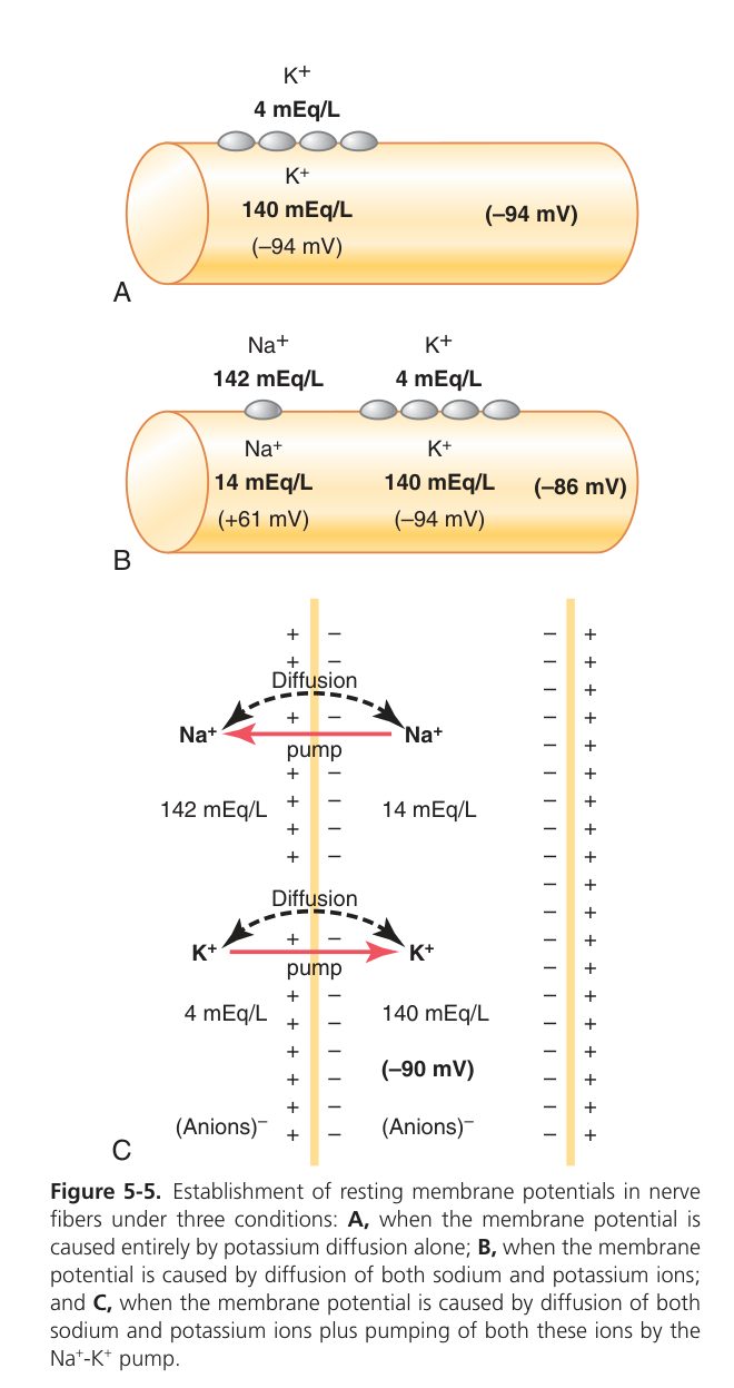
All living cells are polarised: the inside is electrically negative relative to the outside. The resting membrane potential (RMP) is about −70 mV in a typical neuron (and −90 mV in skeletal/cardiac muscle and large nerve fibres, per Guyton).
Two factors create it:
- K⁺ diffusion potential (the main cause): at rest the membrane is far more permeable to K⁺ than to Na⁺. K⁺ leaks out down its concentration gradient, leaving the inside negative, until the electrical pull balances the chemical push → the RMP sits close to Eₖ.
- Na⁺/K⁺ pump: maintains the gradients and adds a few mV of negativity (electrogenic).
| Ion | ECF (mM) | ICF (mM) | Equilibrium potential (37°C) |
|---|---|---|---|
| K⁺ | 4 | 140 | Eₖ ≈ −94 mV |
| Na⁺ | 142 | 14 | Eₘₐ ≈ +61 mV |
| Cl⁻ | 103–108 | 4 | E₊ₗ ≈ −70 to −90 mV |
| Ca²⁺ | ~1.2 (ionised) | ~0.0001 | E₊ₐ ≈ +120 mV |
The Nernst equation gives the equilibrium potential of a single ion (the voltage that exactly balances its concentration gradient):
The Goldman–Hodgkin–Katz (GHK) equation gives the actual membrane potential when several ions contribute, each weighted by its permeability (P):
Here is the clinical scenario that makes RMP theory life-and-death. A patient is brought in after kidney failure: serum K⁺ is 7 mM instead of 4. What happens? The K⁺ gradient (in/out) is smaller, so less K⁺ leaves, so the inside is less negative — the RMP drifts from −90 toward −70 mV. At first this is dangerous because the cell is now closer to threshold — it can fire too easily, the heart turns irritable. But push K⁺ even higher and the RMP sits so depolarised that voltage-gated Na⁺ channels go into their inactivated state and refuse to open at all. Now the heart cannot fire and arrests in diastole. This biphasic curve — first more excitable, then dead — is exactly why severe hyperkalaemia kills.
Raising ECF [K⁺] (e.g. 4 → 8 mM) reduces the K⁺ concentration gradient → less K⁺ leaves → Eₖ becomes less negative → the membrane depolarises (RMP rises toward 0, e.g. −90 → −70 mV).
Effect on excitability is biphasic: mild hyperkalaemia brings RMP closer to threshold → increased excitability; severe/sustained hyperkalaemia keeps the membrane depolarised so voltage-gated Na⁺ channels become inactivated → the cell cannot fire → decreased excitability (cardiac arrest in diastole).
• What ion mainly sets the RMP and why? → K⁺ — resting permeability is highest for K⁺
• Nernst at 37 °C? → Eion = (61/z) · log₁₀([out]/[in])
• Effect of high ECF K⁺ on RMP? → depolarises (RMP becomes less negative)
• Mild vs severe hyperkalaemia effect on excitability? → mild ↑, severe ↓ (Na⁺ channels inactivated)
The Action Potential
The cell sits at rest, slightly negative inside, holding its battery charged. Then a stimulus arrives — a touch on the skin, a transmitter at a synapse — and pushes the membrane just past a threshold around −55 mV. In that instant the cell does something dramatic: it reverses its own polarity in under a millisecond, then resets, then is ready to do it again. That is the action potential, and it is how every nerve in your body says anything to anything.
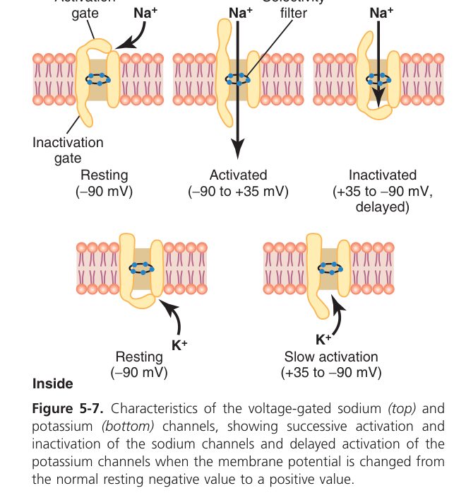
The trick is built into a single channel: the voltage-gated Na⁺ channel. When the membrane depolarises to threshold, these channels snap open and Na⁺ floods in. The inrush makes the membrane more positive, which opens more Na⁺ channels — a runaway positive feedback that drives the voltage all the way up toward +30 mV. This is the upstroke. Then two things end it: the Na⁺ channels enter an inactivated state (closed-and-locked) and a slower set of voltage-gated K⁺ channels opens, letting K⁺ flood out. K⁺ efflux carries positive charge away — the membrane plunges back toward rest. The K⁺ channels stay open a fraction too long, briefly making the cell more negative than baseline (after-hyperpolarisation), then everyone resets and the cell is ready to fire again.
Notice what makes the AP "all-or-none": the upstroke is regenerative, so once it starts it must complete. There is no half-AP. Strength of stimulus is therefore not encoded in the size of one spike but in the frequency of spikes — a loud sound makes auditory neurons fire faster, not bigger.
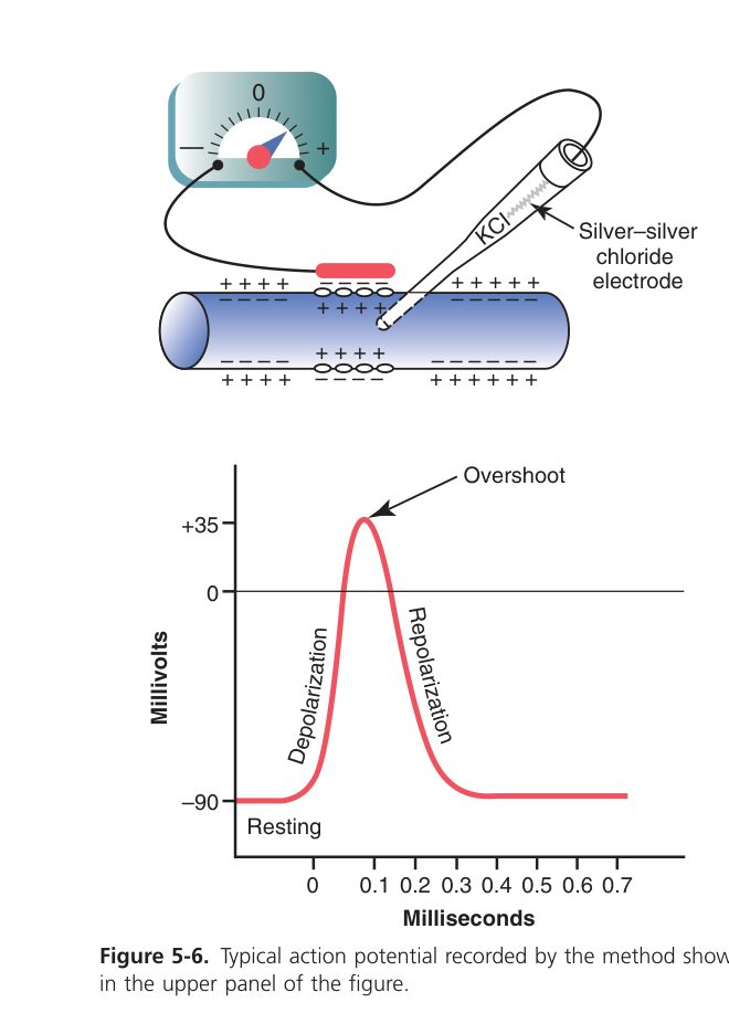
An action potential (AP) is a rapid, all-or-none, self-propagating reversal of membrane potential. Threshold ≈ −55 mV (TMU slide states ≈ −50 mV; varies by cell).
| Phase | Voltage change | Ion movement | Channel |
|---|---|---|---|
| Resting / polarised | −70 to −90 mV | K⁺ leak; pump active | K⁺ leak channels |
| Depolarisation (upstroke) | → toward +30 mV | Na⁺ influx (regenerative, positive feedback) | Voltage-gated Na⁺ channels open (fast) |
| Repolarisation | back toward rest | Na⁺ channels inactivate; K⁺ efflux | Na⁺ inactivation + delayed voltage-gated K⁺ |
| After-hyperpolarisation | briefly < RMP | K⁺ channels still open (overshoot) | Slow-closing K⁺ channels |
The Na⁺/K⁺ pump later restores the tiny amounts of Na⁺/K⁺ that moved — but it is not what repolarises the membrane (K⁺ efflux does that).
- All-or-none: once threshold is reached, the AP fires at full, fixed amplitude and shape; a stronger stimulus does not make a bigger AP. Stimulus intensity is encoded by AP frequency (frequency code), not size.
- Absolute refractory period (ARP): during the spike, Na⁺ channels are open then inactivated → no second AP is possible, however strong the stimulus. The ARP sets the maximum firing frequency and ensures one-way propagation.
- Relative refractory period (RRP): during after-hyperpolarisation a stronger-than-normal stimulus can fire an AP.
“Na⁺ in = up, K⁺ out = down.” Depolarisation = Na⁺ rushing IN; repolarisation = K⁺ flowing OUT. Absolute refractory = Na⁺ channels inactivated (the ‘closed-and-locked’ state, distinct from the resting closed state).
| Feature | Local (graded) potential | Action potential |
|---|---|---|
| Stimulus | Subthreshold | Threshold or above |
| Amplitude | Graded (varies with stimulus) | All-or-none (constant) |
| Summation | Yes (temporal & spatial) | No (cannot summate) |
| Propagation | Decremental (dies out, electrotonic) | Non-decremental (regenerated, constant) |
| Refractory period | None | Present (absolute + relative) |
| Ion basis | Few channels (e.g. partial Na⁺) | Voltage-gated Na⁺ then K⁺ |
| Examples | EPSP, IPSP, receptor & end-plate potential | Nerve impulse, muscle AP |
• Upstroke = which ion in? → Na⁺ influx (Na⁺ channels open)
• Repolarisation = which ion out? → K⁺ efflux
• What makes the absolute refractory period? → Na⁺ channels are inactivated, cannot reopen
• How is stimulus intensity coded? → AP frequency, not amplitude (all-or-none)
• Local potential vs AP — one key difference? → local potential is graded & summates, AP does not
Conduction of the Impulse
An AP at one patch of axon does not just sit there — it sends positive charges flowing forward inside the axon (and backward, but the backward direction is in refractory period and cannot re-fire). When those currents reach the next segment of membrane they push it past threshold, and that segment now fires its own full-size AP. The signal is regenerated at every step, so it never weakens with distance — unlike a passive electrical signal that fades like ripples on water, the AP is more like a fuse that re-ignites itself segment by segment.
The AP propagates because the depolarised patch acts as a current source: local circuit (eddy) currents flow forward inside the axon, depolarising the next segment to threshold, which then fires its own AP — so the AP is continuously regenerated and conducted without decrement.
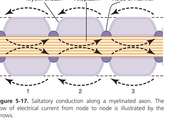
If every patch of membrane has to fire its own AP, conduction is slow and expensive. So most vertebrate nerves wrap the axon in myelin — an insulating fatty sheath made by Schwann cells (in the periphery) or oligodendrocytes (in the CNS). Myelin stops the AP from happening across most of the axon by physically blocking ion flow; the only places left where the axon can fire are the bare gaps between myelin segments — the nodes of Ranvier. So the AP "jumps" from node to node, depolarising the next node by local current flow. This is saltatory conduction (Latin saltare, to leap). It is many times faster than continuous conduction, and far cheaper energetically because only a tiny fraction of the axon's membrane actually depolarises — less Na⁺ in, less K⁺ out, less pump work to restore.
This is also why demyelinating disease is so disabling. Take away the myelin (multiple sclerosis, Guillain–Barré) and the impulse can no longer jump — it has to crawl. Conduction slows, blocks, or fails altogether; depending on where the affected nerves go, the patient gets weakness, numbness, blindness, paralysis. The damage is to the wrapping, not to the wire itself.
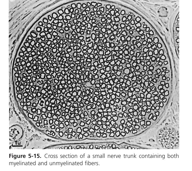
| Feature | Unmyelinated fibre | Myelinated fibre |
|---|---|---|
| Mode | Continuous (point-to-point) | Saltatory (jumps node to node) |
| AP site | Whole membrane | Only at nodes of Ranvier |
| Velocity | Slower | Faster (myelin insulates & ↑ resistance) |
| Energy cost | Higher (more ions move) | Lower (fewer ions, less pump work) |
Conduction velocity also rises with axon diameter (larger = lower internal resistance = faster).
Demyelinating disease — multiple sclerosis (CNS) and Guillain–Barré syndrome (PNS) — strips myelin, so saltatory conduction fails and impulses slow or block → weakness, numbness, paralysis. Local anaesthetics (lidocaine) block voltage-gated Na⁺ channels → no AP can be generated → loss of sensation; small unmyelinated pain fibres are blocked first.
• What does saltatory conduction mean? → the AP jumps from node of Ranvier to node, skipping myelin
• Why does myelin speed conduction? → insulates internodes; only nodes fire → faster + cheaper
• Conduction velocity depends on? → myelination + axon diameter (bigger = faster)
• Two demyelinating diseases? → multiple sclerosis (CNS), Guillain–Barré (PNS)
Cell Physiology complete
Transport, signalling, membrane & action potentials mastered. Next: Nerve & Muscle — NMJ and contraction.