Skin & Appendages
Skin Overview
Skin is the largest organ of the body, and when you really look at it on a slide, you see it is built like a two–layer sandwich riding on a fatty cushion. On top sits the epidermis, a keratinised stratified squamous epithelium derived from ectoderm; it is the only part of the skin that is actually epithelium, and it is avascular. Beneath it lies the dermis, a tough connective–tissue layer of mesodermal origin that carries the blood vessels, nerves and almost everything else of value. Both layers rest on a third, technically extra–cutaneous layer, the hypodermis (subcutaneous tissue, superficial fascia), made of loose connective tissue plus adipose — the thermal blanket and shock absorber.
Two functional flavours of skin matter for the histology exam. Thick skin lines the palms and soles: it has all five epidermal strata (including the famous stratum lucidum), a heavy stratum corneum, dermal ridges that give you fingerprints, plenty of eccrine sweat glands, but no hair follicles and no sebaceous glands. Thin skin covers the rest of the body: thinner epidermis, only four obvious strata, but rich in hair, sebaceous glands and a variety of sweat glands. When a slide shows you palmar epidermis as thick as the dermis underneath, you have already named the location.
Skin does five jobs at once — barrier (keratin + lipid against water loss and microbes), thermoregulation (sweat + dermal vasculature), sensation (receptors and free nerve endings), immune surveillance (Langerhans cells, dermal dendritic cells) and endocrine/vitamin D synthesis. Keep this functional list in mind: nearly every micro–structure you will be asked to identify maps onto one of those five jobs.
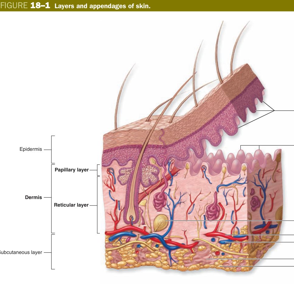
Think of skin as a brick wall on a leather pad on a duvet. The bricks are keratinocytes glued by lipid mortar (epidermis). The leather pad is the dermis — thin, tough, full of plumbing. The duvet is the hypodermis — soft fat for insulation. Hair, glands and nails are tunnels and pipes drilled down from the bricks into the leather.
Recall Skin = epidermis (keratinised stratified squamous, ectoderm, avascular) + dermis (CT, mesoderm, vascular). Hypodermis = loose CT + fat, not skin proper. Thick skin (palms/soles): 5 layers, no hair, no sebaceous; thin skin (rest): 4 layers, has hair + sebaceous. Five jobs: barrier, thermoregulation, sensation, immunity, vitamin D.
Epidermis — the 5 Layers (essay-grade)
The epidermis is an assembly line: a new keratinocyte is born at the bottom, walks slowly upward over about four weeks, and is shed dead from the top as a flat keratin flake. As it climbs it changes shape and protein content so dramatically that pathologists named each step a separate stratum. You can read the slide deep–to–superficial like the chapters of a short biography.
From deep → superficial the keratinocytes form five strata. All five are present only in thick skin; in thin skin the lucidum is absent or unrecognisable. The order is sacred — learn it as one continuous word.
| Layer (deep→superficial) | Key features |
|---|---|
| Stratum basale (germinativum) | Single layer of cuboidal/columnar cells on basement membrane; mitotically active (renews the epidermis); contains stem cells, melanocytes, Merkel cells; anchored by hemidesmosomes; K5/K14 keratins |
| Stratum spinosum | Several layers of polyhedral cells joined by desmosomes → “spiny” / prickle appearance on fixation; tonofilaments; Langerhans cells lurk here |
| Stratum granulosum | 3–5 layers of flat cells full of basophilic keratohyalin granules (filaggrin precursor) + lamellar bodies (Odland bodies; lipid for water barrier) |
| Stratum lucidum | Thin, clear, pale band of dead flattened cells with eleidin — thick skin only |
| Stratum corneum | Many layers of dead, flat, anucleate keratin-filled squames glued by lipid; continually shed (desquamation) |
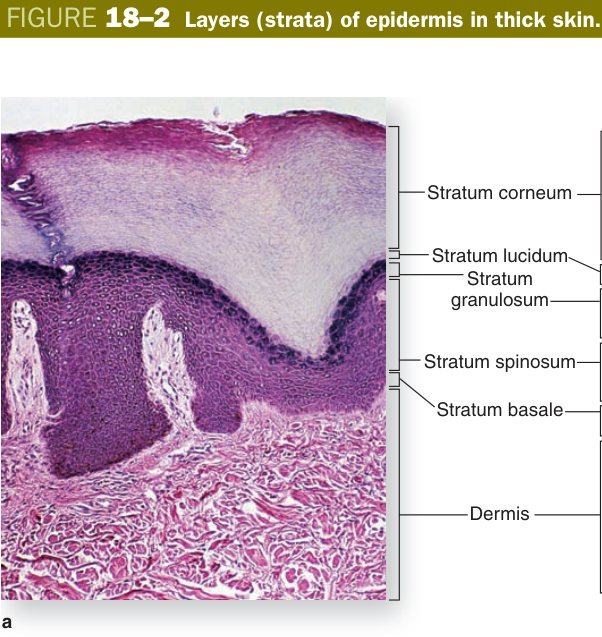
Mark the basale as the only layer that divides. Every other layer is a stage of differentiation, not proliferation. Half the daughters of a basal mitosis stay basal (renewal); the other half migrate outward, lose their nuclei, dump their organelles and end up as armoured plates of keratin. By the time a cell reaches the corneum it is no longer a cell — it is a building material.
The epidermis is a bakery conveyor belt. Dough (basale) is mixed at the bottom. It rises and develops gluten threads (spinosum — tonofilaments). Yeast granules appear (granulosum — keratohyalin). It enters the oven and becomes invisible glass (lucidum). Finally it comes out as a hard crusty bread (corneum) that flakes off the surface.
Deep→superficial: “Basale · Spinosum · Granulosum · Lucidum · Corneum” = “British Soldiers Grew Lush Corn”. Only the basale divides; lucidum = thick skin only.
Recall Five strata, deep→superficial: basale · spinosum · granulosum · lucidum · corneum. Basale = only mitotic layer, K5/K14, hemidesmosomes. Spinosum = desmosomes & spines, K1/K10 switch, Langerhans. Granulosum = keratohyalin + lamellar bodies = waterproof barrier. Lucidum = thick skin only. Corneum = dead anucleate squames. Transit time ~4 weeks.
As a keratinocyte travels from basale to corneum it switches on a sequence of structural proteins as predictable as a recipe. At the bottom it makes a “soft” keratin pair (K5/K14) that lets it divide and stretch. The moment it commits to differentiation in the spinosum it swaps for a “hard” pair (K1/K10) that bundles into tonofilaments and gives the cell its mechanical strength. In the granulosum it begins to build the cornified envelope — loricrin and involucrin are cross–linked under the plasma membrane by transglutaminases, while profilaggrin is cleaved to filaggrin, which then bundles the keratin filaments into the dense matrix you will see as bright pink keratin.
At the same time, granulosum cells dump the contents of their lamellar bodies (Odland bodies) into the intercellular space — glycolipids, sterols and free fatty acids that solidify into the lipid mortar between the corneocytes. This lipid is the waterproof barrier of the skin. Lose the granulosum, and you lose the ability to hold water in: that is exactly what happens in severe burns and in filaggrin–deficient ichthyosis.
| Layer | Key proteins / events |
|---|---|
| Basale | K5/K14 keratins; mitosis via p63 stem cells; half the daughter cells migrate upward |
| Spinosum | Switch to K1/K10 keratins; desmoglein-1/3 in desmosomes holds cells together (target in pemphigus vulgaris) |
| Granulosum | Profilaggrin → filaggrin (aggregates keratin); loricrin & involucrin (cornified envelope precursors); lamellar bodies (Odland bodies) exocytose glycolipids & sterols into intercellular space — the waterproof barrier |
| Lucidum (thick only) | Dead flattened cells; eleidin (degraded keratohyalin); appears clear in H&E |
| Corneum | Anucleate, flat corneocytes; cross-linked cornified envelope; desquamation by serine proteases |
Thick versus thin skin is the second classic compare–and–contrast. Once you understand that thick skin exists because palms and soles must take heavy mechanical wear and produce a lot of cooling sweat, every difference falls out logically: more strata for armour, dermal ridges to lock the epidermis onto the dermis like Velcro, no hair (which would be uncomfortable to grip with), no sebaceous glands (oil + grip do not mix), and a forest of eccrine glands for thermoregulation.
| Feature | Thick skin | Thin skin |
|---|---|---|
| Location | Palms, soles | All other body surfaces |
| Layers | All 5 (including lucidum) | 4 (no lucidum) |
| Stratum corneum | Very thick | Thin |
| Appendages | No hair follicles, no sebaceous glands; many eccrine sweat glands | Hair follicles, sebaceous glands, eccrine & apocrine glands |
| Dermal ridges | Prominent (fingerprints) | Less pronounced |
Pemphigus vulgaris — autoantibodies against desmoglein-1/3 → loss of desmosomal adhesion in spinosum → intra-epidermal blisters (acantholysis); Nikolsky sign positive. Bullous pemphigoid — autoantibodies against hemidesmosomal proteins (collagen XVII / BPAG) at the BM → sub-epidermal blisters (deeper, tenser). Ichthyosis vulgaris — filaggrin mutations → defective desquamation and scaly skin (also a strong risk for atopic dermatitis). Psoriasis — accelerated basal turnover with parakeratosis (retained nuclei in corneum) and Munro microabscesses.
Recall Protein switch: K5/K14 (basale) → K1/K10 (spinosum) → filaggrin + loricrin + involucrin (granulosum) → cross–linked envelope (corneum). Lamellar bodies = lipid mortar = water barrier. Thick skin = palms/soles, all 5 layers, no hair, no sebaceous. Pemphigus vulgaris = anti–desmoglein, intra–epidermal; bullous pemphigoid = anti–hemidesmosome, sub–epidermal.
Non-Keratinocyte Cells of the Epidermis
Keratinocytes are the bulk of the epidermis, but three minority residents do all the interesting non–structural work: melanocytes pigment, Langerhans cells defend, Merkel cells sense. Each has a different birthplace, a different home in the epidermis and a different signature on histology.
Melanocytes are wanderers. Embryologically they come from the neural crest, migrate during development through the dermis and finally take up residence in the stratum basale, where they sit on the basement membrane between keratinocytes. On routine H&E they look like small rounded cells with a pale halo around the nucleus — this halo is partly fixation artefact and partly the fact that their dendritic processes do not stain. Special stains (DOPA reaction, S100, HMB–45, Melan–A) reveal long branching dendrites that reach up into the spinosum, each one delivering pigment to neighbouring keratinocytes.
Inside the melanocyte, the enzyme tyrosinase oxidises tyrosine through DOPA to produce melanin, which is packaged in lysosome–like organelles called melanosomes. There are two pigments: brown–black eumelanin and red–yellow pheomelanin. The ratio is genetically set and tunes your skin and hair colour. Fully packed melanosomes are then transferred to keratinocytes by a unique mechanism called cytocrine secretion — the dendrite tip is essentially phagocytosed by the keratinocyte. Inside the keratinocyte the melanin granules pile up like a cap on the sun–facing side of the nucleus and physically absorb UV photons before they can damage DNA. Skin colour difference is not the number of melanocytes (everyone has roughly the same number) but the size and distribution of melanosomes.
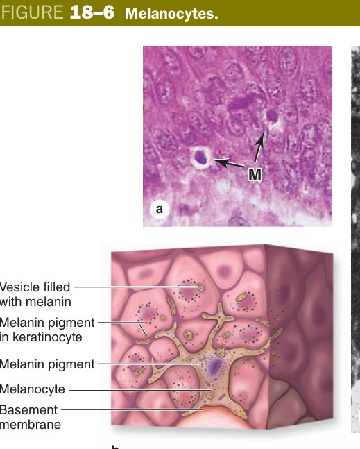
A melanocyte is a UV umbrella factory with a delivery service. It makes black umbrellas (melanosomes), then hands them out through long arms to about 30–40 surrounding keratinocytes (the “epidermal melanin unit”). Each keratinocyte parks its umbrella over the nucleus so DNA does not get sunburnt.
Vitiligo — autoimmune destruction of melanocytes; depigmented patches. Albinism — tyrosinase or related enzyme defect; normal melanocyte numbers but no functional pigment, leading to very high skin–cancer risk. Melanoma — malignant transformation of a melanocyte; check by ABCDE (Asymmetry, Border, Colour, Diameter, Evolution); prognosis driven by Breslow depth.
Recall Melanocyte = neural crest, basal layer, dendritic. Tyrosinase converts tyrosine → melanin in melanosomes. Cytocrine transfer to keratinocytes; melanin caps the nucleus for UV shielding. Eumelanin (brown) vs pheomelanin (red). Vitiligo = autoimmune loss; albinism = tyrosinase defect; melanoma = malignancy, Breslow depth prognostic.
Two cells with similar–sounding names but completely different jobs share the epidermis. Langerhans cells are immune cells: bone–marrow–derived dendritic cells that live mainly in the stratum spinosum, where they patrol the epidermis like security guards. They pick up foreign antigens (microbes, contact allergens), migrate down through the dermis to a draining lymph node and present those antigens on MHC class II to naive T cells. On electron microscopy they have a unique tennis–racquet–shaped granule called the Birbeck granule, which is essentially endocytic. They are the front line in contact dermatitis (think nickel allergy, poison ivy) and are also the cell of origin of Langerhans cell histiocytosis.
Merkel cells, in contrast, are sensory. They sit in the stratum basale, often clustered in “touch domes” on fingertips and lips, and form synapse–like contacts with a flattened terminal of a sensory nerve (the Merkel disc). Together they form a slowly–adapting mechanoreceptor exquisitely sensitive to steady pressure and fine spatial detail — the receptor you use when reading Braille or feeling the shape of a key in your pocket. Their malignant counterpart, Merkel cell carcinoma, is a rare but aggressive neuroendocrine skin tumour associated with Merkel cell polyomavirus.
| Cell | Site | Function |
|---|---|---|
| Langerhans cell | Mainly stratum spinosum | Dendritic antigen-presenting cell (immune surveillance); bone marrow origin; Birbeck granules on EM; MHC II |
| Merkel (tactile) cell | Stratum basale | Slowly-adapting mechanoreceptor for fine touch & pressure; synapse with sensory nerve terminal (Merkel disc); neuroendocrine |
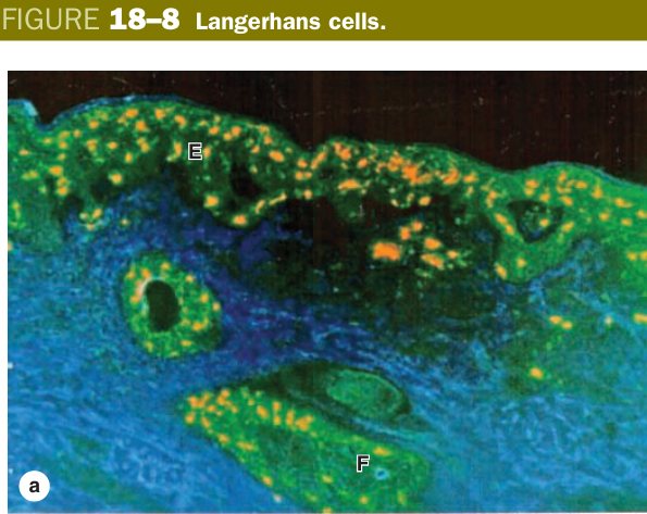
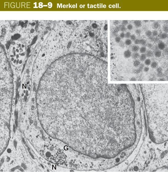
Think of the epidermis as a small village. Keratinocytes are the houses. Melanocytes are the awning–makers (UV shade). Langerhans cells are the patrolling police (immunity). Merkel cells are the doorbell buttons (touch). Knowing the job tells you the cell.
4 epidermal cell types: Keratinocyte (bulk) · Melanocyte (pigment, basale) · Langerhans (immune, spinosum) · Merkel (touch, basale). “Keep Making Light Marks.”
Recall Langerhans = bone marrow origin, spinosum, dendritic APC, MHC II, Birbeck granule, contact dermatitis. Merkel = basal, mechanoreceptor for fine touch, synapses with sensory nerve, neuroendocrine origin, Merkel cell carcinoma (polyomavirus).
Dermis & Sensory Receptors
If the epidermis is the wallpaper, the dermis is the wall. It is the thick, vascular, nerve–rich connective–tissue layer that gives skin its tensile strength, holds the appendages, feeds the avascular epidermis by diffusion and houses every encapsulated sensory receptor of the body wall. It is divided into two layers that you can almost always tell apart at low power by the size of the collagen bundles.
The superficial papillary dermis is a thin sheet of loose connective tissue rich in fine type–III (reticular) fibres. It interdigitates with the epidermis through finger–like dermal papillae that fit into epidermal rete ridges — a clever way to lock the two layers together mechanically and to maximise the surface area for diffusion of nutrients up into the epidermis. Loops of capillaries climb into each papilla. Meissner corpuscles, the receptors for fine discriminative touch, also sit inside these papillae, which is why fingertips can read tiny braille dots.
Beneath this lies the much thicker reticular dermis, a sheet of dense irregular connective tissue dominated by interwoven bundles of type–I collagen plus elastic fibres. The collagen weave runs in dominant directions called Langer's lines; surgeons cut along these lines because incisions parallel to them gape less and scar more cosmetically. The reticular dermis carries the larger vessels, the bases of hair follicles and sweat glands, and the deep mechanoreceptors — Pacinian corpuscles for vibration and pressure, and Ruffini endings for sustained stretch.
| Receptor | Location | Modality |
|---|---|---|
| Free nerve endings | Up into the epidermis | Pain, temperature, itch — unmyelinated Aδ / C fibres |
| Meissner (tactile) corpuscle | Dermal papillae (papillary layer) | Fine/light touch — encapsulated, stacked flattened Schwann cells; rapidly adapting |
| Pacinian (lamellated) corpuscle | Deep reticular dermis / hypodermis | Deep pressure & vibration — large, “onion-skin” concentric lamellae; rapidly adapting |
| Ruffini ending | Reticular dermis | Sustained stretch & skin distortion; slowly adapting |
| Krause end bulb | Mucocutaneous junctions | Cold sensation (classical teaching) |
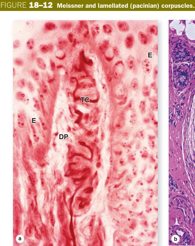
The Pacinian corpuscle is a cut onion in the deep dermis — the nerve sits in the centre and the layered “onion skin” lamellae filter out everything except rapid mechanical changes, so only vibration gets through to the axon. The Meissner corpuscle, by contrast, is a little stack of pancakes tucked up into the dermal papilla where the epidermis can “press” it during fine touch.
Pacinian (lamellated) corpuscle: a large encapsulated mechanoreceptor in the deep dermis/hypodermis; a central unmyelinated nerve terminal surrounded by concentric lamellae of flattened Schwann/connective–tissue cells (“onion”), responding rapidly to deep pressure and vibration.
Recall Dermis = papillary (loose CT, type III, dermal papillae, capillary loops, Meissner) + reticular (dense irregular, type I, Langer's lines, Pacinian, Ruffini). Sensation map: free endings = pain/temp/itch; Meissner = fine touch; Pacinian = vibration/pressure; Ruffini = stretch; Krause = cold.
Skin Appendages
Skin appendages are epidermal invaginations that have burrowed down into the dermis during development — hair follicles, sebaceous glands, sweat glands and nails. Every one of them is lined by epithelium continuous with the surface, which is exactly why they can act as reservoirs of stem cells to reseed the epidermis after a partial–thickness burn. If only the corneum is lost, the basale grows back from below; if the dermis is also destroyed, the appendage epithelium is your last chance.
- Hair follicle: down-growth of epidermis; concentric layers (from in→out: medulla, cortex, cuticle of hair; then internal & external root sheaths); a hair bulb sits on a CT dermal papilla; the arrector pili smooth muscle attaches the follicle to the papillary dermis.
- Sebaceous gland: holocrine — whole cells disintegrate to release oily sebum into the hair follicle; cells have pale, foamy lipid-filled cytoplasm; absent from palms & soles.
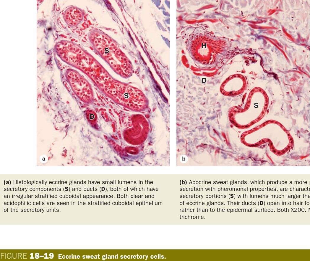
- Eccrine sweat gland: simple coiled tubular merocrine gland; dark (mucoid) + clear (watery) secretory cells + contractile myoepithelial cells; thermoregulation; opens directly to skin surface.
- Apocrine sweat gland: axilla, areola, anogenital; large lumen; opens into hair follicle; active after puberty; pheromonal role; despite the name, in humans secretion is largely merocrine.
- Nail: hard keratin plate produced by the nail matrix at the root; lunula = visible matrix; eponychium (cuticle) seals the proximal nail fold; hyponychium thickens under the free edge.
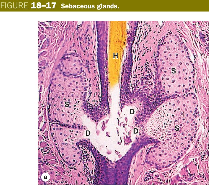
Three secretion modes show up cleanly in the appendages and they are favourite exam fodder. Merocrine (eccrine) means exocytosis with no loss of cytoplasm — the cell is untouched. Apocrine means the apical cytoplasm pinches off with the secretion (classically taught for true apocrine sweat and the lipid component of milk). Holocrine means the entire cell becomes the secretion — this is sebaceous gland behaviour, and the cells must be continuously regenerated from a peripheral basal layer.
Think of three coffee servers. Merocrine is the polite barista who pours coffee from the pot. Apocrine is the server who throws the top half of the cup at you with the coffee in it. Holocrine is the server who climbs into a blender and turns himself into the coffee. Sebaceous = the blender server.
Acne vulgaris — pilosebaceous unit blocked by hyperkeratosis + sebum, colonised by Cutibacterium acnes. Hidradenitis suppurativa — chronic apocrine–follicular inflammation of axilla/groin. Cystic fibrosis — defective CFTR in the eccrine duct cannot reabsorb Cl− → salty sweat (positive sweat chloride test). Alopecia areata — immune attack on hair follicle bulb. Burns are graded by depth; basal layer + appendage epithelium reseed the epidermis after partial-thickness loss. Basal cell carcinoma (pearly nodule, basal–layer origin, sun–exposed) and squamous cell carcinoma (actinic keratosis precursor, keratin pearls on histology) round out the common skin cancers besides melanoma.
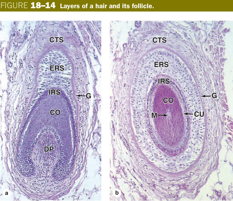
A hair follicle is a cylindrical down–growth of epidermis into the dermis, and on a longitudinal section you can read it like a set of nested tubes. From outside in you cross a connective–tissue sheath, a glassy membrane (a thickened basement membrane), the external root sheath (which is continuous with surface epidermis and is the stem–cell reservoir), the internal root sheath in three sub–layers (Henle's, Huxley's and the IRS cuticle), and finally the hair shaft itself with its cuticle, cortex and medulla.
All of these layers are born from the same place: the matrix at the bottom of the bulb, a cap of rapidly dividing keratinocytes sitting on the dermal papilla. The dermal papilla is a small lump of vascularised connective tissue that signals to the matrix — it is the inductive switch for the entire follicle. Knock out the papilla and the hair stops growing; surgical hair transplants succeed only when the papilla travels with the graft.
| Layer (outer→inner) | Derivation & features |
|---|---|
| Connective tissue sheath | Dermal origin; glassy membrane (thickened BM); contains blood vessels |
| External root sheath (ERS) | Continuous with surface epidermis; non-keratinising (glycogen-rich); the bulge region of the ERS = stem cell niche (attachment of arrector pili) |
| Internal root sheath (IRS) | From matrix cells; 3 layers: Henle's layer (outermost, single layer) → Huxley's layer → IRS cuticle (interlocks with hair cuticle); disappears at the sebaceous gland duct opening |
| Hair shaft | From outer→inner: cuticle (overlapping dead squames) → cortex (hard keratin, melanin granules from melanocytes in the bulb) → medulla (loose vacuolated cells, absent in fine hair) |
| Hair bulb | Base of follicle; matrix cells (rapidly dividing, give rise to hair + IRS) rest on the dermal papilla (CT, highly vascular; induces hair growth) |
Every hair lives a three–stage life cycle. Anagen is active growth, lasting years for scalp hairs and weeks for eyebrows — that ratio determines maximum length. Catagen is a brief regression in which the follicle shrinks and the bulb retracts upward. Telogen is the resting phase; the old shaft is held loosely until a new anagen pushes it out. About 85–90% of scalp hairs are in anagen at any moment; physiological or emotional shocks can synchronise many hairs into telogen and produce diffuse shedding (telogen effluvium) months later.
IRS (outer → inner): “Henle Has IRS” — Henle's → Huxley's → IRS Cuticle (HHC). The IRS cuticle and hair cuticle interdigitate like zipper teeth, anchoring the hair shaft in the follicle.
Recall Hair follicle layers out→in: CT sheath / glassy BM / ERS (with bulge stem cells) / IRS (Henle → Huxley → cuticle) / hair shaft (cuticle/cortex/medulla). Matrix on dermal papilla = growth engine. Cycle: anagen (growth) → catagen (regression) → telogen (rest). Arrector pili = smooth muscle attached at bulge.
The eccrine sweat gland is the most abundant of all skin appendages and the master thermoregulator. On a section you will see two very different epithelial profiles for one gland: a deep secretory coil sitting in the deep dermis or hypodermis, and a duct that climbs up through the dermis, corkscrews into the epidermis and opens directly on the surface as a sweat pore. They differ in epithelium, function and staining.
The secretory coil is a simple columnar/cuboidal tube with three cell types: pale clear cells rich in mitochondria that secrete the watery primary fluid (an isotonic NaCl solution), darker mucoid (dark) cells with apical glycoprotein granules, and underneath them flat myoepithelial cells that contract on sympathetic command to squirt the primary secretion into the duct. The duct then becomes a two–layered stratified cuboidal epithelium, stains slightly darker, and actively reabsorbs Na+ (and Cl− follows) from the lumen so that the final sweat reaching the skin is hypotonic.
| Portion | Epithelium | Cell types & function |
|---|---|---|
| Secretory coil (deep dermis/hypodermis) | Simple columnar/cuboidal | Dark cells (columnar, mucoid, apical glycoprotein granules) + clear cells (pale, pyramidal, mitochondria–rich; secrete isotonic water & electrolytes) + myoepithelial cells (contractile, squeeze secretion into duct) |
| Duct (dermis → epidermis) | Stratified cuboidal (2 layers, no myoepithelial cells) | Actively reabsorbs Na+ (and Cl−) from the primary secretion → final sweat is hypotonic; under aldosterone control |
The eccrine gland is a desalination plant in reverse. The coil produces salty water (isotonic). The duct then pulls the salt back inside the body so you do not lose precious sodium when you sweat for hours — what reaches the surface is mostly water with a little salt. In cystic fibrosis the salt–pulling pump (CFTR) is broken, so the sweat stays salty — that is what the sweat chloride test measures.
Eccrine: merocrine secretion (exocytosis); found all over body; small lumen; opens directly onto skin surface; thermoregulation; sympathetic cholinergic innervation (an exception). Apocrine: developmentally apocrine (apical pinch–off; in humans much of the actual secretion is merocrine); axilla, areola, anogenital; large lumen; opens into hair follicle; active after puberty; odour produced by bacterial action on the lipid–rich secretion.
Recall Eccrine gland = simple coiled tubular merocrine. Coil (simple cub/columnar) = clear + dark + myoepithelial cells → isotonic primary secretion. Duct (stratified cuboidal, 2 layers) = reabsorbs Na+/Cl− → final sweat hypotonic. CF = defective Cl− reabsorption → salty sweat → sweat chloride test. Cholinergic sympathetic innervation.
TMU Exam Drill
📝 Open the full TMU Question Bank — 20 MCQ + 6 terms + 5 essays →
Authentic Tianjin Medical University past-paper questions (2021 Final & the multi-section Final with answer key) mapped to this unit, in the real exam format. Click Show answer to self-test.
□ Single best answer
- A. basal layer (stratum basale)
- B. spinous layer (stratum spinosum)
- C. granular layer (stratum granulosum)
- D. clear layer (stratum lucidum)
- E. cornified layer (stratum corneum)
□ Fill in the blank
- (fill base→surface)
Skin complete
Epidermal layers, melanocyte/Langerhans/Merkel, dermal receptors & appendages mastered. Last: General Embryology.