General Embryology
Overview & Timeline
Human development between fertilisation and the end of week 8 is the entire embryonic period — the slice of life in which a single diploid cell becomes a recognisable little human with a beating heart, looped gut, paddle-shaped limbs and a folded neural tube. Everything you will study in systemic histology, anatomy and pathology has its origin here, in a sequence so tightly choreographed that a one-day delay in any step usually means malformation rather than catch-up.
The sequence is worth memorising as a single sentence before any of the detail: fertilisation → cleavage → morula → blastocyst → implantation → bilaminar disc (week 2) → trilaminar disc by gastrulation (week 3) → organogenesis and body folding (weeks 4–8), all of it supported by the fetal membranes and the developing placenta. Every event in this unit slots onto that spine.
The geography is just as important as the timing. Fertilisation happens in the lateral third of the oviduct (the ampulla), cleavage happens as the conceptus drifts down the tube, implantation happens in the upper posterior wall of the uterus, and from there the placenta grows outward into the decidua basalis. You can almost trace a finger across a pelvic diagram — ovary, ampulla, uterine cavity, posterior wall — and watch the first week unfold.
Picture the oviduct as a one-way conveyor belt that picks up the oocyte at the fimbriae, fertilises it at the ampulla, then carries the conceptus toward the uterus while it divides. The conveyor takes about five days — by the time the belt empties into the uterus, you already have a hollow blastocyst ready to burrow in.
| Time | Event |
|---|---|
| Day 0 | Fertilization in the ampulla of the oviduct → zygote |
| Day 3 | Morula (~16 blastomeres) |
| Day 4–5 | Blastocyst (blastocoele, trophoblast, inner cell mass) |
| Day 5–12 | Implantation into the endometrium |
| Week 2 | Bilaminar germ disc (epiblast + hypoblast); amnion & yolk sac |
| Week 3 | Gastrulation → trilaminar disc (ecto/meso/endoderm); notochord |
| Week 4–8 | Germ-layer differentiation, neural tube, somites, body folding |
A handy memory frame the lecture deck repeats is “week of ones, twos, threes”: in week 1 you have one big single-cell zygote multiplying into a single hollow blastocyst, in week 2 you have a bilaminar disc made of two layers (epiblast and hypoblast), and in week 3 you finish gastrulation and have three germ layers. After that, weeks 4–8 are organogenesis and the embryo is at maximum risk from teratogens.
Fertilization
Fertilisation is the fusion of a capacitated spermatozoon with a secondary oocyte in the ampulla of the oviduct. It is the single event that restores the diploid chromosome number, determines genetic sex (because the sperm carries either an X or a Y) and triggers cleavage. Miss any of those three consequences in an exam answer and you have not really defined fertilisation.
A few hours before fertilisation, the dominant follicle has ruptured and discharged its oocyte into the peritoneal cavity, where the fimbriae of the oviduct sweep it into the ampulla. The oocyte is, at this moment, a secondary oocyte arrested in metaphase II — it has already completed meiosis I (extruding the first polar body in the ovary) but will only finish meiosis II if and when a sperm fuses with it. If no sperm arrives within roughly 24 hours, the oocyte dies and is shed at the next menstruation.
The sperm, meanwhile, has had to swim through the cervical mucus, the uterine cavity and most of the oviduct. Only a few hundred of the original hundreds of millions ever reach the ampulla, and none of them are competent to fertilise on arrival — they must first be capacitated.
Think of the oocyte as a sealed package wrapped in two layers — an outer halo of follicular cells (the corona radiata) and a thick glassy shell (the zona pellucida). The sperm must first earn a key (capacitation), then crack the wrapping (acrosome reaction with acrosin), then trip an alarm so no other sperm gets in (the cortical/zona reaction).
Capacitation is the functional maturation of sperm inside the female reproductive tract, lasting roughly a day. As sperm move through the uterus and oviduct, surface glycoproteins added in the epididymis are stripped away and membrane cholesterol is redistributed. Only after this stripping can the sperm bind the zona pellucida and undergo the acrosome reaction. Sperm freshly ejaculated cannot fertilise — they have to spend time inside the female tract first, which is why in-vitro fertilisation requires artificial capacitation media.
The acrosome reaction is triggered when a capacitated sperm binds to the zona pellucida glycoprotein ZP3. The acrosomal cap fuses with the sperm plasma membrane and releases its hydrolytic enzymes — chiefly acrosin, plus hyaluronidase — which dissolve a path through the corona radiata and the zona pellucida. The sperm corkscrews through and its head finally fuses with the oocyte plasma membrane.
The instant the sperm and oocyte membranes fuse, the oocyte responds with the cortical reaction (sometimes called the zona reaction at the level of the zona pellucida). Cortical granules immediately under the oolemma exocytose their contents into the perivitelline space; their enzymes denature ZP3, harden the zona and prevent any further sperm from binding. This is the block to polyspermy, and without it the resulting zygote would carry extra paternal chromosomes and die.
Once polyspermy is blocked, the oocyte completes meiosis II, releasing the second polar body and leaving a haploid female pronucleus. The sperm head decondenses into a haploid male pronucleus. The two pronuclei migrate together, their nuclear envelopes break down and the maternal and paternal chromosomes line up on a single mitotic spindle — the moment the diploid number is restored is the moment we call zygote formation.
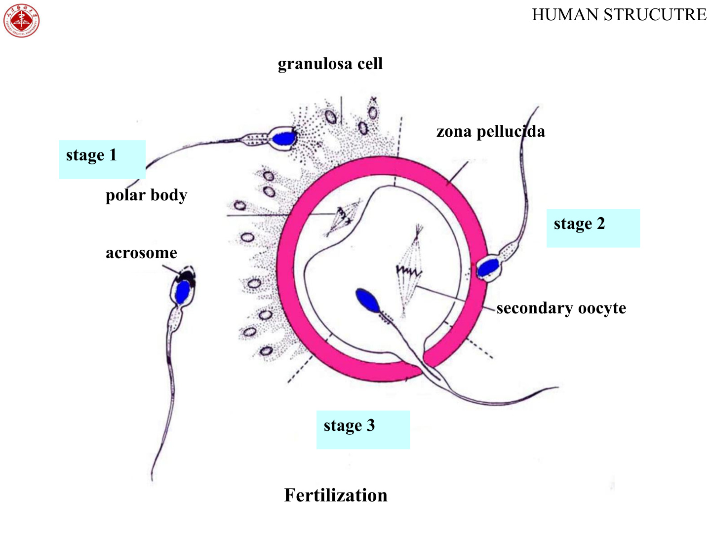
Fertilization: the process by which a capacitated spermatozoon fuses with a secondary oocyte in the ampulla, restoring the diploid chromosome number, determining genetic sex, and initiating cleavage. (Significance: restores diploid karyotype + integrates parental genes; determines sex; triggers development.)
Ectopic pregnancy — if fertilisation happens normally but the conceptus stalls in the tube, implantation occurs in the ampulla rather than the uterus. The thin tubal wall cannot accommodate a growing placenta and ruptures around 6–8 weeks with catastrophic intraperitoneal haemorrhage. Risk factors include prior pelvic inflammatory disease (chlamydia) and tubal surgery.
Cleavage → Morula → Blastocyst
Cleavage is a peculiar form of mitosis: the zygote divides repeatedly but the total volume of the embryo does not increase, because the whole process happens inside the rigid zona pellucida. Each daughter cell — called a blastomere — therefore ends up smaller than its parent. The first cleavage produces two cells by about 24–30 hours after fertilisation, the second produces four, and so on as the conceptus drifts down the oviduct toward the uterus.
By day 3, around the time the conceptus reaches the uterine cavity, there are about sixteen tightly packed blastomeres forming a solid mulberry-like ball called the morula (Latin morum, mulberry). At this stage the outer cells begin to flatten against each other and form tight junctions — a process called compaction — while the inner cells stay rounded. This is the first time the embryo shows obvious cell differentiation: the outer cells are committed to becoming trophoblast, the inner cells to becoming the embryo proper.
The blastocyst forms when fluid pumped into the morula by the outer cells coalesces into one big central bubble — the blastocoele. The outer cells flatten into a balloon wall (the trophoblast) and the inner cells cluster at one pole (the inner cell mass). Picture a soap bubble with a knot of foam stuck inside at one end.
By day 4–5, with the embryo now floating free in the uterine cavity, the small intercellular spaces inside the morula fuse into a single fluid-filled cavity, the blastocoele. The conceptus now has two clearly distinct cell populations: a flat outer wall, the trophoblast, which will form the placenta and extra-embryonic membranes, and an eccentric clump on one pole, the inner cell mass (embryoblast), which will form the embryo proper plus a few extra-embryonic accessories.
Before implantation can begin, the blastocyst has to escape from the zona pellucida — a process called hatching, around day 5–6. The zona thins and ruptures, the naked blastocyst emerges, and only now can the trophoblastic surface actually adhere to the endometrium. The pole of the blastocyst nearest the inner cell mass — the embryonic pole — is the one that attaches first, ensuring the embryo proper is buried deep in the decidua and the trophoblast spreads outward into it.
- Cleavage: the specialised mitosis of the zygote; daughter cells = blastomeres. It occurs inside the zona pellucida, so each blastomere shrinks as their number rises.
- Morula (day 3): ~16 closely-packed blastomeres forming a solid ball with obvious cell differentiation.
- Blastocyst (day 4–5): the morula enters the uterine cavity; at ~100 cells small spaces fuse into a single blastocoele; the zona pellucida disintegrates. Wall = flat trophoblast; an eccentric inner cell mass (embryoblast) projects into the cavity.
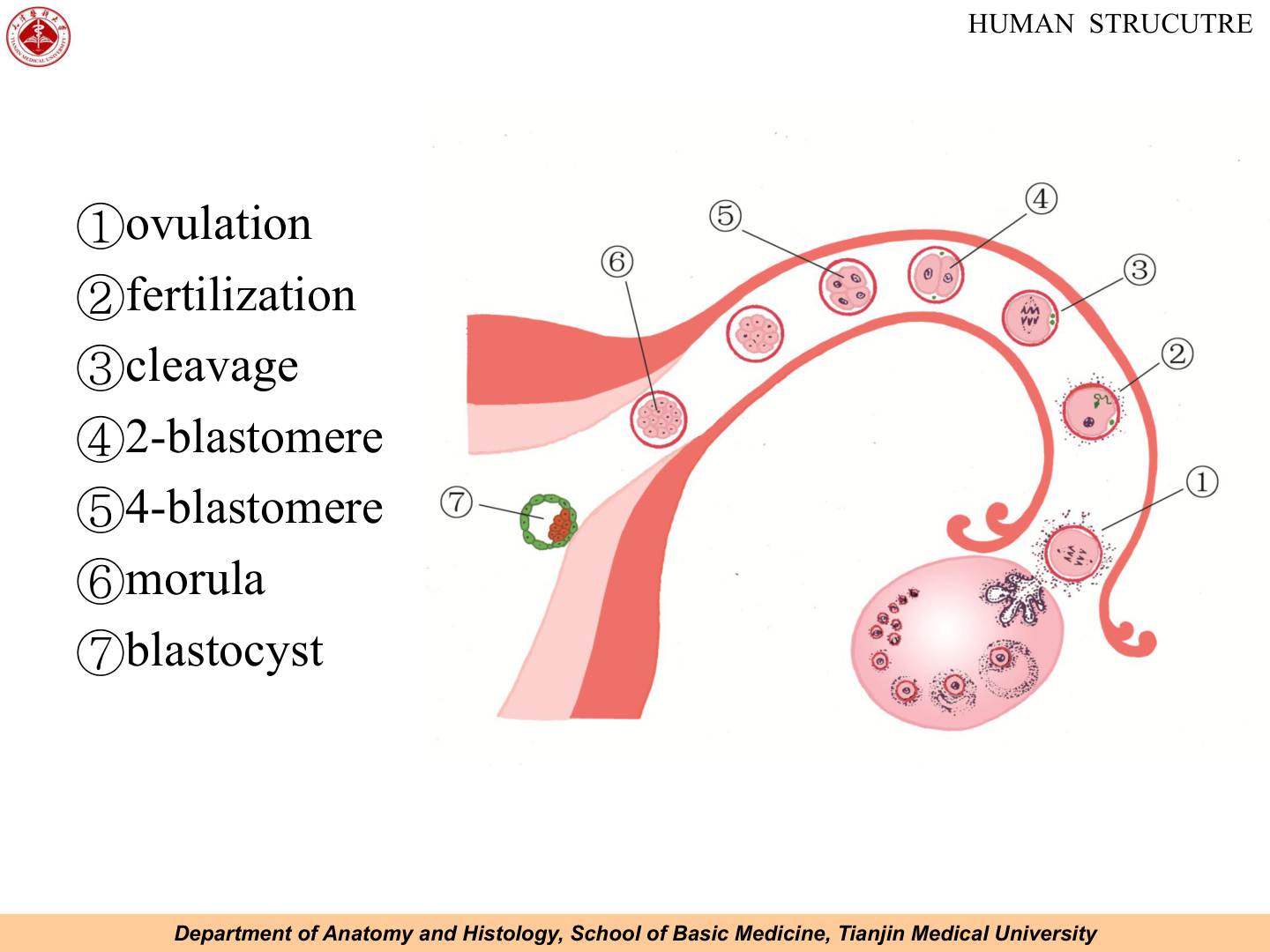
“Zygote → Morula (16, day 3) → Blastocyst (100, day 4–5).” Blastocyst = first stage with a cavity (blastocoele) + two cell populations (trophoblast vs inner cell mass).
Monozygotic twinning depends on when one zygote splits. A split at the two-cell stage (days 1–3) produces two completely separate amnions and chorions (dichorionic/diamniotic). A split inside the blastocyst at days 4–8 gives one chorion but two amnions (monochorionic/diamniotic) — the type at risk of twin-twin transfusion. Splits after day 13 give conjoined twins.
Implantation
Implantation is the process by which the hatched blastocyst embeds itself into the secretory-phase endometrium. It begins on day 5–6 and is complete by about day 11–12, by which time the conceptus is entirely buried below the uterine epithelium and the overlying surface has healed over. The normal site is the upper posterior wall of the uterine body or the fundus, where the endometrium is thickest and best perfused.
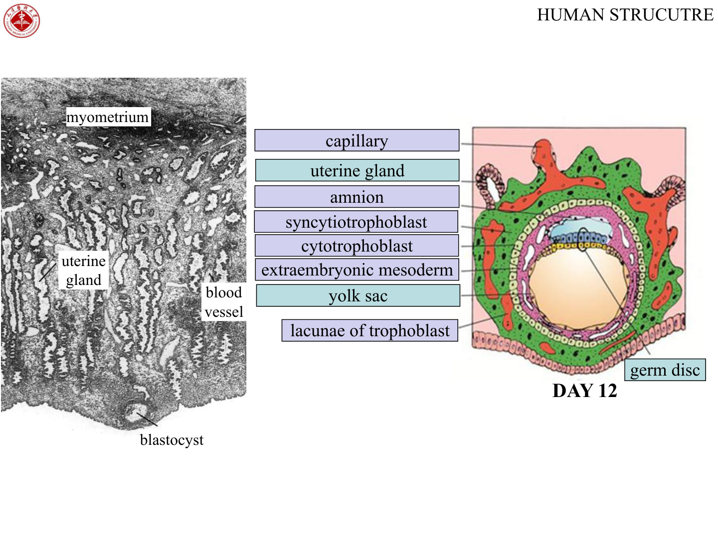
The polar trophoblast — that is, the part of the trophoblast directly overlying the inner cell mass — adheres first and starts to invade. The cells of the contacting trophoblast almost immediately differentiate into two layers: an inner, mitotically active cytotrophoblast with discrete cell boundaries, and an outer, multinucleate syncytiotrophoblast formed by the fusion of cytotrophoblast cells as they push outward into the maternal tissue.
The syncytiotrophoblast is the invasive front. It secretes proteolytic enzymes that erode the endometrium and the walls of maternal capillaries; within its substance small spaces called lacunae appear and quickly fill with maternal blood. By the end of week 2 these lacunae have connected to maternal spiral arteries and veins, establishing the first primitive utero-placental circulation. The syncytiotrophoblast also secretes human chorionic gonadotropin (hCG) from day 8 onward — the hormone that rescues the corpus luteum from regression and that the home pregnancy test detects.
Picture the blastocyst as a seed and the endometrium as freshly turned soil. The syncytiotrophoblast is the advancing root tip, dissolving its way forward; the cytotrophoblast behind it is the cambium, dividing to keep the root growing. The maternal blood in the lacunae is groundwater seeping into the gaps the root has opened.
The endometrium responds to the invading conceptus with the decidua reaction: stromal cells swell, accumulate glycogen and lipid in their cytoplasm, and become rounded decidual cells, while uterine glands expand their secretion and the vascular bed dilates further. The whole functional layer of the endometrium is now called the decidua. By location it is divided into three named regions — the decidua basalis lying deep to the conceptus (which will form the maternal part of the placenta), the decidua capsularis covering it on the luminal side, and the decidua parietalis lining the rest of the uterine cavity.
Implantation only succeeds when timing, site and signalling all line up. If the conceptus stalls in the oviduct, you get an ectopic pregnancy — tubal implantation in the ampulla being the commonest variety. The thin tubal wall cannot expand to house a growing placenta, so the pregnancy ruptures and bleeds. If the blastocyst implants too low in the uterus, near the internal os, the resulting placenta covers the cervical opening — the obstetric emergency known as placenta praevia, which presents as painless third-trimester bleeding.
| Layer | Features |
|---|---|
| Syncytiotrophoblast (outer) | Cells fused; no cell boundaries; invasive; develops lacunae that fill with maternal blood; secretes hCG |
| Cytotrophoblast (inner) | Cuboidal cells with clear boundaries; divide continuously to feed the syncytiotrophoblast |
- Decidua reaction: the secretory-phase endometrium thickens further, its blood supply & gland secretion increase, and stromal cells enlarge (glycogen + lipid) into decidual cells.
- Decidua regions: decidua basalis (deep to embryo), decidua capsularis (over embryo), decidua parietalis (rest of uterus).
- Normal site = upper body/fundus of uterus; abnormal (ectopic) sites include the oviduct — dangerous.
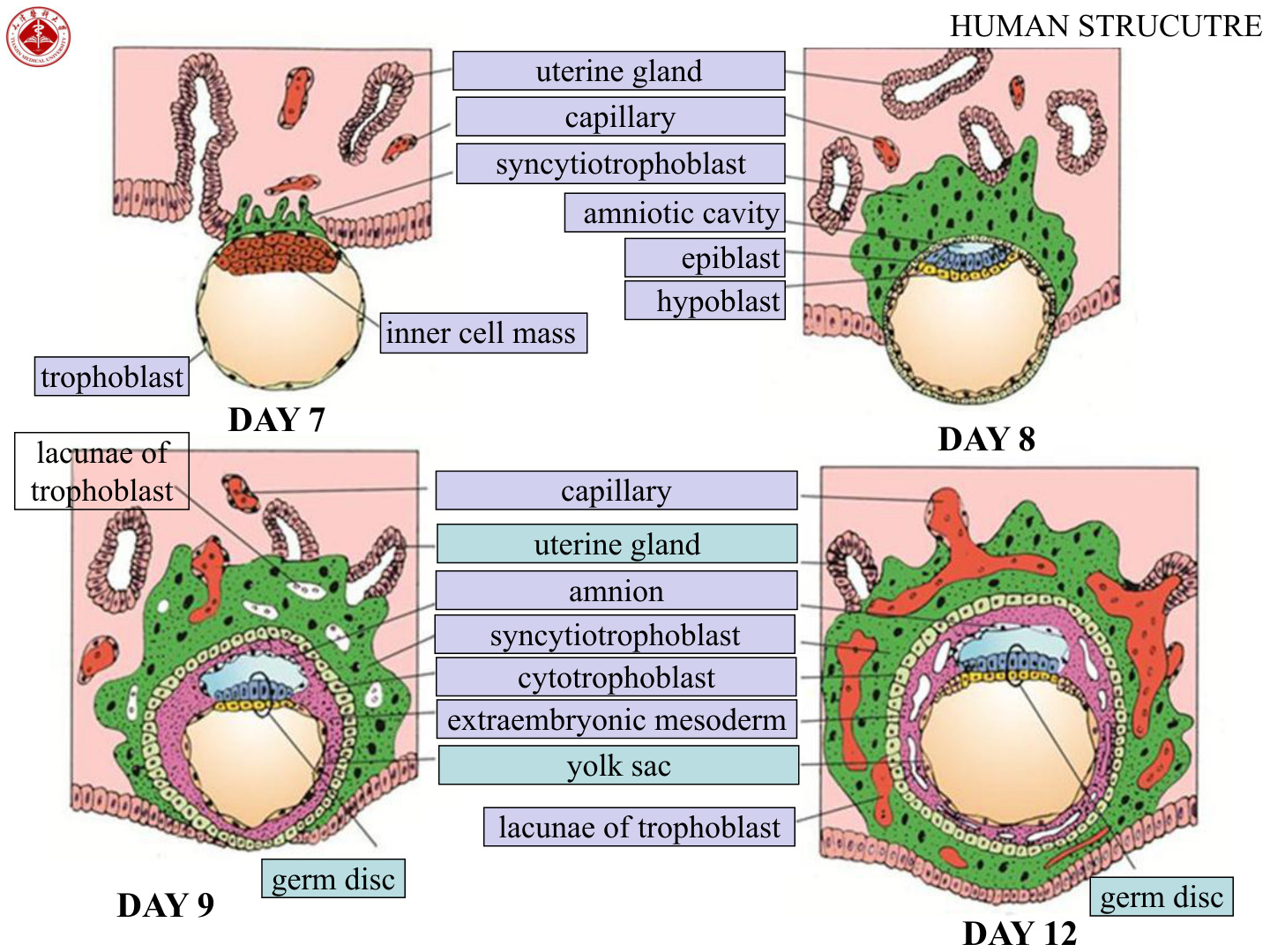
Ectopic pregnancy (most often tubal) = implantation outside the uterine body — can rupture and haemorrhage. Placenta praevia = implantation low over the internal os. Hydatidiform mole is an abnormal trophoblastic proliferation, classically with very high hCG and grape-like villi on ultrasound; complete moles (46,XX, all paternal) carry a risk of progression to choriocarcinoma.
Formation of the Germ Layers
Through the second week — the “week of twos” — the inner cell mass reorganises itself into a flat two-layered plate called the bilaminar germ disc. The dorsal cells become tall columnar epiblast, the ventral cells become small cuboidal hypoblast. Together they form a disc only two cells thick, suspended between two new fluid-filled cavities.
On the dorsal side of the disc, small spaces open up among the epiblast cells and fuse to form the amniotic cavity, whose roof is a thin epithelium of amnioblasts secreting amniotic fluid. On the ventral side, hypoblast cells migrate around the inside of the blastocoele to line a second cavity, the primary yolk sac; this is later remodelled into the secondary (definitive) yolk sac. Between the trophoblast and these two cavities a loose tissue called extra-embryonic mesoderm appears, and within it cavities coalesce to form the chorionic cavity. The bilaminar disc ends up suspended in the chorionic cavity by a stalk of extra-embryonic mesoderm — the connecting (body) stalk — which will later become the core of the umbilical cord.
By the end of week 2 the trophoblast itself has finished differentiating into the inner cytotrophoblast and outer syncytiotrophoblast; primary chorionic villi (cytotrophoblast cores covered by syncytiotrophoblast) are beginning to project from the chorionic plate into the maternal lacunae. Everything is in place for gastrulation to begin in week 3.
- The inner cell mass splits into a dorsal epiblast and a ventral hypoblast = the bilaminar disc.
- Amniotic cavity: a fluid-filled space appears within the epiblast (roofed by amnioblasts) → amniotic fluid.
- Yolk sac: hypoblast cells grow ventrally to line a cavity of simple squamous cells; the hypoblast forms its roof.
- Extraembryonic mesoderm + the body (connecting) stalk suspend the disc within the chorionic cavity.
The bilaminar disc is a pizza-base squeezed between two balloons — the amniotic cavity above (epiblast roof) and the yolk sac below (hypoblast floor). Both balloons hang inside a bigger room (the chorionic cavity) from a single rope (the connecting stalk).
Gastrulation is the central event of human embryology — the conversion of a two-layered disc into a three-layered disc — and it is the most important week of pregnancy a teratogen can hit. Week 3 is also when the basic body axis is laid down: cranial vs caudal, dorsal vs ventral, left vs right.
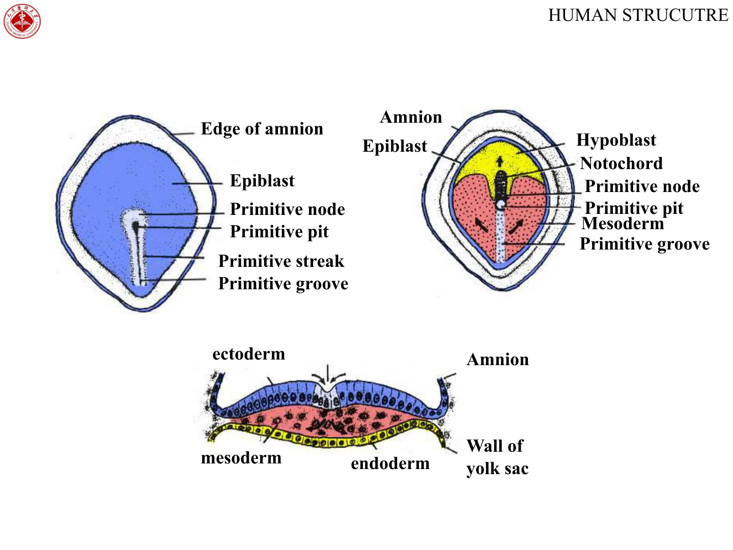
It begins with the appearance of the primitive streak, a midline thickening of the epiblast at the caudal end of the disc that elongates cranially. Its cranial end expands into the primitive node, and along its length runs a shallow primitive groove with a deeper primitive pit in the node. The streak is the embryo’s assembly line: epiblast cells stream toward the midline, dive down through the groove, and ingress between the two existing layers.
The first wave of ingressing epiblast cells displaces the hypoblast altogether and replaces it with a new layer of definitive endoderm. A second wave inserts itself between the new endoderm and the remaining epiblast to form mesoderm. The epiblast cells that never moved are now renamed ectoderm. Three germ layers, all from the epiblast — remember that, because it explains how a single layer of cells eventually gives rise to the entire body.
From the primitive pit a cord of cells migrates cranially in the midline, sandwiched between ectoderm and endoderm, to form the notochord. The notochord defines the primary body axis and, crucially, induces the overlying ectoderm to thicken into the neural plate — the start of neurulation. Where the disc has only two layers because the notochord stops short — cranially the oropharyngeal membrane (future mouth) and caudally the cloacal membrane (future anus) — ectoderm and endoderm remain in direct contact.
- Primitive streak = a thickening of the epiblast; its cranial end expands into the primitive node. A midline primitive groove + central primitive pit form.
- Epiblast cells migrate through the streak: one group becomes endoderm (displacing hypoblast), another becomes mesoderm (between the layers); the remaining epiblast is renamed ectoderm.
- By the end of week 3 the trilaminar disc (all 3 layers, all from epiblast) is complete.
- Notochord: cells from the primitive pit migrate cranially as a midline cord between ecto- and endoderm — the primary body axis & inducer of the neural plate. Oropharyngeal & cloacal membranes mark future mouth and anus.
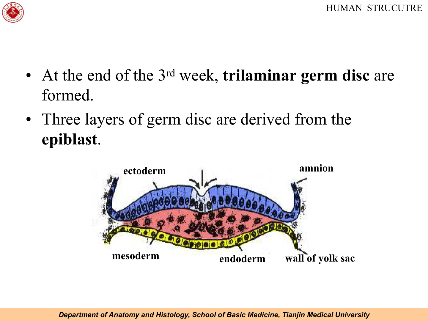
All three germ layers come from the epiblast. “Endo first, Meso between, Ecto stays on top.” Gastrulation = week 3 via the primitive streak.
Picture the primitive streak as a factory slot. Epiblast cells walk along the surface, drop through the slot, and are sorted into the basement (endoderm) or the in-between floor (mesoderm). Whatever stays upstairs becomes ectoderm. The notochord is the central pillar laid down through the slot — it tells everything above to start building a nervous system.
If the primitive streak fails to regress completely, remnants can grow into a sacrococcygeal teratoma, the commonest tumour of the newborn — a midline mass at the lower back containing all three germ layers. Gastrulation defects also explain caudal regression syndrome, seen with maternal diabetes.
Germ-Layer Differentiation & Body Folding (wk 4–8)
Once the three germ layers exist, the rest of embryology is essentially a story of who-becomes-what. The fastest way to learn it is to anchor a small number of derivatives in each layer and then add detail as you meet the organ systems later in the course.
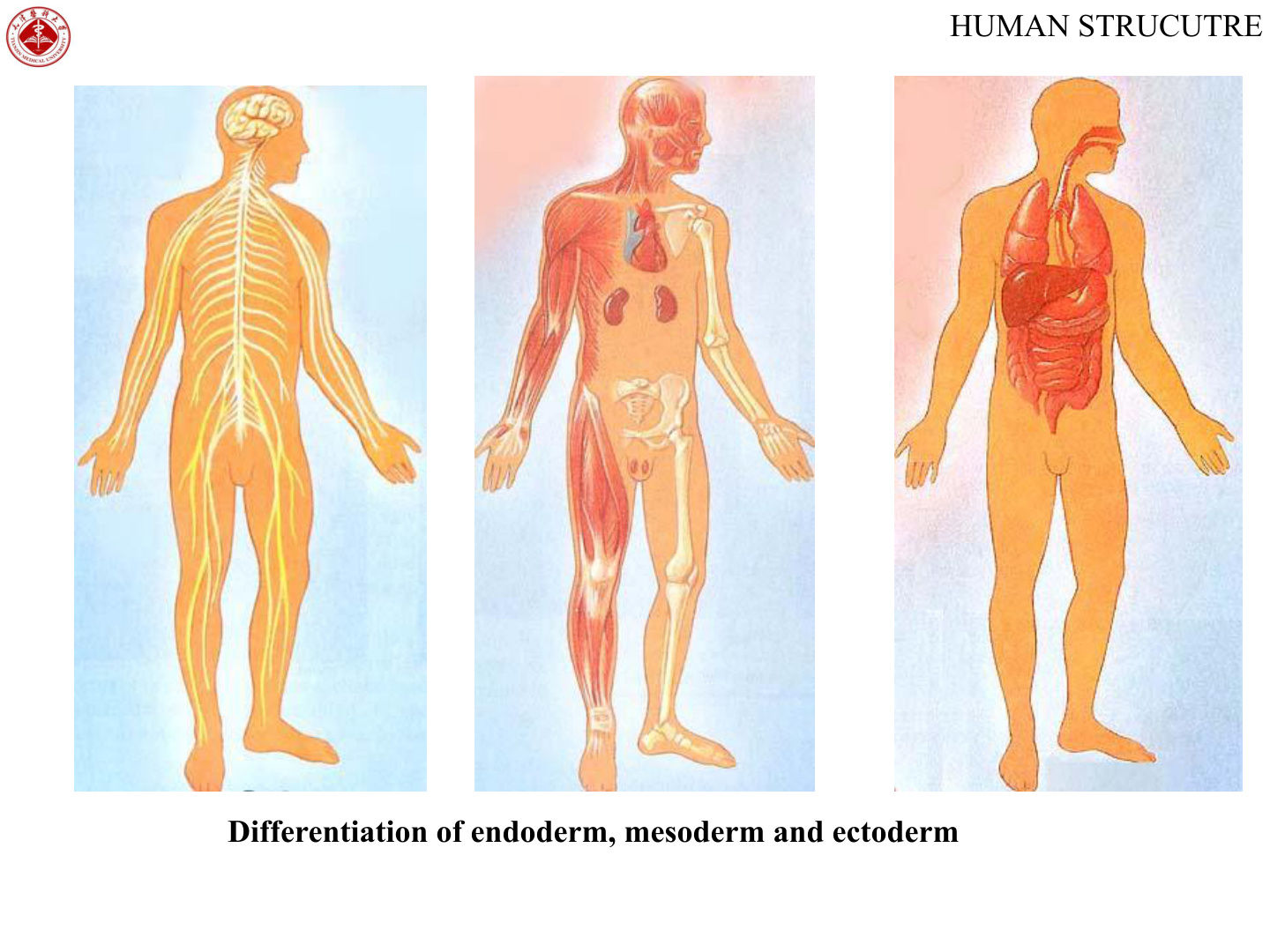
Ectoderm splits early into two populations: surface ectoderm and neuroectoderm. Surface ectoderm gives you the epidermis and its appendages (hair, nails, sweat and sebaceous glands, mammary glands), the lens of the eye, the inner ear epithelium, the anterior pituitary (Rathke pouch) and tooth enamel. Neuroectoderm folds into the neural tube — the entire CNS, retina and posterior pituitary — while the cells at the crest of the closing folds delaminate as neural crest. Neural crest is the great wanderer of the embryo: it goes on to form peripheral ganglia (sensory, sympathetic, parasympathetic, enteric), Schwann cells, melanocytes, the adrenal medulla, the parafollicular C cells of the thyroid, odontoblasts, aorticopulmonary septum and most of the cartilage and bone of the pharyngeal arches.
Mesoderm immediately organises itself paraxially, intermediately and laterally on either side of the notochord. Paraxial mesoderm segments into somites — each somite gives a sclerotome (vertebrae and ribs), a myotome (skeletal muscle) and a dermatome (dermis of the back). Intermediate mesoderm forms the urogenital system: kidneys, ureters, gonads and their ducts. Lateral plate mesoderm splits into a parietal (somatic) sheet attached to the body wall and a visceral (splanchnic) sheet attached to the gut; the cavity between them is the intra-embryonic coelom (future pericardial, pleural and peritoneal cavities), and from these lateral plates come the heart, smooth muscle and connective tissue of the gut, blood vessels, blood cells and the serous membranes. The notochord itself does not vanish — remnants persist as the nucleus pulposus of each intervertebral disc.
Endoderm lines the inside of everything that handles food, air or urine. It forms the epithelium of the entire gastrointestinal tract from the pharynx to the upper anal canal, the lining of the respiratory tract, the parenchymal cells of the liver and pancreas, the follicular cells of the thyroid, the parathyroids, the thymic epithelium, the epithelium of the bladder and most of the urethra, and the epithelial lining of the auditory tube and tympanic cavity.
| Germ layer | Major derivatives |
|---|---|
| Ectoderm | Surface: epidermis & appendages, lens, inner-ear epithelium, anterior pituitary, tooth enamel · Neural tube: CNS, retina, posterior pituitary · Neural crest: PNS ganglia, Schwann cells, melanocytes, adrenal medulla, thyroid C cells, aorticopulmonary septum, pharyngeal-arch cartilage, odontoblasts |
| Mesoderm | Paraxial / somites: vertebrae & ribs (sclerotome), skeletal muscle (myotome), dermis (dermatome) · Intermediate: kidneys, gonads, ducts · Lateral plate: heart, blood vessels, blood, smooth muscle & CT of gut, serous membranes · Notochord → nucleus pulposus |
| Endoderm | Epithelial lining of GI tract, respiratory tract, bladder & most of urethra; parenchyma of liver, pancreas, thyroid, parathyroids; thymic epithelium; auditory tube & tympanic-cavity lining |
Neurulation begins in week 3 when the notochord induces the overlying midline ectoderm to thicken into the neural plate. The lateral edges of the plate lift up as neural folds, the centre sinks as the neural groove, and the folds meet in the midline to zip the neural tube closed. Closure begins at the future cervical region around day 22 and proceeds in both directions; the anterior neuropore closes around day 25, the posterior neuropore around day 27.
If the anterior neuropore fails to close, the brain never forms properly and you get anencephaly. If the posterior neuropore fails to close, you get a spina bifida — ranging from mild occulta (a vertebral defect only) through meningocele (meninges herniate) to myelomeningocele (cord and meninges herniate, with neurological deficit). Periconceptional folate supplementation dramatically reduces the incidence of all these defects, which is why folic acid is started before pregnancy is even confirmed.
Somites are blocks that bud off the paraxial mesoderm in a strict cranio-caudal sequence from day 20 onward — about three pairs a day, so somite count is used to age very early embryos. Each somite differentiates into a sclerotome (forms the vertebra and ribs), a myotome (forms skeletal muscle of that segment) and a dermatome (forms the dermis of the overlying skin and explains why sensory innervation of the skin is segmented).
- Neurulation: notochord induces ectoderm → neural plate → folds → neural tube; neural crest peels off the crests.
- Somites: paraxial mesoderm segments cranio-caudally; sclerotome (vertebrae), myotome (muscle), dermatome (dermis).
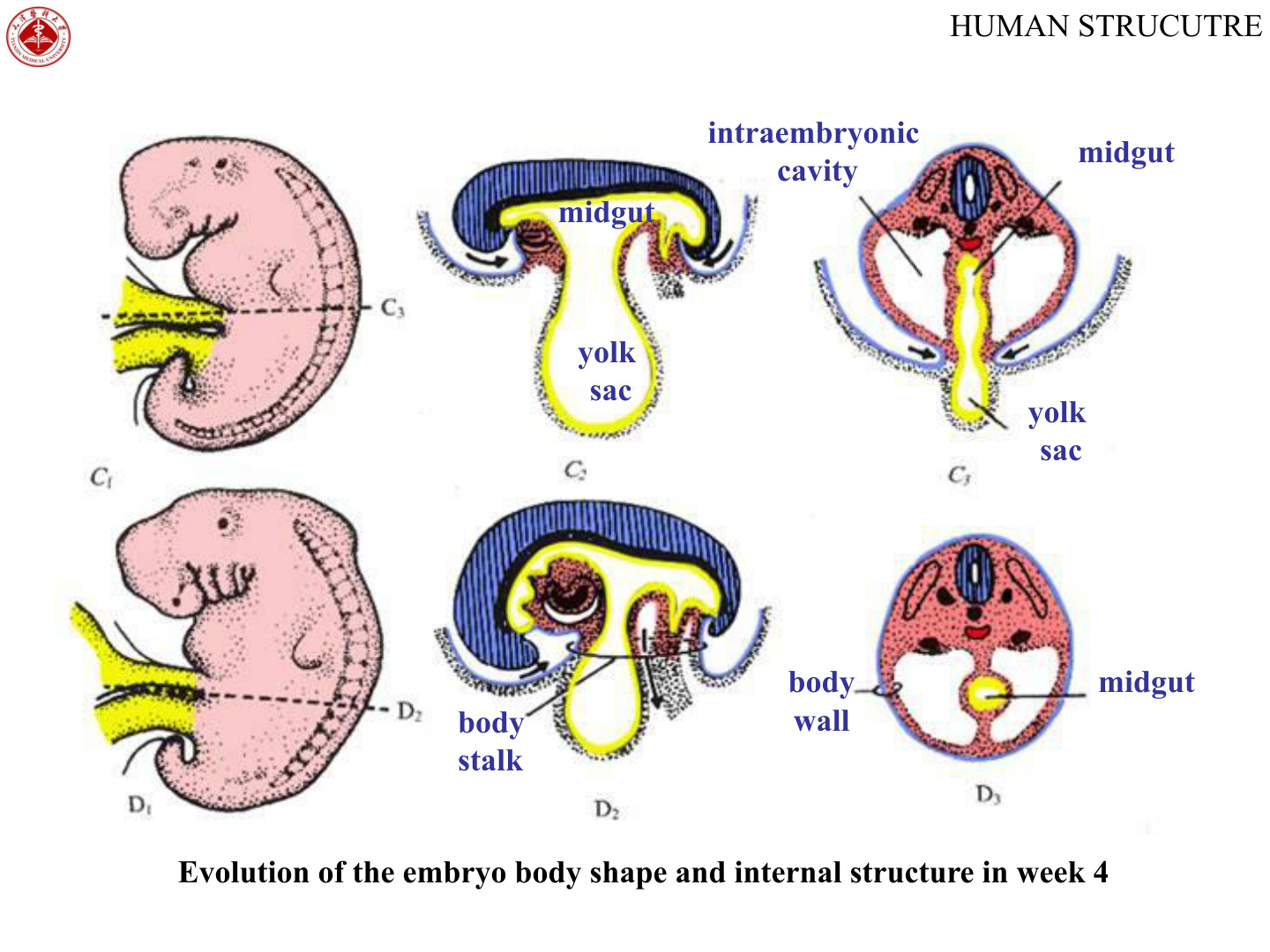
- Body folding: the flat disc folds ventrally (head, tail, and paired lateral folds) into a cylindrical body that bulges into the amniotic cavity; the body stalk + yolk sac fuse, covered by amnion, to form the umbilical cord.
Neural tube closure is like zipping a sleeping bag from the middle outward in both directions — one slider runs cranially toward the head, the other caudally toward the sacrum. If the cranial slider jams, the head end stays open (anencephaly); if the caudal slider jams, the foot end stays open (spina bifida).
Through week 4 the flat trilaminar disc folds itself into a tube-within-a-tube. Two folds happen at right angles to each other. Cephalo-caudal folding pulls the head end and tail end ventrally, tucking the amniotic cavity around the embryo. Lateral folding pulls the two sides ventrally as well, so that the embryo lifts off the yolk sac like a sandwich folding around a filling.
The result is a recognisable cylindrical embryo with the gut tube running down its core. The narrow communication between this gut tube and the residual yolk sac is the vitelline (yolk) stalk. The connecting stalk, the vitelline stalk and the allantois (a small endodermal diverticulum into the connecting stalk) all become enclosed by amnion as the embryo folds — and that bundle is the umbilical cord.
The primitive gut tube is divided into three regions by its blood supply. The foregut (coeliac trunk) becomes the pharynx, oesophagus, stomach, proximal duodenum, liver, gallbladder, pancreas and the lower respiratory tract. The midgut (superior mesenteric artery) becomes the distal duodenum through the proximal two-thirds of the transverse colon; during weeks 6–10 it temporarily herniates into the umbilical cord as “physiological umbilical herniation”, rotates 270° and returns. The hindgut (inferior mesenteric artery) becomes the distal one-third of the transverse colon down to the upper anal canal; its terminal cloaca is divided by the urorectal septum into the rectum dorsally and the urogenital sinus ventrally.
Failure of the midgut to return cleanly after physiological herniation gives omphalocele (gut herniates into the cord, covered by peritoneum and amnion). Failure of body-wall folding lateral to the cord gives gastroschisis (bowel through a defect to the right of the cord, no covering membrane). Failure of the urorectal septum to descend gives imperforate anus with rectovesical or rectovaginal fistulas. The whole VACTERL constellation (Vertebral, Anal, Cardiac, TracheoEsophageal, Renal, Limb) clusters together because the affected structures all arise in week 4 folding.
Fetal Membranes & Placenta
The fetal membranes are everything the conceptus produces outside its own body to keep itself alive in the uterus: the chorion, the amnion, the residual yolk sac, the allantois and the umbilical cord that ties the embryo to them. All except the inner-cell-mass derivatives count as extra-embryonic, and all of them are discarded at delivery.
The chorion is the outermost membrane — trophoblast (cytotrophoblast + syncytiotrophoblast) on the outside with extra-embryonic mesoderm lining the inside. Together these form the chorionic plate from which the chorionic villi grow. Villi develop in three stages of complexity: primary villi are solid cytotrophoblast covered in syncytiotrophoblast, secondary villi add an extra-embryonic mesodermal core, and tertiary villi contain fetal capillaries within that core — tertiary villi are the gas-exchanging structures of the placenta. The pole of the chorion facing the decidua basalis grows lush villi (chorion frondosum), while the rest of the chorion, facing the decidua capsularis, has its villi regress to become the smooth chorion laeve.
The amnion is the innermost membrane — a single layer of amniotic epithelium on a thin layer of avascular extra-embryonic mesoderm — enclosing the amniotic fluid that bathes the fetus. The fluid cushions mechanical insults, allows fetal movement (and so muscle and joint development), maintains a stable temperature and chemical environment, and provides a sample of fetal cells for amniocentesis. Too little (oligohydramnios) compresses the fetus (Potter sequence: pulmonary hypoplasia, limb deformities, characteristic facies) and usually reflects renal agenesis or chronic leakage. Too much (polyhydramnios) suggests inability to swallow it — oesophageal atresia, anencephaly or maternal diabetes.
The yolk sac is small and largely vestigial in humans, but it is far from useless: it is the site of the earliest blood-cell formation in the third week and, crucially, the source of the primordial germ cells that migrate later into the gonadal ridge. The allantois is a small endodermal diverticulum from the hindgut into the connecting stalk; its blood vessels become the umbilical vessels, and its lumen is eventually obliterated to form the urachus — the future median umbilical ligament.
The umbilical cord wraps all of these together. It contains two umbilical arteries (carrying deoxygenated blood from fetus to placenta) and one umbilical vein (carrying oxygenated blood back to the fetus), embedded in a gel of mucoid connective tissue called Wharton jelly, all covered by an outer sleeve of amnion. A single umbilical artery (SUA) is found in about 1% of cords and is associated with renal and other anomalies.
- Chorion = trophoblast + extraembryonic mesoderm, forming the chorionic plate + stem villi (primary → secondary → tertiary, when fetal capillaries appear) + villi. The pole facing the decidua basalis grows lush villi (chorion frondosum → placenta); the rest becomes smooth (chorion laeve).
- Amnion: a sac of amniotic epithelium + a little avascular extraembryonic mesoderm, enclosing the amniotic fluid that cushions the fetus.
- Yolk sac (early blood cells & primordial germ cells) & allantois (becomes urachus → median umbilical ligament) largely regress.
- Umbilical cord: body stalk + yolk-sac stalk covered by amnion; 2 arteries + 1 vein in Wharton jelly.
Picture the fetus as an astronaut on a spacewalk. The amnion is the inner pressurised spacesuit. The chorion is the outer hull holding the suit against the wall (decidua). The umbilical cord is the tether carrying oxygen and food in and waste out — arteries take used air back to the recycler (placenta) and a single vein brings fresh air back to the astronaut.
The placenta is the disc-shaped organ where fetal villi dipped in maternal blood do all the work of a lung, a gut and a kidney for the fetus. By term it weighs about 500–600 g and is about 20 cm in diameter, with a smooth fetal surface (covered by amnion, with the cord attached centrally or eccentrically) and a rough maternal surface divided by septa into 15–20 bulges called cotyledons.
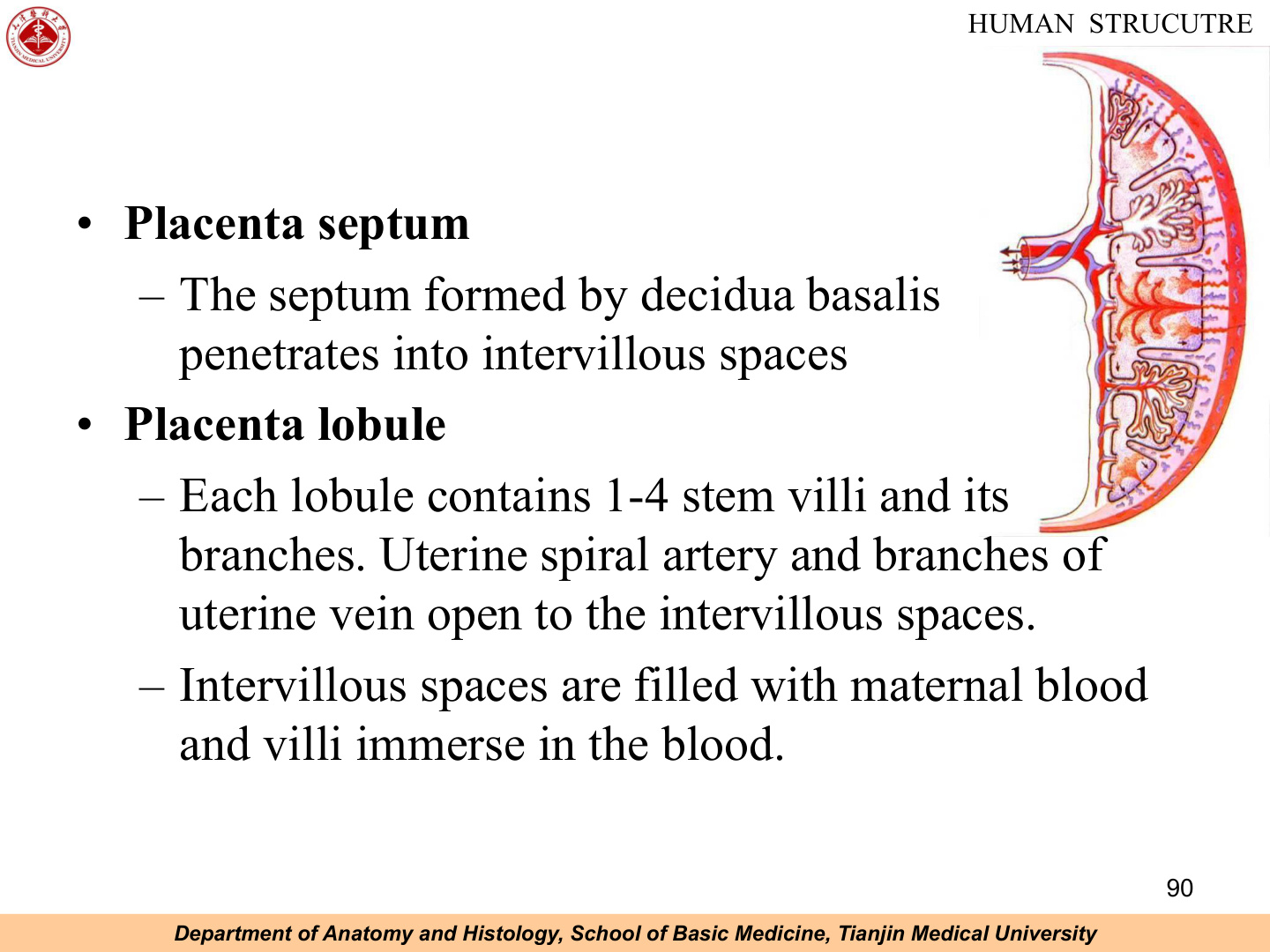
The fetal part is the chorion frondosum: the chorionic plate from which the stem villi grow, with their many-branched terminal villi floating in maternal blood. The maternal part is the decidua basalis; placental septa from the basalis project up into the chorionic plate, incompletely subdividing the space between them into compartments and giving the lobulated cotyledon pattern.
Between the chorionic plate and the basal plate is the intervillous space, filled with maternal blood. Maternal blood arrives via the spiral arteries of the endometrium, jets into the intervillous space, percolates over the villi (where exchange happens), and drains away through endometrial veins. Fetal blood, by contrast, never leaves its own vessels: the umbilical arteries feed the villous capillaries, and oxygenated blood returns through the umbilical vein. The two bloodstreams never mix; everything passes by diffusion or transport across the placental barrier.
That barrier in early pregnancy has four layers (counting from maternal side): (1) syncytiotrophoblast, (2) cytotrophoblast with its basement membrane, (3) loose connective tissue of the villous core, and (4) endothelium of the fetal capillary with its basement membrane. As pregnancy advances, the cytotrophoblast layer thins out and almost disappears, so by the third trimester the barrier is mainly syncytiotrophoblast applied directly to fetal capillary endothelium — thin and efficient.
The placenta is also a major endocrine organ. The syncytiotrophoblast secretes human chorionic gonadotropin (hCG) in the first trimester to maintain the corpus luteum (which keeps progesterone going until the placenta itself can make enough). It also secretes human placental lactogen (hPL / hCS) — structurally similar to growth hormone, which contributes to the maternal insulin resistance of pregnancy and makes glucose available to the fetus — together with rising amounts of oestrogen and progesterone, eventually taking over hormonal maintenance of pregnancy from the corpus luteum.
- Fetal part = chorion frondosum (smooth fetal surface, covered by amnion, umbilical cord attached centrally/eccentrically).
- Maternal part = decidua basalis (rough surface; placental lobules / cotyledons visible). Placental septa from the decidua basalis partly divide the intervillous spaces, which are filled with maternal blood bathing the villi.
- Circulation: maternal blood from uterine spiral arteries → intervillous spaces → (exchange across villi) → uterine veins. Fetal blood: umbilical arteries → villous capillaries → (exchange) → umbilical vein to the fetus. The two bloods never mix.
Placental barrier = the layers separating maternal & fetal blood, from maternal side inward: (1) syncytiotrophoblast → (2) cytotrophoblast + its basement membrane → (3) connective tissue (villous core) → (4) endothelium + basement membrane of the fetal capillary. (Later it thins as cytotrophoblast regresses.)
| Placental function | Detail |
|---|---|
| Material exchange | O₂ & nutrients to fetus; CO₂ & metabolites to mother; immunoglobulin G (passive immunity) |
| Endocrine (syncytiotrophoblast) | hCG (maintains corpus luteum; from week 2, peaks ~week 8), human placental lactogen (maternal insulin resistance), progesterone & oestrogen |
| Barrier | Excludes many infections & large molecules; admits some drugs/viruses — teratogens act most weeks 3–8 |
Picture the placenta as a hand of villi dangling into a warm bath of maternal blood. The fingertips are tertiary villi, each with a tiny capillary inside. Maternal blood is the bathwater spraying in from spiral arteries and draining back through veins. Fetal blood runs only inside the fingers — the two never mix, but oxygen, sugar and CO₂ cross the skin freely.
hCG is the basis of the pregnancy test. The barrier admits some drugs/viruses (e.g. rubella, alcohol, valproate, warfarin, ACE inhibitors, retinoic acid) — teratogens act most during weeks 3–8 (organogenesis). TORCH infections (Toxoplasma, Other, Rubella, CMV, HSV) cross the placenta. Hydatidiform mole = abnormal trophoblastic proliferation; complete mole (46,XX, all paternal) carries a risk of progression to choriocarcinoma. Placental abruption = premature separation of the placenta from the decidua basalis — painful third-trimester bleeding.
TMU Exam Drill
📝 Open the full TMU Question Bank — 20 MCQ + 6 terms + 5 essays →
Authentic Tianjin Medical University past-paper questions (2021 Final & the multi-section Final with answer key) mapped to this unit, in the real exam format. Click Show answer to self-test. Note: embryology does not appear in the past papers; these are TMU-deck-format practice items.
□ Explain the following terms
🎉 All 19 units complete
The full TMU Histology & Embryology course is live. Back to the unit hub to revise.