Female Reproductive System
Ovary Overview
Picture the ovary as an almond–sized fruit with a tough rind and a juicy interior. When you scan a section under the microscope, the most striking feature is the eccentric arrangement: the cortex, where all the follicles live, sits as a wide band around the periphery, while the medulla — loose connective tissue stuffed with large blood vessels, lymphatics and nerves — fills the centre. This is opposite to most parenchymal organs, where the secretory tissue is central; in the ovary, the action is at the surface.
On the outside you see a single layer of cells classically called the germinal epithelium. The name is a 19th–century misnomer — we now know oocytes do not arise from it. It is simply a simple squamous to cuboidal mesothelium continuous with the peritoneal lining. Just beneath it lies a dense, hypocellular collar of collagen called the tunica albuginea, the “white coat” that gives the cut ovary its pale glistening surface.
Drop into the cortex itself and the stroma takes over: spindle–shaped fibroblast–like cells embedded in a delicate matrix, with follicles at every stage of development scattered through it. Some are dormant primordials hugging the tunica; others are large antral follicles bulging toward the surface; some are atretic ghosts; and at the right point in the cycle you will spot a corpus luteum or a pale fibrous corpus albicans. The cortex is therefore a histological time machine — every phase of follicular life is visible simultaneously.
Think of the ovary like an orchard. The trees (follicles) all grow at the rind (cortex) because that is where they get harvested — ovulation has to break through the surface. The trunk in the middle (medulla) is just plumbing: blood and nerves feeding the orchard.
The germinal epithelium misnomer matters clinically: most ovarian carcinomas (serous, mucinous, endometrioid) actually arise from this surface mesothelium or its inclusion cysts trapped in the cortex. Because the ovary's surface ruptures and reseals at every ovulation, the surface epithelium is repeatedly injured and repaired — possibly explaining why incessant ovulation (early menarche, late menopause, nulliparity) is a risk factor for ovarian cancer.
The “germinal epithelium” is the favoured origin of epithelial ovarian carcinoma — the most common and most lethal ovarian cancer. Polycystic ovary syndrome (PCOS) shows numerous arrested subcapsular antral follicles under a thickened tunica albuginea (the “string of pearls” on ultrasound).
Follicle Development (essay-grade)
A follicle is the ovary's functional unit: one oocyte plus the somatic cells that nurse, protect and hormonally support it. Folliculogenesis is a slow, wasteful journey — a fetal ovary holds 6–7 million primary oocytes, a newborn ~1 million, puberty ~400,000, and across a reproductive lifetime only about 400 actually ovulate. Every other follicle dies by atresia. Understanding the histological stages means understanding how a single dormant oocyte is gradually wrapped in glycoprotein, then layered with granulosa cells, hollowed out by an antrum, and finally cradled by a steroidogenic theca before bursting through the ovarian surface.
The stages run in a strict morphological sequence — primordial → primary (unilaminar then multilaminar) → secondary (antral) → mature (Graafian). Each step is recognised by counting and shape: how many granulosa layers, are they flat or cuboidal, is the zona pellucida visible, is there an antrum, is there a theca? Once you internalise this checklist, you can stage any follicle in a slide in seconds.
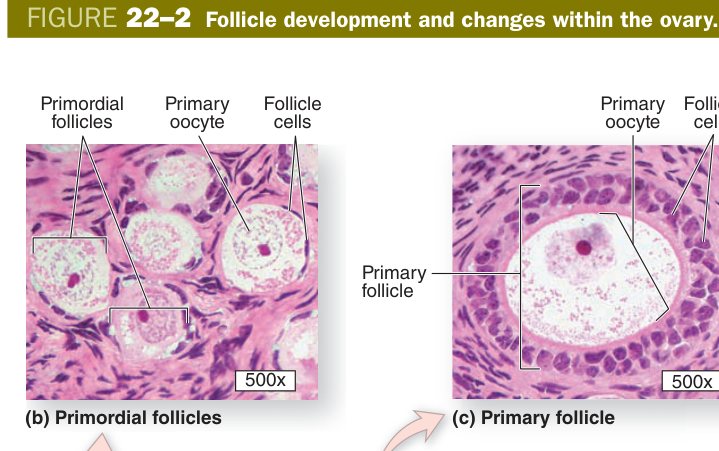
The primordial follicle is the resting form, the “sleeping” reserve sitting in the outer cortex just under the tunica albuginea. The primary oocyte inside it has been arrested in prophase of meiosis I since fetal life and may wait there 12 to 50 years before being recruited. Around it lies a single layer of flat (squamous) follicular cells resting on a thin basal lamina. There is no zona pellucida yet, no theca, no antrum — just oocyte and a wisp of flat cells.
When the follicle is recruited (the trigger is poorly understood and partly FSH–independent at this stage), the flat cells thicken into cuboidal cells and the oocyte begins to grow. This is the unilaminar primary follicle. As the granulosa cells proliferate into multiple layers, a glassy, eosinophilic glycoprotein shell appears between the oocyte and the innermost granulosa — the zona pellucida. Simultaneously, stromal cells outside the basal lamina condense into the theca folliculi. You now have a multilaminar primary follicle.
| Stage | Oocyte | Surrounding cells |
|---|---|---|
| Primordial | Primary oocyte (arrested in meiosis I) | Single layer of flat (squamous) follicular cells |
| Unilaminar primary | Primary oocyte enlarging | Single layer of cuboidal follicular cells |
| Multilaminar primary | + zona pellucida appears (glycoprotein coat) | Several layers of granulosa cells; theca forms from stroma |
Think of the zona pellucida as the oocyte's wedding dress — it only appears when the follicle has “committed” to multilaminar growth, and later it is the very layer the sperm has to drill through. Squamous → cuboidal → stratified is the same Goldilocks rule you used in epithelium: the cells get bigger and more layered as the workload (steroid + protein synthesis) climbs.
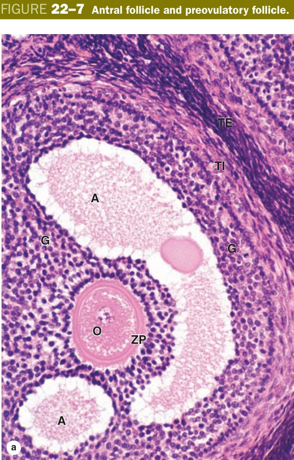
This is the follicle the TMU examiners love. The defining histological event is the appearance of liquor folliculi — a transudate–like fluid rich in oestrogen, hyaluronan and growth factors — that first appears as small lakes between granulosa cells (call–Exner bodies), then coalesces into a single, large, eccentric cavity called the follicular antrum. The moment that single antrum forms, the follicle is by definition secondary (antral). Once you see an antrum, you can drop the words “primary” and “multilaminar” from your vocabulary.
The fluid pushes the oocyte to one side, where it sits perched on a hill of granulosa cells called the cumulus oophorus. The innermost granulosa cells, directly hugging the zona pellucida, form the corona radiata — they send fine cytoplasmic processes through the zona to communicate with the oocyte via gap junctions. The rest of the granulosa cells form the wall of the antrum, the stratum granulosum (membrana granulosa), sitting on a basement membrane that is famously avascular: no blood vessel ever crosses into the granulosa layer until after ovulation.
Outside the basement membrane, the theca has now differentiated into two layers. The inner theca interna is highly vascular and steroidogenic — the cells are large, polygonal and full of smooth ER and lipid droplets, the classic look of an androgen–producing endocrine cell. Under LH, they secrete androgens (mostly androstenedione), which diffuse across the basement membrane into the granulosa cells. The granulosa cells, under FSH, express aromatase and convert those androgens into oestradiol. This is the classic “two–cell, two–gonadotropin” model — theca + LH makes the substrate, granulosa + FSH makes the product. The outer theca externa is a fibromuscular sheath of spindle cells and smooth muscle that helps squeeze the follicle at ovulation.
When you describe a secondary follicle in an essay, walk the examiner from inside outward in one breath:
- Primary oocyte — arrested in meiosis I.
- Zona pellucida — thick glycoprotein layer hugging the oocyte.
- Corona radiata — granulosa cells directly around the zona pellucida.
- Cumulus oophorus — the mound of granulosa cells anchoring the oocyte to the wall.
- Follicular antrum — cavity filled with liquor folliculi.
- Stratum granulosum (membrana granulosa) — the granulosa-cell wall, on a basement membrane.
- Theca interna — vascular, endocrine; secretes androgens (aromatised to oestrogen by granulosa cells).
- Theca externa — outer fibrous CT + smooth muscle.
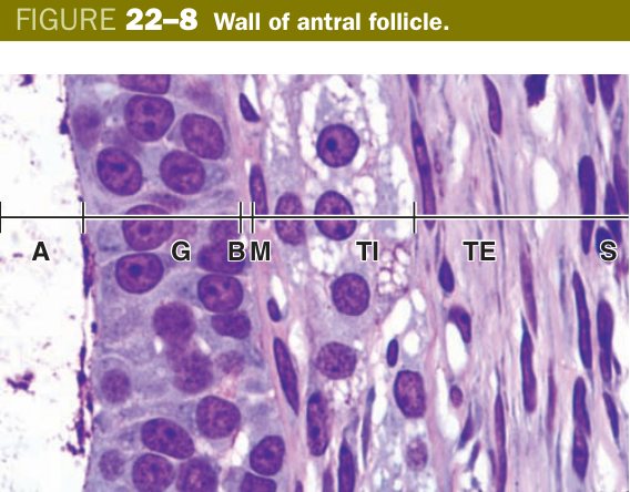
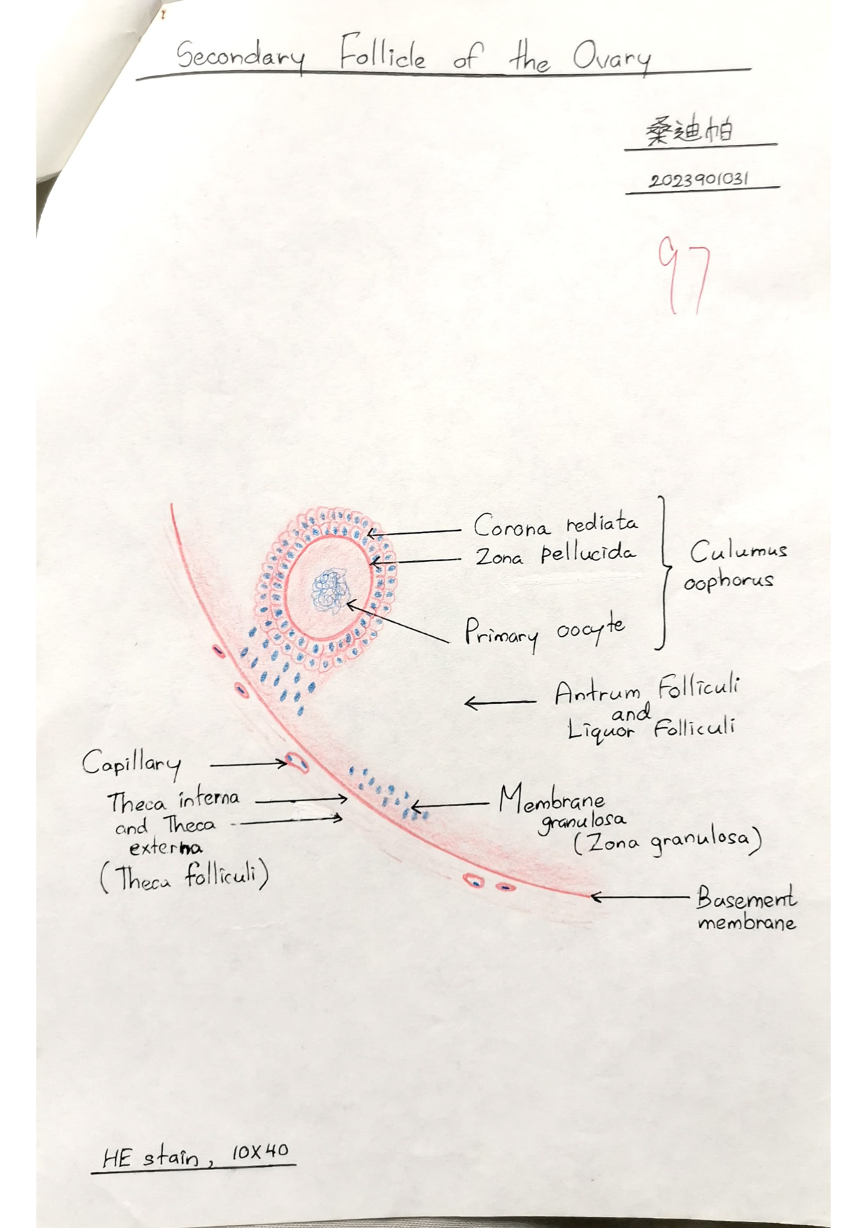
Secondary follicle: a follicle with a single fluid-filled antrum, containing a primary oocyte surrounded by zona pellucida and corona radiata, attached by the cumulus oophorus to the stratum granulosum; enclosed by theca interna (endocrine, androgen-secreting) and theca externa (fibrous/muscular).
The antral follicle is a water balloon with a stowaway. The balloon's wall is granulosa, the water is liquor folliculi, and the oocyte is the passenger riding on the cumulus mound. The theca outside is the protective rubber band that tightens during ovulation. The two–cell model is a tiny chemistry factory: theca brings the raw androgen, granulosa is the aromatase machine that finishes it as oestrogen.
A single antral follicle wins the dominance race each cycle, swelling to 15–25 mm and bulging from the ovarian surface like a blister. This is the mature (Graafian) follicle, named after Regnier de Graaf who first described it in 1672. The antrum is now huge, the granulosa wall is relatively thin (because the same cells are stretched over a larger sphere), and the cumulus oophorus + oocyte sit perched on one wall, sometimes almost free–floating in the fluid.
Hours before ovulation, the mid–cycle LH surge finally lets the primary oocyte resume meiosis. It completes meiosis I, extrudes the first polar body and becomes a secondary oocyte, but immediately re–arrests — this time in metaphase of meiosis II. Meiosis II only completes if a sperm fertilises the egg; otherwise the oocyte degenerates within ~24 hours. The LH surge also weakens the follicular wall by upregulating proteases (plasmin, collagenase) in the granulosa and theca externa, raises intrafollicular pressure modestly, and triggers the cumulus to detach.
At ovulation (around day 14 of an idealised 28–day cycle), the thinned wall ruptures and the secondary oocyte, still wrapped in its zona pellucida and corona radiata, is swept out with a gush of antral fluid. The fimbriae of the oviduct sweep it into the ampulla. What is left behind in the ovary will become the corpus luteum. Most other recruited follicles in that cycle — the ones that did not win — undergo atresia, a controlled apoptotic involution that leaves shrunken, hyalinised “atretic follicles” identifiable by a thickened glassy zona pellucida persisting after the oocyte is gone.
The LH surge is the starter pistol: it tells the oocyte “finish meiosis I, now,” and it tells the follicular wall “digest yourself.” Within ~36 hours the gun fires the egg. The oocyte itself is in no hurry to finish meiosis II — it would rather wait for a sperm to wake it up than complete the division pointlessly.
- Mature (Graafian) follicle: very large antrum bulging on the ovarian surface; the oocyte (on its cumulus) completes meiosis I just before ovulation → secondary oocyte (arrested in metaphase of meiosis II).
- Ovulation (~day 14, triggered by the LH surge): the wall ruptures and the secondary oocyte + zona pellucida + corona radiata are released into the oviduct. Meiosis II completes only if fertilised.
Stages: Primordial → Primary → Secondary (antral) → Mature. “Antrum = secondary.” Theca interna = incrine (endocrine, makes androgen); theca externa = structural.
Mittelschmerz = mid–cycle pelvic pain from the ovulatory rupture irritating the peritoneum. A failed rupture with persistent fluid → follicular cyst. PCOS = arrested antral follicles never reach ovulation under chronically high LH/low FSH; anovulation + hyperandrogenism. The egg released is a secondary oocyte arrested in metaphase II — fertilisation (sperm entry) is the only physiological trigger that lets it finish meiosis II.
Corpus Luteum & Corpus Albicans
After the Graafian follicle has emptied at ovulation, what is left in the ovary is a deflated bag — collapsed wall, broken basement membrane, a small central blood clot (the corpus haemorrhagicum). Under the continuing influence of LH, this ruin reorganises within hours into one of the body's most spectacular temporary endocrine glands: the corpus luteum, the “yellow body” named for the carotenoid pigment in its luteinised cells.
The transformation is called luteinisation and involves three big changes: the basement membrane breaks down, blood vessels invade the granulosa layer for the first time (the granulosa was avascular until now), and the two follicular cell types hypertrophy and turn into steroid factories. The product is a folded, sausage–shaped structure with a central clot and two histologically distinct cell populations.
| Cell | Origin | Position & LM | Secretes |
|---|---|---|---|
| Granulosa lutein cells | Granulosa cells | Large, pale, central; bulk of the gland | Progesterone (+ oestrogen) |
| Theca lutein cells | Theca interna cells | Smaller, darker, peripheral (in folds) | Oestrogen (+ progesterone) |
Under the microscope, the granulosa lutein cells make up about 80% of the gland and look like classic steroid–secreting cells: large (30–50 µm), polygonal, with pale foamy cytoplasm packed with smooth ER and lipid droplets, central round nuclei. They occupy the bulk of the folded wall. The smaller, darker theca lutein cells sit at the periphery and in the folds between granulosa lutein cells, often hugging blood vessels. Both populations are now vascularised by capillaries that have invaded from the old theca interna — essential, because they are dumping their steroid output straight into the bloodstream.
The corpus luteum is the follicle in retirement. Same staff (granulosa + theca interna), new job description: instead of growing an egg, they now run a progesterone factory for two weeks to prepare the uterus for a possible pregnancy. Yellow colour = lipid + carotenoid = classic steroid–cell signature.
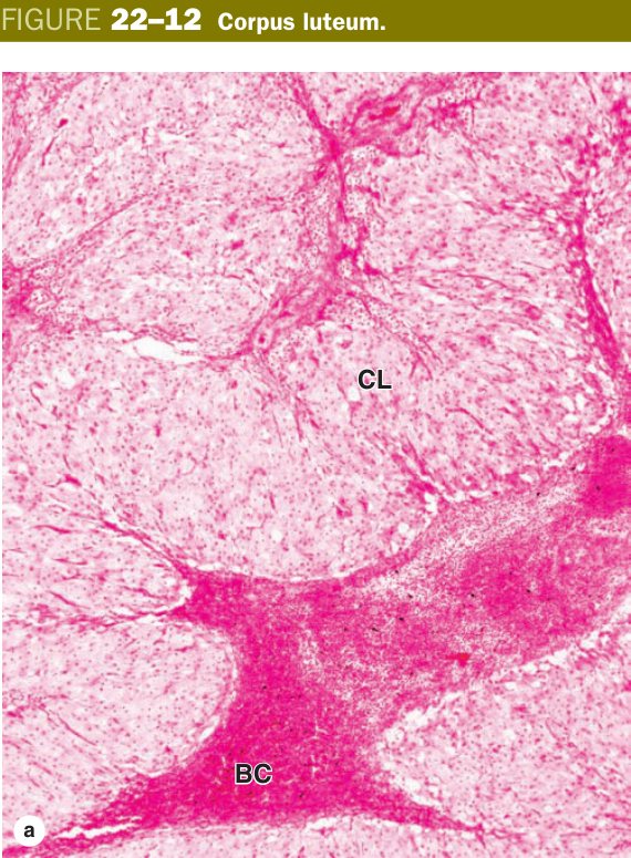
The corpus luteum has two very different destinies depending on whether the released oocyte gets fertilised and an embryo implants. If no implantation happens, LH support fades and the gland involutes after about 10–14 days; this is the corpus luteum of menstruation. The cells undergo apoptosis, the parenchyma is replaced by hyalinised connective tissue, and a pale white fibrous scar — the corpus albicans — is left in the cortex, gradually shrinking over months but often persisting for years as a histological marker of past ovulations.
If, however, the conceptus implants around day 6–7, the trophoblast starts secreting human chorionic gonadotropin (hCG). hCG binds the same LH receptor on lutein cells and rescues the corpus luteum: it enlarges into the corpus luteum of pregnancy, reaching ~5 cm in early gestation, and pumps out progesterone to maintain the decidua until the placenta takes over steroid production around weeks 8–12. After that handover the corpus luteum of pregnancy involutes too, eventually leaving a large corpus albicans.
- Corpus luteum of menstruation: if no fertilisation, persists ~10–14 days then degenerates.
- Corpus luteum of pregnancy: rescued by hCG, enlarges and persists for months.
- Corpus albicans: the degenerated corpus luteum — a pale dense-CT scar.
Urine pregnancy tests detect β–hCG — the very signal rescuing the corpus luteum. Corpus luteum cyst = persistent fluid–filled corpus luteum that may rupture and bleed (acute pelvic pain in early pregnancy, mimics ectopic). Luteal phase deficiency → inadequate progesterone → early miscarriage. After ~weeks 8–12 of pregnancy, the placenta makes enough progesterone that ovarian luteectomy no longer threatens the pregnancy.
Uterus & Menstrual Cycle
The uterus is a thick–walled muscular organ whose job description changes radically every four weeks: build a vascular nest, hold it, then tear it down and start over. Its wall has three layers — an inner mucosa, a thick muscular middle, and a thin outer covering.
The endometrium is the mucosa. Its surface is a simple columnar epithelium of ciliated and non–ciliated secretory cells, dipping into the underlying lamina propria as simple tubular uterine glands. The lamina propria stroma is unusually cellular and is dominated by progesterone–sensitive stromal fibroblasts that will later become decidual cells if pregnancy occurs. Crucially, the endometrium is divided into two functional zones: a deep stratum basale (basal layer), supplied by short, straight basal arteries and containing the bottoms of the glands — this layer is the regenerative reservoir and is never shed; and a superficial stratum functionale (functional layer), supplied by progesterone–sensitive spiral arteries — this is the layer that thickens, secretes, and is sloughed at menstruation.
The myometrium is a thick mass of smooth muscle arranged in three to four ill–defined, interwoven layers. The middle layer, the stratum vasculare, is richest in blood vessels. During pregnancy the myometrium undergoes spectacular hyperplasia and hypertrophy — smooth muscle cells multiply and lengthen up to tenfold, allowing the uterus to grow from a 50–g pear to a 1–kg organ that can fill the abdomen. At term, oxytocin–driven coordinated contractions expel the fetus.
The perimetrium is the outermost covering — mostly a serosa (mesothelium + thin CT) where it faces the peritoneal cavity, and an adventitia where the uterus is fused to surrounding structures (e.g. against the bladder).
Endometrium has a shedding upstairs and a permanent basement downstairs. The spiral arteries are the upstairs plumbing — sensitive to progesterone withdrawal, they constrict and starve the functional layer so it falls off. The basal arteries are the basement plumbing — insensitive to hormone withdrawal, they keep the regenerative crew alive.
The menstrual cycle is the cyclic remodelling of the stratum functionale driven by the ovarian steroid hormones, which are themselves driven by the hypothalamic–pituitary–ovarian axis. The nominal cycle is 28 days; day 1 is conventionally the first day of bleeding.
Menstrual cycle: the cyclic (~28-day) structural changes in the functional layer of the endometrium in a non-pregnant woman, driven by the hypothalamo–pituitary–ovarian hormones; comprises the menstrual, proliferative and secretory phases.
| Phase | Days | Ovary & hormone | Endometrium |
|---|---|---|---|
| Menstrual | 1–4 | Corpus luteum gone → progesterone & oestrogen fall | Functional layer sheds (bleeding) |
| Proliferative (follicular) | 5–14 | Growing follicles → oestrogen | Functional layer regrows; straight glands; spiral arteries lengthen |
| Secretory (luteal) | 15–28 | Corpus luteum → progesterone | Coiled, sawtooth glands full of glycogen secretion; oedematous; thickest |
In the proliferative (follicular) phase, growing antral follicles secrete oestrogen, which drives the basal layer to rebuild the functional layer from scratch. The glands at this stage are straight, narrow tubular with crowded basal nuclei and pseudostratification — classic “proliferative” histology. Spiral arteries lengthen and become slightly coiled. The endometrium reaches a thickness of about 2–3 mm by ovulation.
After ovulation, the corpus luteum's progesterone drives the dramatic secretory (luteal) phase. The glands become extravagantly coiled and sawtoothed, their epithelial cells fill with sub–nuclear glycogen vacuoles on day 17–18 (the histological marker that ovulation has happened), then dump glycogen and mucin into the lumen as luminal secretion later in the phase. The stroma becomes oedematous and pre–decidual, the spiral arteries grow even more tortuous, and the endometrium peaks at 5–7 mm — a glycogen–rich, fluffy bed ready to receive a blastocyst.
If no implantation occurs, the corpus luteum dies and progesterone falls. The spiral arteries respond to progesterone withdrawal by intense vasoconstriction, ischaemia kills the functional layer, the vessels then transiently dilate, the necrotic tissue desquamates, and bleeding begins — this is the menstrual phase. The basal layer, fed by progesterone–insensitive basal arteries, is spared and immediately starts regenerating the surface even before bleeding ends. The cycle restarts.
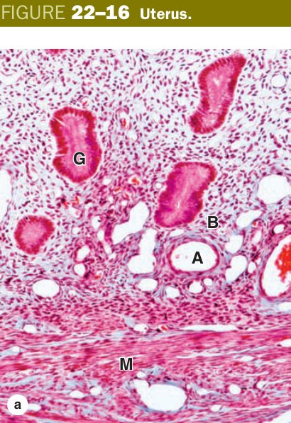
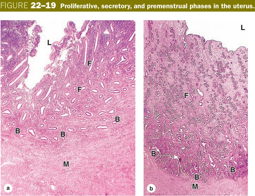
Straight glands = oestrogen = proliferative. Coiled, sawtooth, glycogen–rich glands = progesterone = secretory. The sub–nuclear glycogen vacuole on day 17–18 is the histopathologist's proof of ovulation — it can only appear if there's a working corpus luteum.
Endometrial dating: an endometrial biopsy in the secretory phase can be dated to within ~2 days by gland and stromal morphology — useful for infertility workup. Endometriosis = ectopic endometrium (ovary, peritoneum) that still cycles — bleeds, scars, “chocolate cysts.” Adenomyosis = endometrium within the myometrium → bulky tender uterus. Endometrial hyperplasia → adenocarcinoma arises from unopposed oestrogen (anovulation, obesity, HRT without progestin, tamoxifen) and presents as postmenopausal bleeding. Leiomyoma (fibroid) is the most common pelvic tumour — benign smooth muscle of the myometrium.
Oviduct & Cervix
The oviduct is a 10–12 cm muscular tube with four segments: the funnel–shaped infundibulum with its finger–like fimbriae sweeping the ovarian surface; the wide, thin–walled ampulla, where fertilisation almost always occurs; the narrow muscular isthmus; and the intramural (uterine) segment piercing the uterine wall.
Its mucosa is famously thrown into elaborate branching folds, most exuberant in the ampulla, almost simple in the isthmus — cut the ampulla in cross–section and it looks like a labyrinth of papillae. The epithelium is simple columnar with two cell types whose proportions shift across the cycle: ciliated cells dominate near ovulation (oestrogen–driven, beat toward the uterus) and non–ciliated (peg) secretory cells produce a nutritive fluid that supports the oocyte and capacitates sperm. The muscularis is two layers (inner circular, outer longitudinal) of smooth muscle whose peristaltic waves help shepherd the conceptus toward the uterus. The serosa covers it externally.
The oviduct is a moving sidewalk in a labyrinth. The fimbriae are the airport–style hands sweeping the egg in; the cilia are the conveyor belt; the peg cells stock the snack bar. The ampulla is the wide lounge where sperm meets egg — and unfortunately where ectopic pregnancies most often implant.
Ectopic pregnancy — ~95% are tubal, of which most are in the ampulla; the thin–walled tube cannot stretch and ruptures around 6–8 weeks, causing life–threatening haemoperitoneum. Pelvic inflammatory disease (Chlamydia, Neisseria) scars the folds, traps the blastocyst, and is the leading risk factor for ectopic + tubal infertility (hydrosalpinx).
The cervix is the dense, fibrous neck of the uterus: its wall is mostly dense collagen with only a little smooth muscle — very different from the muscular body. The lumen is lined by two completely different epithelia. The upper endocervical canal bears simple columnar mucus–secreting epithelium with deep branching mucous glands; the consistency of the mucus changes across the cycle (thin and sperm–friendly at ovulation, thick and hostile in the luteal phase). The lower ectocervix facing the vagina is covered by non–keratinised stratified squamous epithelium, continuous with the vaginal lining and adapted to mechanical and chemical wear.
The abrupt junction between these epithelia is the squamocolumnar junction, and the dynamic zone where columnar epithelium is gradually replaced by squamous (squamous metaplasia) is called the transformation zone. Its position shifts across life — everted onto the ectocervix during reproductive years, retreating into the canal after menopause. This is the single most clinically important piece of histology in the female tract.
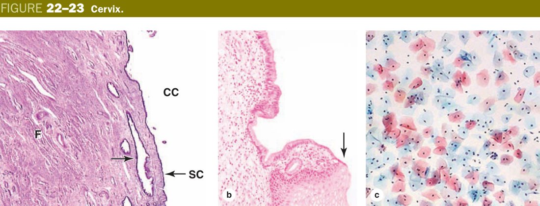
The transformation zone is a border crossing where two epithelial nations meet. Border crossings get the most traffic and the most trouble — that is exactly why HPV finds it so easy to set up CIN there. The Pap smear is, literally, a sample taken from the border.
Almost all cervical squamous cell carcinoma arises in the transformation zone, driven by oncogenic HPV 16/18, progressing through CIN I → II → III → invasive carcinoma. The Pap smear samples this zone; HPV vaccination prevents it. Occluded endocervical mucous glands form retention Nabothian cysts — benign and common.
The vagina has no glands of its own; its mucosa is non–keratinised stratified squamous epithelium, thickened and glycogen–rich under oestrogen. Resident lactobacilli ferment that glycogen to lactic acid, dropping the vaginal pH to ~4 and protecting against pathogens. Lubrication comes from the cervix and from the Bartholin glands at the vestibule. Underneath lie a vascular lamina propria, an inner circular + outer longitudinal smooth muscle layer, and an adventitia.
The mammary gland is a modified apocrine sweat gland organised as 15–20 compound tubuloalveolar lobes in a fibrofatty stroma. In the resting (non–pregnant) state it is mostly duct system with little secretory tissue. In pregnancy, oestrogen + progesterone + prolactin drive massive alveolar proliferation. Lactation uses two secretory modes simultaneously: lipid droplets leave the apex of the alveolar cell wrapped in plasma membrane (apocrine secretion), while protein (casein, α–lactalbumin) is released by classical exocytosis (merocrine secretion). The early milk (colostrum) is rich in secretory IgA. Around each alveolus, contractile myoepithelial cells respond to oxytocin from suckling to eject the milk into the ducts.
Milk fat = apocrine (cell skin pinched off). Milk protein = merocrine (clean exocytosis). Same alveolar cell, two doors. Myoepithelial cells are the “squeeze” muscle — without them, oxytocin has nothing to act on.
Vaginal: Lactobacillus loss → bacterial vaginosis. Breast benign: fibroadenoma (mobile, young), fibrocystic change. Breast malignant: invasive ductal carcinoma (most common) and lobular carcinoma; DCIS is the in–situ precursor; Paget disease of the nipple = malignant cells invading nipple epidermis; inflammatory carcinoma = dermal lymphatic invasion (peau d'orange). Lactational mastitis is usually Staph aureus.
At day 6–7, the blastocyst implants in the secretory endometrium. Its trophoblast splits into an inner cytotrophoblast (mitotic stem layer) and an outer syncytiotrophoblast (multinucleate, invasive, hormone–producing). The syncytiotrophoblast erodes maternal vessels to create lacunae and secretes β–hCG, which rescues the corpus luteum.
By term, the placenta is organised into chorionic villi bathed in maternal blood within the intervillous space. At full term the cytotrophoblast layer thins out, leaving the placental barrier as essentially: syncytiotrophoblast → thin connective tissue → fetal capillary endothelium. The fetal surface clusters villi into cotyledons.
Hydatidiform mole: complete (46 paternal, no embryo, “bunch of grapes”) vs partial (69 chromosomes, some fetal tissue) — very high β–hCG. Choriocarcinoma = malignant syncytiotrophoblast, spectacularly high β–hCG, lung metastases. Placenta praevia = implantation over the cervical os (painless bleeding). Placental abruption = premature separation, painful bleeding.
TMU Exam Drill
📝 Open the full TMU Question Bank — 20 MCQ + 6 terms + 5 essays →
Authentic Tianjin Medical University past-paper questions (2021 Final & the multi-section Final with answer key) mapped to this unit, in the real exam format. Click Show answer to self-test.
□ True or false
□ Explain the following terms
□ Structure essay
Female reproductive complete
Follicle stages, secondary-follicle essay, corpus luteum & menstrual cycle mastered. Next: Skin & Appendages.