Male Reproductive System
Testis Overview
Picture the testis as a tightly packed warehouse of tubing. A thick fibrous capsule, the tunica albuginea, wraps the whole organ and sends connective-tissue septa inward, partitioning the parenchyma into roughly 250 pyramidal lobules. Each lobule is stuffed with one to four coiled seminiferous tubules — this is the actual sperm factory floor.
Between the tubules sits the loose interstitial tissue, where you will find blood vessels, lymphatics, nerves, and the eosinophilic clusters of Leydig cells that make testosterone. So a single low-power view of the testis already tells a story of two compartments: tubules doing meiosis on the inside, and Leydig cells doing steroidogenesis on the outside.
Finished sperm exit the tubules through a short, straight segment (tubuli recti), drain into the anastomosing channels of the rete testis, then leave the testis through 10–15 efferent ductules that pierce the tunica albuginea and lead into the head of the epididymis. From the epididymis the pathway continues as vas deferens → ejaculatory duct → urethra. Memorise that road map — every duct question in the exam lives on it.
Think of the testis as a fruit: the tunica albuginea is the rind, the septa are the white pith dividing segments, and each segment (lobule) is packed with coiled noodles (seminiferous tubules) bathed in a thin sauce of Leydig-cell-rich interstitium.
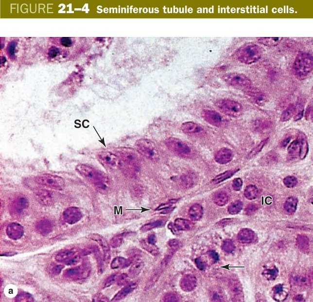
Seminiferous Tubule (essay-grade)
If the examiner asks you for one essay this year, it will be this one. Walk through the tubule wall from outside in, so you can answer in the same order regardless of how the question is phrased.
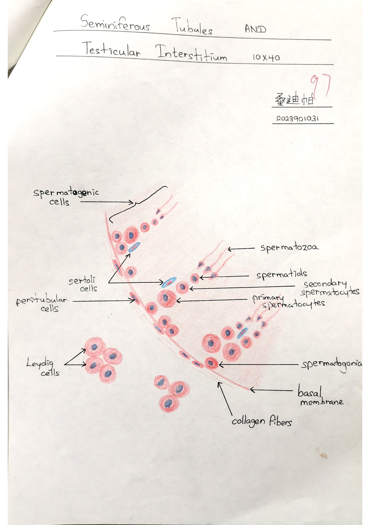
The tubule is bounded by a limiting (peritubular) membrane made of collagen fibrils plus 3–5 layers of contractile myoid cells — these gently squeeze the tubule to nudge immotile sperm toward the rete. Just deep to that lies the basement membrane, and resting on it sits the seminiferous (germinal) epithelium, a thick stratified-looking arrangement of two cell populations: spermatogenic cells climbing through meiosis, and Sertoli cells that scaffold the whole process.
Read the seminiferous epithelium like a vertical timeline: the youngest cells sit on the basement membrane, the most mature cells dangle into the lumen. Spermatogenesis takes roughly 74 days in the human, and the ladder runs in a fixed order.
Spermatogonia are the diploid stem cells on the basement membrane. Type A-dark cells are the reserve stem pool, type A-pale cells are the renewing pool, and type B cells are the ones committed to enter meiosis. Once a type B divides, its daughters become primary spermatocytes — these are the largest germ cells you see, and because they spend ~22 days in meiotic prophase (pachytene), their thick condensed chromatin makes them unmistakable.
Secondary spermatocytes are produced fleetingly — they finish meiosis II within hours, so you rarely catch them on a slide. Their daughters, the spermatids, are small haploid cells near the lumen. Spermatids do not divide; instead they undergo spermiogenesis — a remodelling in which the Golgi assembles the acrosome, the centriole grows the flagellum, the manchette of microtubules shapes the head, mitochondria spiral into the midpiece, and excess cytoplasm is shed as a residual body (phagocytosed by Sertoli cells). The product is the spermatozoon, released into the lumen carrying either 23+X or 23+Y.
| Stage | Feature |
|---|---|
| Spermatogonia | Diploid stem cells on the basement membrane (A-dark reserve, A-pale renewing, B committed) |
| Primary spermatocytes | Largest germ cells; long meiotic prophase — often seen with pachytene chromatin |
| Secondary spermatocytes | Short-lived (meiosis II finishes in hours) — rarely seen |
| Spermatids | Small, near the lumen; undergo spermiogenesis (no division) |
| Spermatozoa | Mature haploid sperm released into the lumen (23+X or 23+Y) |
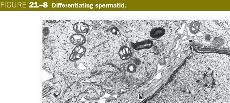
Think of the seminiferous epithelium as an escalator going up to the lumen. Stem cells step on at the bottom (basement membrane); each step up, they shrink, divide, and discard cytoplasm; what falls off the top is a fully aerodynamic sperm.
Sequence: spermatogonia → primary spermatocyte → secondary spermatocyte → spermatid → spermatozoon. Direction: basal → luminal. Primary spermatocytes = biggest cells on the slide.
Sertoli cells are the unsung directors of the whole tubule. Each one is a tall, irregular, columnar cell that stretches all the way from the basement membrane to the lumen, with deep lateral indentations cradling the developing germ cells at every stage of meiosis. The nucleus is pale and ovoid with a prominent nucleolus — once you recognise that pale nucleus near the basement membrane, you have found a Sertoli cell.
Their jobs are several. They nourish the germ cells (germ cells have no direct blood supply once they leave the basal compartment). They phagocytose residual bodies shed during spermiogenesis. They carry FSH receptors and respond by secreting androgen-binding protein (ABP), which traps testosterone inside the tubule at concentrations 100× serum; they also secrete inhibin (feedback to the pituitary) and, in the foetus, anti-Müllerian hormone that regresses the Müllerian duct system.
Most importantly, neighbouring Sertoli cells weld themselves together with occluding tight junctions, creating the blood-testis barrier. This barrier splits the epithelium into a basal compartment (containing spermatogonia and early primary spermatocytes) and an adluminal compartment (containing later spermatocytes, spermatids and sperm). The point of the barrier is immunological: meiotic and post-meiotic cells express novel surface antigens the immune system has never seen, so they would be attacked as foreign. Sertoli tight junctions hide them.
- Tall columnar cells spanning basement membrane → lumen, with an irregular outline embracing the germ cells; pale ovoid nucleus + prominent nucleolus.
- Functions: support & nourish germ cells; form the blood-testis barrier (Sertoli–Sertoli tight junctions) dividing basal & adluminal compartments; phagocytose residual bodies; secrete androgen-binding protein, inhibin & anti-Müllerian hormone.
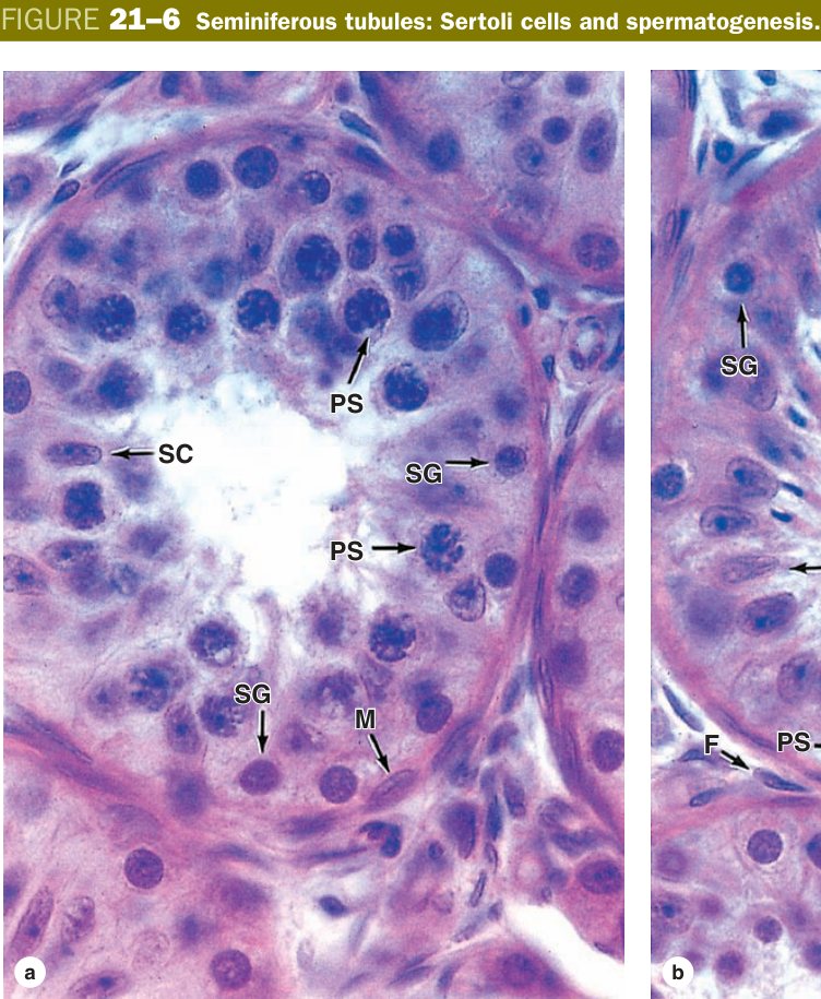
Sertoli cells are the nannies plus security guards of the tubule. They feed the children (germ cells), clean up after them (eat residual bodies), and lock the playroom door (tight junctions) so the immune system police never see what is going on inside.
Blood-testis barrier: formed by tight junctions between adjacent Sertoli cells, dividing the epithelium into a basal compartment (spermatogonia) and an adluminal compartment (spermatocytes onward); it isolates the developing (genetically unique, haploid) germ cells from the immune system.
Trauma or infection (e.g. mumps orchitis, vasectomy with rupture) can break the barrier, exposing germ-cell antigens to lymphocytes and generating anti-sperm antibodies — a recognised cause of immunological infertility.
The mature spermatozoon is built like a miniature submarine: a sharp warhead, a power core, and a long whip. Total length about 60 µm, of which the head is only ~5 µm.
The head houses the haploid nucleus, whose DNA is so tightly packed (histones replaced by protamines) that it stains deeply and uniformly. The anterior two-thirds of the head is covered by the acrosomal cap, a Golgi-derived lysosome-like vesicle loaded with hyaluronidase, acrosin and other hydrolases that digest the corona radiata and zona pellucida at fertilisation.
Behind the head comes the short neck (connecting piece, proximal centriole), then the midpiece — this is where the action lives. The midpiece contains the central axoneme (the universal 9+2 microtubule arrangement) surrounded by outer dense fibres, all wrapped in a helical mitochondrial sheath that supplies ATP for tail-beating. The principal piece is the longest segment, where mitochondria are replaced by a tough fibrous sheath. The end piece is the bare axoneme tip.
| Region | Structure | Function |
|---|---|---|
| Head | Flattened oval; nucleus with highly condensed chromatin (protamines); anterior 2/3 covered by the acrosomal cap (Golgi-derived; acrosin, hyaluronidase) | Carries haploid genome; acrosome enzymes dissolve zona pellucida at fertilisation |
| Neck | Short; proximal centriole; connecting piece | Articulates head to flagellum |
| Midpiece | Axoneme (9+2) + outer dense fibres wrapped by a helical mitochondrial sheath | ATP generation for flagellar movement — the engine room |
| Principal piece | Longest segment; axoneme + outer dense fibres + fibrous sheath (replaces mitochondria) | Propulsion |
| End piece | Short terminal segment; axoneme only, no fibrous sheath | Terminal flagellar tip |
Head = warhead with a chemical drill (acrosome). Midpiece = mitochondrial battery pack. Principal + end piece = the whip. The whole thing exists to deliver 23 chromosomes through one cell membrane.
H-N-M-P-E → Head → Neck → Midpiece (Mitochondria) → Principal piece → End piece. Axoneme = 9+2 throughout.
Leydig (Interstitial) Cells
If Sertoli cells are the foremen inside the tubule, Leydig cells are the chemical plant outside. They sit in clusters in the loose interstitial connective tissue between tubules, almost always huddled next to capillaries — the testosterone they make has to reach the bloodstream and the adjacent tubule fast.
On light microscopy they are large polygonal cells with a round nucleus, one or two nucleoli, and a strikingly eosinophilic foamy cytoplasm. The foaminess is real ultrastructure: abundant smooth endoplasmic reticulum, tubular-cristae mitochondria and lipid droplets — the classic ultrastructural signature of a steroid-secreting cell. In humans only, Leydig cells contain odd rod-shaped crystals of Reinke in the cytoplasm; nobody knows what they do, but examiners love them.
Functionally, Leydig cells carry LH receptors. LH from the anterior pituitary drives them to convert cholesterol into testosterone, which then diffuses into the adjacent seminiferous tubule (where Sertoli-secreted ABP holds it at very high local concentrations to support spermatogenesis) and into the bloodstream (where it masculinises the body and feeds back negatively onto the hypothalamus and pituitary).
- Lie in the interstitial tissue between seminiferous tubules, usually in groups.
- LM: large, polygonal; round nucleus with 1–2 nucleoli; acidophilic cytoplasm.
- EM: typical steroid-secreting ultrastructure (abundant SER, tubular mitochondria, lipid droplets, Reinke crystals).
- Function: secrete testosterone under LH control.
Leydig cells are tiny steroid factories. Anywhere in the body you see eosinophilic cells full of SER + lipid droplets — adrenal cortex, theca interna, corpus luteum, Leydig — you should think “this cell makes a steroid”.
Klinefelter syndrome (47,XXY): small firm testes with hyalinised tubules (spermatogenic failure) but paradoxically hyperplastic Leydig cells — yet testosterone is still low because the cells are dysfunctional. Result: infertility, gynaecomastia, eunuchoid habitus.
Ducts & Accessory Glands
Now you are following sperm out of the testis. As they leave the rete (cuboidal epithelium, irregular anastomosing lumina), they enter 10–15 efferent ductules. The signature of these ductules under the microscope is a scalloped, wavy lumen — because the lining alternates between groups of tall ciliated columnar cells and groups of short non-ciliated cuboidal cells. The ciliated tufts beat sperm toward the epididymis; the short cells absorb most of the testicular fluid.
The epididymis is a single, immensely coiled tube about 6 m long folded into a comma along the posterior testis (head, body, tail). Its lining is uniform pseudostratified columnar epithelium with very long, branched, non-motile stereocilia (which are actually giant microvilli, not true cilia). Two cell types — tall principal cells with stereocilia, and basal cells at the basement membrane. A smooth muscle coat surrounds it, thin in the head and progressively thicker toward the tail. Functionally, sperm mature here (acquire forward motility and the ability to fertilise) and are stored in the tail until ejaculation. Travel time is roughly 12 days.
The ductus (vas) deferens is the muscular tube that pushes sperm from the tail of the epididymis through the spermatic cord into the ejaculatory duct. Its epithelium is similar pseudostratified columnar with short stereocilia, but the wall is famous: three layers of smooth muscle — inner longitudinal, middle circular, outer longitudinal. That sympathetic-driven peristaltic burst at ejaculation comes from this thick muscle coat.
| Structure | Epithelium | Key Feature |
|---|---|---|
| Efferent ductules (10–15 tubules) | Alternating tall ciliated columnar + short non-ciliated cuboidal cells → scalloped / wavy luminal outline | Cilia move sperm toward epididymis; short cells absorb fluid; thin smooth muscle layer |
| Epididymis | Pseudostratified columnar with long stereocilia (non-motile microvilli); principal + basal cells | Sperm maturation (~12 days) + storage in tail; progressively thicker smooth muscle coat |
| Ductus (vas) deferens | Pseudostratified columnar + three-layered smooth muscle (inner long + middle circular + outer long) | Rapid peristaltic propulsion of sperm at ejaculation |
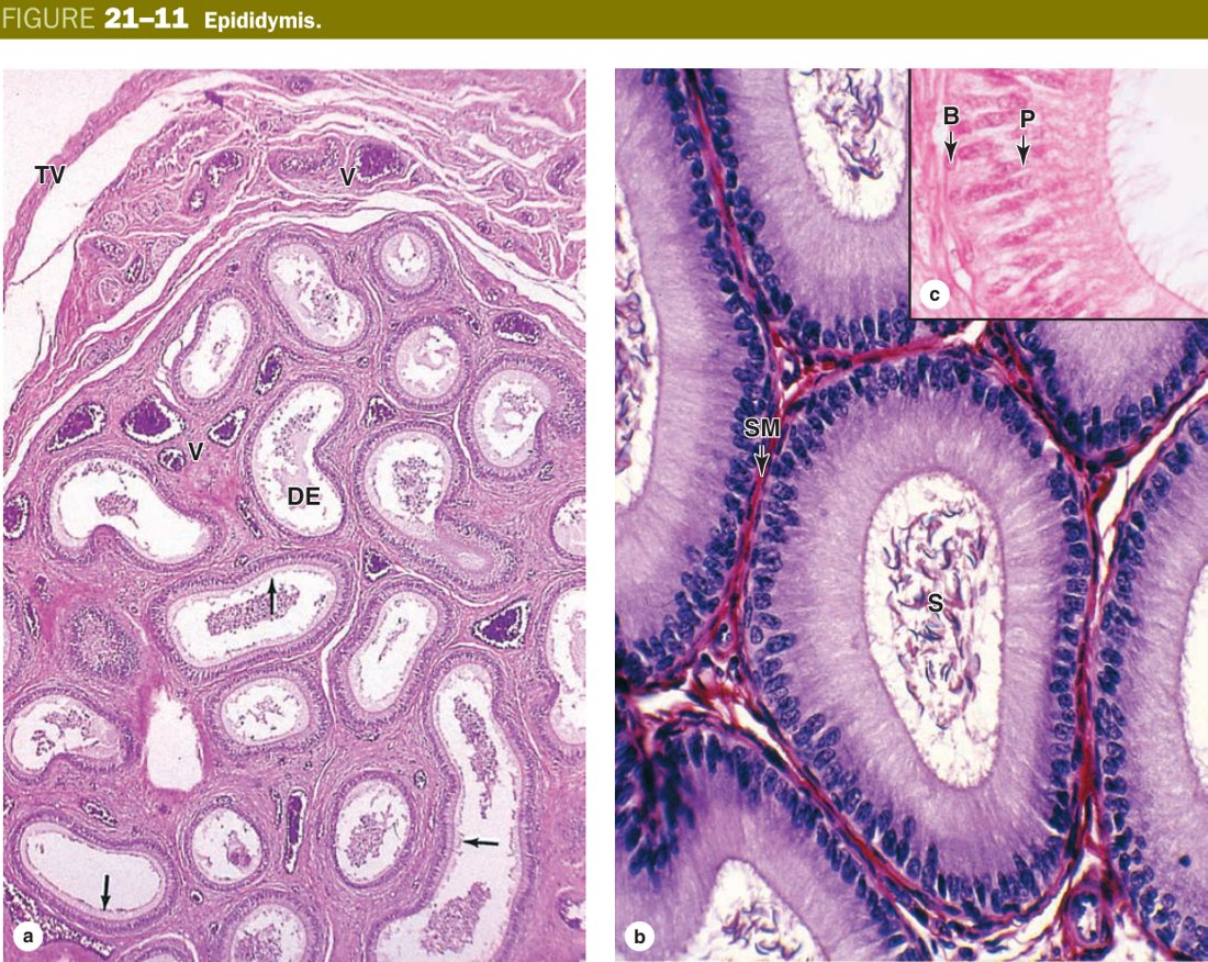
Three ducts, three textures. Efferent = wavy lumen (alternating cell heights). Epididymis = even lumen with fuzzy stereocilia. Vas deferens = tiny lumen, enormous three-layer muscle wall — the muscle is the diagnosis.
Epididymitis in young men is usually Chlamydia trachomatis or Neisseria gonorrhoeae; in older men, E. coli from urinary reflux. Vasectomy = bilateral surgical interruption of the ductus deferens.
Only ~5–10% of ejaculate is actually sperm. The rest is the contribution of three pairs (or one) of accessory glands. Learn each by its histology and its secretion.
The seminal vesicle is a paired, coiled pouch whose mucosa is so elaborately folded that on cross-section it looks like a honeycomb. The lining is pseudostratified columnar epithelium studded with yellow lipochrome pigment granules. It contributes about 60–70% of semen volume: viscous, alkaline, fructose-rich fluid (the sperm energy source), prostaglandins, fibrinogen and seminal-vesicle-specific coagulation proteins that gel the ejaculate.
The prostate is a walnut-sized fibromuscular organ surrounding the proximal urethra. It contains 30–50 tubuloalveolar glands arranged in three concentric zones (McNeal zones): the peripheral zone (~70% of the volume — the seat of prostate adenocarcinoma), the central zone (~25%), and the small transition zone surrounding the urethra (~5% — the seat of benign prostatic hyperplasia). The glandular lumina often contain concentric eosinophilic lamellated concretions called corpora amylacea; they increase in number with age and have no clinical importance, but they are a giveaway on slides. The prostate adds ~30% of semen volume: thin, slightly acidic fluid containing PSA (a serine protease that liquefies the coagulum after a few minutes), citric acid, fibrinolysin and zinc.
The paired bulbourethral (Cowper) glands are small compound tubuloalveolar mucous glands lying within the urogenital diaphragm. Their clear pre-ejaculatory secretion lubricates the urethra and neutralises residual urinary acidity ahead of the sperm.
| Gland | Histology | Secretion & Function |
|---|---|---|
| Seminal vesicle | Elaborate, highly folded mucosa giving a honeycomb appearance; epithelium = pseudostratified columnar with yellow lipochrome pigment granules; fibromuscular wall | ~60–70% of semen: fructose (energy), prostaglandins, citric acid, fibrinogen, coagulation proteins |
| Prostate | 30–50 tubuloalveolar glands in a fibromuscular stroma; simple to pseudostratified columnar epithelium; lumina often contain laminated corpora amylacea; McNeal zones (peripheral 70% / central 25% / transition 5%) | ~30% of semen: PSA (liquefies coagulum), citric acid, fibrinolysin, zinc (antibacterial) |
| Bulbourethral (Cowper's) glands | Compound tubuloalveolar; mucus-secreting columnar epithelium; fibromuscular capsule | Pre-ejaculatory secretion: neutralises urethral acidity, lubricates urethra |
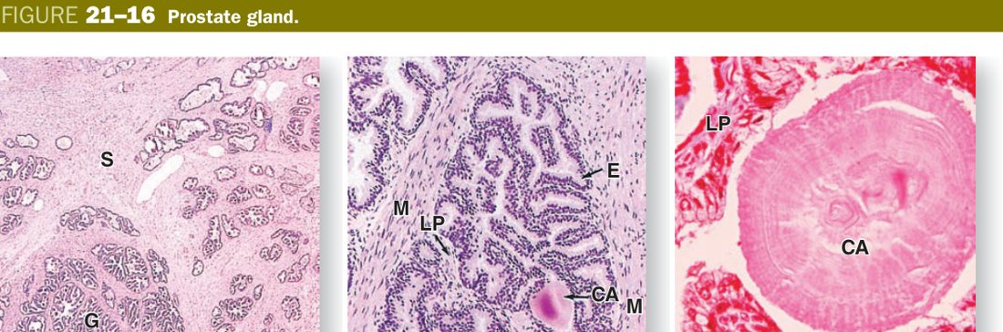
Zone ↔ disease: peripheral zone → cancer (palpable on DRE, picked up by PSA, scored by Gleason), transition zone → BPH (squeezes the urethra). Remember it as “cancer hides at the edge, BPH crushes the middle”.
BPH: transition-zone hyperplasia → urinary frequency, hesitancy, retention. Prostate adenocarcinoma: peripheral zone, palpated as a hard nodule, raised serum PSA, graded with the Gleason system. Corpora amylacea themselves are benign age-related concretions.
A note on the penis: three cylinders of erectile tissue (two dorsal corpora cavernosa + one ventral corpus spongiosum containing the spongy urethra), each wrapped in a tunica albuginea. Erection is a parasympathetic event — nerve-released nitric oxide raises smooth-muscle cGMP, helicine arteries dilate, cavernous sinuses engorge, and venous outflow is mechanically compressed. Sildenafil works here by blocking phosphodiesterase-5, keeping cGMP elevated.
Cryptorchidism — undescended testis stays at body-core temperature, arrests spermatogenesis and carries a markedly raised risk of seminoma. Testicular torsion — twisted spermatic cord chokes off blood supply; surgical emergency. Varicocele — dilated pampiniform plexus, classic “bag of worms”, left-sided in >85% because of the right-angle drainage into the left renal vein; impairs fertility through heat. Hydrocele — serous fluid in the tunica vaginalis. Seminoma — uniform sheets of clear “fried-egg” cells with a lymphocytic infiltrate; radiosensitive. Erectile dysfunction from vascular causes responds to PDE5 inhibitors.
TMU Exam Drill
📝 Open the full TMU Question Bank — 20 MCQ + 6 terms + 5 essays →
Authentic Tianjin Medical University past-paper questions (2021 Final & the multi-section Final with answer key) mapped to this unit, in the real exam format. Click Show answer to self-test.
□ True or false
□ Structure essay
- Seminiferous (germinal) epithelium: spermatogenic cells — spermatogonia, primary spermatocytes, secondary spermatocytes, spermatids, spermatozoa (basal→luminal) — plus Sertoli (supporting) cells.
- Basement membrane.
- Limiting membrane: collagen fibrils + contractile myoid cells.
- Interstitium between tubules contains Leydig cells.
Male reproductive complete
Seminiferous tubule, spermatogenesis, Sertoli & Leydig mastered. Next: Female Reproductive.