Eye & Ear
The Eye — Three Tunics
The eyeball is built like a layered ball — three concentric coats wrapped around a transparent core. Each coat has one dominant job: the outer fibrous tunic keeps the shape, the middle vascular tunic (the uvea) brings blood and pigment, and the inner neural tunic (the retina) does the actual seeing. If you fix those three jobs in your head, every histological detail downstream slots into place.
Anteriorly the fibrous coat becomes transparent (cornea); posteriorly it stays opaque and white (sclera). The vascular coat splits into three regional specialisations — choroid behind, ciliary body in the middle, iris in front — each one a variation on the same theme of pigmented, vessel-rich connective tissue with added muscle. The retina is most elaborate posteriorly and thins to a non-photoreceptive pigmented strip as it sweeps forward over the ciliary body and iris.
| Tunic | Components |
|---|---|
| Fibrous (outer) | Cornea (anterior, transparent) + sclera (white, opaque) |
| Vascular (uvea, middle) | Choroid + ciliary body + iris |
| Neural (inner) | Retina |
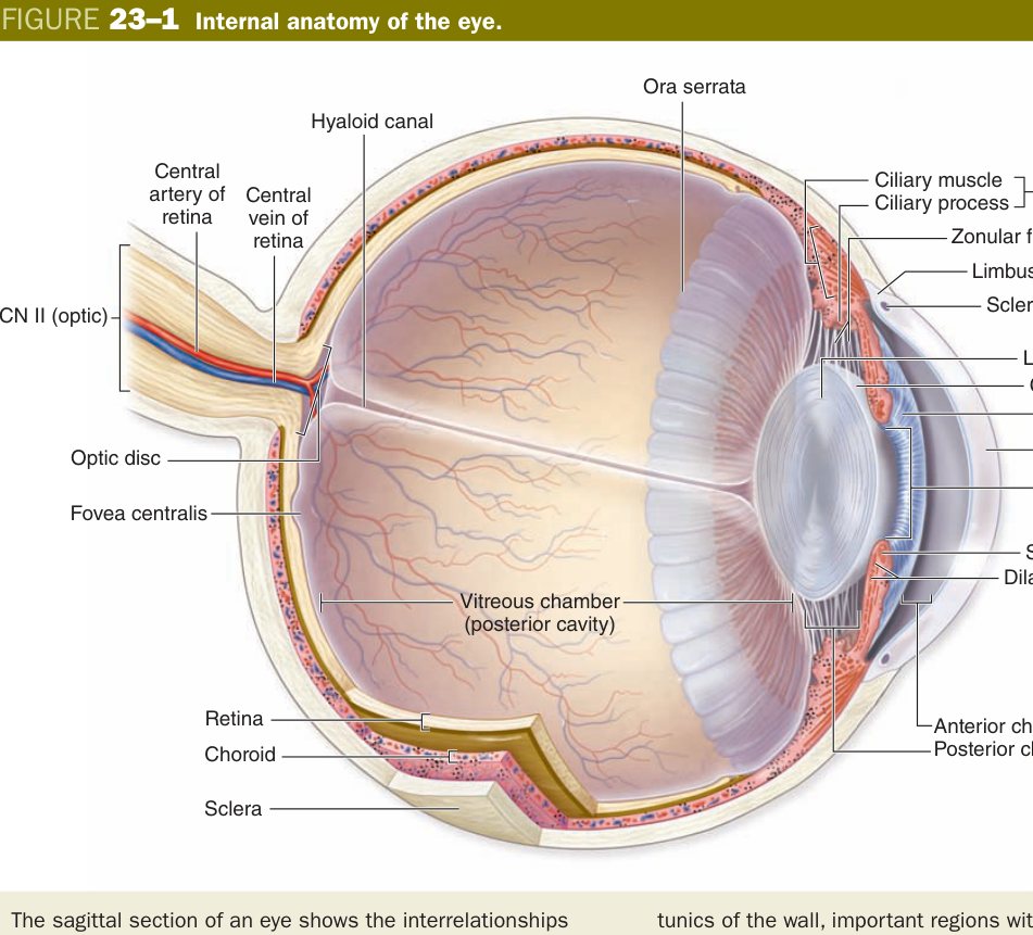
Think of the eye as a camera body. The sclera is the rigid plastic shell. The cornea is the fixed front element of the lens system. The iris is the aperture diaphragm. The lens is the autofocus element. The retina is the digital sensor. The choroid is the matt-black interior paint that absorbs stray light so it doesn't bounce around and fog the image.
The cornea has to do two contradictory things at once: be tough enough to take a finger-poke without bursting, and be perfectly transparent so light can pass through unscattered. Evolution solves this with five carefully stacked layers, each contributing one piece of the puzzle. Walking through them from front to back is the single most exam-friendly drill in this unit.
The corneal epithelium is non-keratinised stratified squamous — the same architecture as the mouth or oesophagus, kept moist by tears. It is densely innervated with free nerve endings (why a corneal abrasion is so painful) and it regenerates briskly from limbal stem cells at the corneo-scleral junction. If you destroy the limbus — chemical burn, severe contact-lens damage — the conjunctival epithelium grows over the cornea instead and vision is lost.
Under the epithelium sits Bowman membrane: an acellular condensation of type I collagen and proteoglycans. It has no cells, so it does not regenerate. Once breached, it scars. The bulk of the cornea is the stroma (substantia propria) — sixty to seventy parallel lamellae of type I collagen embedded in keratan-sulphate-rich ground substance, with quiet flat fibroblasts (keratocytes) tucked between sheets. Transparency depends on the lamellae being precisely spaced (less than the wavelength of visible light) and the stroma being kept slightly dehydrated.
Posteriorly you meet Descemet membrane, which is simply the very thick basement membrane of the endothelium — and it gets thicker with every decade of life. Behind it lies the corneal endothelium: a single layer of squamous-to-cuboidal cells whose ion pumps actively shift water out of the stroma into the aqueous humour. These cells do not divide in adults. You are born with a fixed budget, and you lose them slowly over a lifetime.
Imagine a stack of glass sheets. If you keep them dry and perfectly parallel, light passes straight through. Add a drop of water between two sheets and it goes hazy. The corneal endothelium is a tiny bilge pump on the back wall, working all day to keep the glass-sheet stroma dry. When the pump fails (Fuchs dystrophy), the cornea swells and clouds — classic morning blur that clears as the day goes on and tears osmotically pull water out.
| Layer | Structure |
|---|---|
| Corneal epithelium | Non-keratinized stratified squamous; no melanocytes; many free nerve endings; regenerates from limbal stem cells |
| Bowman's membrane (anterior limiting) | Acellular; type I collagen + ground substance; no regeneration |
| Stroma (substantia propria) | 60–70 regular collagen lamellae + keratocytes; avascular & transparent; the thickest layer |
| Descemet's membrane (posterior limiting) | Thick basement membrane of endothelium; thickens with age |
| Endothelium | Single layer of squamous/cuboidal cells; pumps fluid out to keep cornea clear; non-replicating in adults |
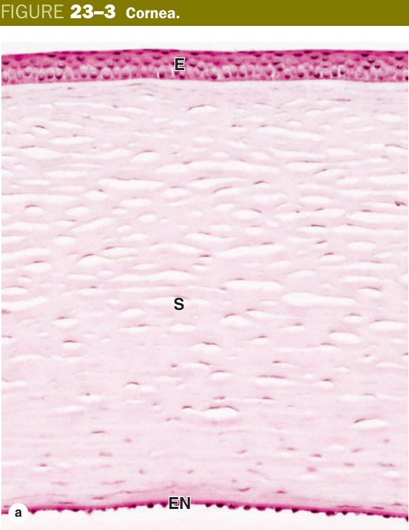
“Eat Bread, Stay During Exam” → Epithelium, Bowman's, Stroma, Descemet's, Endothelium. Transparency needs the stroma avascular and the endothelium pumping water out.
Keratoconus — progressive thinning and conical bulging of the stroma. Fuchs endothelial dystrophy — endothelial cell loss with stromal oedema, treated by Descemet membrane endothelial keratoplasty. A corneal abrasion heals because the epithelium regenerates from the limbus; a deeper corneal ulcer that breaches Bowman scars.
The lens is one of the strangest tissues in the body: avascular, almost entirely cellular yet transparent, and grown like an onion from the outside in, layer upon layer, for your whole life. It hangs behind the iris suspended by a ring of zonular fibres anchored to the ciliary body, and it bends light just enough to throw a sharp image on the retina.
Wrap the whole lens in a thick, glassy capsule — this is a type IV collagen basement membrane (the thickest in the body) and it is what the cataract surgeon hangs the artificial intraocular lens off. Under the anterior capsule sits a single layer of cuboidal lens epithelium. At the equator these epithelial cells stop being cuboidal, elongate enormously, lose their nuclei and organelles, and become packed with crystallin proteins. They are now lens fibres: hexagonal, anucleate, packed so tightly there is essentially no extracellular space, and connected by gap junctions for nutrient sharing because nothing else can get in.
New fibres are laid down on top of old ones every year of life, like growth rings. Old fibres get pushed centrally to form the nucleus of the lens. With age the central crystallins crosslink, harden, and yellow — this is presbyopia (loss of accommodative range) and, taken further, cataract.
The aqueous humour is a tiny river that nourishes everything in the avascular front of the eye. It is born at the ciliary processes, flows downstream through the posterior chamber, ducks under the iris through the pupil, fills the anterior chamber, and finally drains out at the corner where iris meets cornea via the trabecular meshwork into the canal of Schlemm. Dam the river at the corner and pressure rises behind — that is glaucoma.
| Component | Structure |
|---|---|
| Capsule | Thick acellular basement membrane (thickest in the body); type IV collagen; encloses the whole lens |
| Anterior epithelium | Simple cuboidal beneath the anterior capsule; at the equator elongates into new lens fibres throughout life |
| Lens fibres | Hexagonal, elongated, anucleate cells packed with crystallin proteins; gap junctions; layered like onion-rings |
Produced by the non-pigmented epithelium of ciliary processes → fills the posterior chamber (between iris and lens) → flows through the pupil → enters the anterior chamber (between iris/lens and cornea) → drains via the trabecular meshwork into the canal of Schlemm → episcleral veins. Blockage → raised IOP → glaucoma.
The middle coat (uvea) is the pigmented, vessel-dense layer that sits between the tough sclera and the delicate retina. It does three jobs in three regions: behind, the choroid feeds the outer retina; in the middle, the ciliary body shapes the lens and makes aqueous; in front, the iris opens and closes the pupil.
The choroid is loose, melanocyte-rich connective tissue packed with vessels. Its innermost capillary plexus — the choriocapillaris — is what nourishes the avascular outer retina (photoreceptors, RPE) by diffusion across Bruch membrane, a thin five-layered sandwich separating choroid from RPE. In age-related macular degeneration this membrane develops drusen and may be breached by abnormal vessels.
The ciliary body is a wedge of smooth muscle (the ciliary muscle, parasympathetic, CN III) with finger-like ciliary processes on its inner surface. Each process is covered by a double layer of epithelium: an outer pigmented layer continuous with the RPE, and an inner non-pigmented layer that actively secretes aqueous humour. Zonular fibres run from the ciliary processes to the lens equator. When the ciliary muscle contracts (near vision), the ciliary body moves forward and inward, zonules slacken, and the elastic lens rounds up for near focus — accommodation.
The iris is the muscular curtain in front of the lens. Its loose anterior stroma carries the melanocytes that decide eye colour. Two opposing smooth-muscle systems set pupil size: the circular sphincter pupillae at the pupil margin (parasympathetic, miosis) and the radial dilator pupillae built from myoepithelial cells (sympathetic, mydriasis). Posteriorly, two layers of densely pigmented epithelium block any light from passing through the iris itself.
| Structure | Histology | Function |
|---|---|---|
| Choroid | Melanocyte-rich vascular CT; choriocapillaris; Bruch membrane separates from RPE | Feeds outer retina; absorbs stray light |
| Iris stroma | Loose CT with melanocytes & vessels; no anterior epithelium | Eye colour; defines pupil aperture |
| Sphincter pupillae | Ring of smooth muscle at pupil margin (parasympathetic / CN III) | Constricts pupil (miosis) |
| Dilator pupillae | Myoepithelial cells (modified pigmented epithelium; sympathetic) | Dilates pupil (mydriasis) |
| Posterior pigmented epithelium | Two layers of densely pigmented epithelium | Light barrier |
| Ciliary body | Ciliary muscle (smooth, parasymp) + ciliary processes with outer pigmented + inner non-pigmented epithelium | Accommodation; non-pigmented epithelium secretes aqueous |
Cataract: opacification of lens fibres (denatured crystallins) — commonest cause of reversible blindness worldwide. Open-angle glaucoma: trabecular meshwork resistance rises silently → high IOP → optic disc cupping. Closed-angle glaucoma: a narrow anterior chamber angle physically blocks drainage — painful, acute, sight-threatening. Uveitis: inflammation of choroid, ciliary body or iris.
Retina — Ten Layers, Four Principal Cells
The retina is the only part of the central nervous system you can examine directly, through the pupil with an ophthalmoscope. Embryologically it is an outpouching of the diencephalon, and that ancestry shows up in its architecture: an outer pigmented epithelium derived from the outer layer of the optic cup, and an inner neural retina (nine layers) derived from the inner layer. The tiny potential space between them is where retinal detachments happen — it is a developmental cleft that never fully closes.
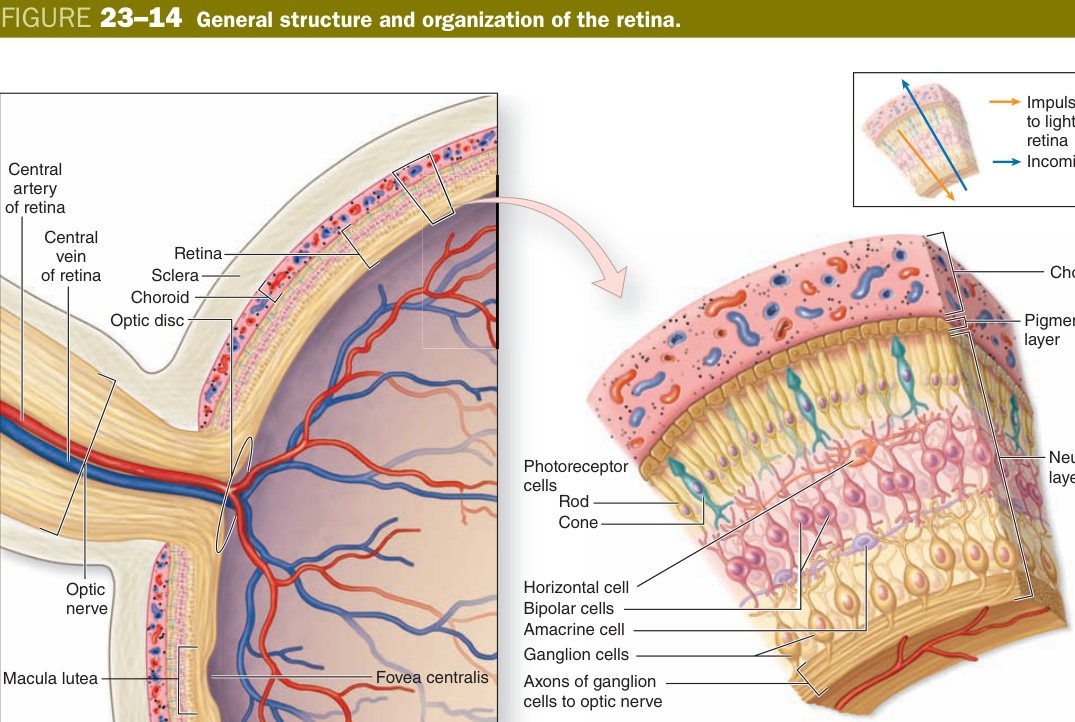
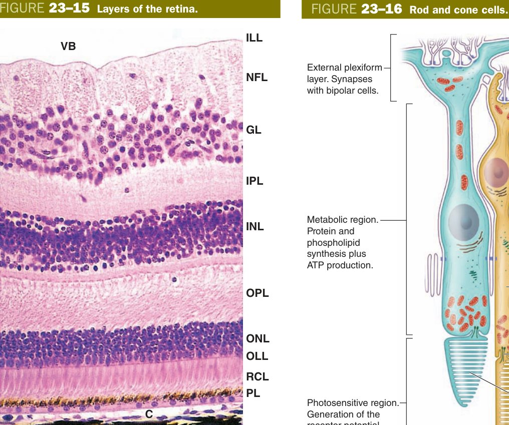
Examined under the microscope, the neural retina shows ten layers running from outside (toward the choroid) inward (toward the vitreous). The trick is to realise the photoreceptors point outward, away from the light. Light therefore passes through almost the entire retina, including the ganglion cell axons, before it strikes the photoreceptor outer segments at the back. The fovea is the one place where evolution has pushed those overlying layers aside so light hits the cones directly — which is why the fovea has the sharpest vision.
| Layer (outer → inner) | What is there |
|---|---|
| 1. Retinal pigment epithelium (RPE) | Cuboidal melanin-laden cells; phagocytose shed outer segments; recycle 11-cis-retinal; tight junctions form blood-retina barrier |
| 2. Photoreceptor outer & inner segments | Rod and cone discs (outer segment) + organelle-rich inner segment |
| 3. External limiting membrane | Junctional band where Müller cell processes meet photoreceptors |
| 4. Outer nuclear layer | Cell bodies of rods and cones |
| 5. Outer plexiform layer | Synapses: photoreceptor → bipolar & horizontal cells |
| 6. Inner nuclear layer | Cell bodies of bipolar, horizontal, amacrine, & Müller cells |
| 7. Inner plexiform layer | Synapses: bipolar → ganglion & amacrine cells |
| 8. Ganglion cell layer | Cell bodies of ganglion cells |
| 9. Nerve fibre layer | Unmyelinated ganglion-cell axons running toward optic disc |
| 10. Internal limiting membrane | Basement membrane of Müller cells at the vitreal surface |
Four principal cell types carry the signal from light to brain — pigment epithelium, photoreceptor, bipolar, ganglion — with horizontal, amacrine and Müller cells acting as lateral integrators and glia. Signal flow: photoreceptor → bipolar → ganglion → optic nerve.
| Cell | Structure / function |
|---|---|
| Pigment epithelium (RPE) | Simple cuboidal with melanin + phagosomes; absorbs stray light; phagocytoses shed photoreceptor discs; recycles 11-cis-retinal; tight junctions form outer blood-retina barrier |
| Photoreceptors | Outer segment (membranous discs with visual pigment) + inner segment + cell body + axon. Rods (1 type, rhodopsin) — scotopic, peripheral. Cones (S/M/L, photopsin) — photopic + colour, concentrated at fovea. Synapse with bipolar cells |
| Bipolar cells | Inner-nuclear-layer interneurons connecting photoreceptors → ganglion cells |
| Ganglion cells | Multipolar neurons; their unmyelinated axons converge at the optic disc to form the optic nerve (CN II) |
The retina is wired backwards. Light has to pass through ganglion cells, bipolar cells and a tangle of axons before it reaches the photoreceptors at the very back. Why? Because the RPE behind the photoreceptors needs to be in close contact with them — it phagocytoses worn-out disc tips every morning and shuttles vitamin A back. At the fovea, evolution worked around the wiring by sliding the overlying layers aside, creating a pit so light goes straight onto cones. That tiny pit is where you read this sentence.
The macula lutea is the yellow-pigmented central retina, about 5 mm across. At its centre is the fovea centralis, a 1.5 mm depression containing only cones (no rods, no overlying bipolars or ganglion cells). The optic disc, slightly nasal to the fovea, is where ganglion axons exit and central retinal vessels enter — no photoreceptors live there, so it is the physiological blind spot.
Age-related macular degeneration: dry form — drusen accumulate in Bruch membrane; wet form — choroidal neovascularisation through Bruch driven by VEGF (treated with anti-VEGF injections). Retinitis pigmentosa: inherited rod degeneration begins peripherally (night blindness, tunnel vision). Retinal detachment: separation between RPE and photoreceptors — the embryonic cleft re-opens. Diabetic retinopathy: microvascular damage → microaneurysms, exudates, neovascularisation. Glaucoma: ganglion-cell axon loss at the optic disc.
The Ear — Three Divisions
The ear is two organs in one piece of bone: a hearing organ (cochlea) and a balance organ (vestibular apparatus), sharing a common fluid system. Sound has to be collected from the air, mechanically transmitted across an air gap, and then converted into electrical signals inside fluid. The three divisions of the ear correspond exactly to those three steps: collect, transmit, transduce.
The external ear (auricle + external auditory meatus) collects and funnels sound. The auricle is built around elastic cartilage covered by thin skin; the meatus is a tube lined by keratinised stratified squamous epithelium with ceruminous glands (modified apocrine sweat glands that secrete earwax) and short hairs. At the bottom of the tube sits the tympanic membrane — three layers: outer keratinised squamous (skin), middle radial+circular collagen, inner respiratory-type mucosa.
The middle ear is an air-filled box in the temporal bone holding three tiny ossicles — malleus, incus, stapes — that lever vibrations from the tympanic membrane across to the oval window of the inner ear. The pharyngotympanic (Eustachian) tube, lined by pseudostratified ciliated epithelium, connects the middle ear to the nasopharynx and equalises pressure (the click when you yawn or swallow on a plane).
The internal ear is a fluid-filled labyrinth carved into the petrous temporal bone. A bony labyrinth filled with perilymph (ECF-like: high Na+, low K+) houses a delicate membranous labyrinth filled with endolymph (ICF-like: high K+, low Na+). The cochlear part of the membranous labyrinth handles hearing; the utricle, saccule and three semicircular ducts handle balance.
Picture a Russian doll: a fluid-filled bag (membranous labyrinth, endolymph) floating inside a fluid-filled bony cavity (bony labyrinth, perilymph). Endolymph is inside the bag and is unique among extracellular fluids because it is high K+, low Na+ — like the inside of a cell. Perilymph is around the bag and looks like ordinary CSF/ECF. The high-K+ endolymph is what powers hair-cell transduction.
| Division | Parts |
|---|---|
| External | Auricle (elastic cartilage + skin), external auditory meatus (keratinised squamous + ceruminous glands + hair), tympanic membrane |
| Middle | Tympanic cavity + ossicles (malleus, incus, stapes) + Eustachian tube (pseudostratified ciliated) |
| Internal | Bony labyrinth (perilymph) housing membranous labyrinth (endolymph): cochlea (hearing) & utricle/saccule/semicircular ducts (balance) |
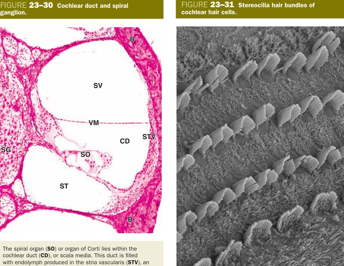
Cut the cochlea across and you see three stacked fluid-filled tubes spiralling together up the cochlear axis. The top and bottom tubes (scala vestibuli and scala tympani) contain perilymph and are continuous at the apex of the cochlea through a tiny opening called the helicotrema. Sandwiched between them is the scala media (cochlear duct), a triangular tube of membranous labyrinth filled with endolymph and carrying the organ of Corti on its floor.
Reissner's (vestibular) membrane — only two cell layers thick — forms the roof of the scala media and separates endolymph from perilymph above. The floor of the scala media is the basilar membrane: an acellular fibrous sheet on which the organ of Corti rests. The basilar membrane is narrow and stiff at the base of the cochlea (high-frequency tuning) and wide and floppy at the apex (low-frequency tuning) — the anatomical basis of place-coding.
On the outer wall of the cochlear duct sits the stria vascularis, a thickened epithelium with three cell layers (marginal, intermediate, basal) and the unusual property of containing intraepithelial blood vessels — the only epithelium in the body to do so. It pumps K+ into the cochlear duct and is solely responsible for producing the high-K+ endolymph. Damage the stria (loop diuretics, aging, autoimmune injury) and hearing fails.
In the central bony pillar of the cochlea (the modiolus) sit the bipolar neurons of the spiral ganglion. Their peripheral processes contact hair cells; their central processes form the cochlear division of CN VIII.
| Space | Fluid | Boundary |
|---|---|---|
| Scala vestibuli (above) | Perilymph (high Na+, low K+ — like ECF) | Above Reissner's (vestibular) membrane |
| Scala media / cochlear duct (middle) | Endolymph (high K+, low Na+ — like ICF) | Between Reissner's membrane (above) and basilar membrane (below); the membranous labyrinth |
| Scala tympani (below) | Perilymph | Below the basilar membrane; ends at the round window |
| Structure | Histology & Role |
|---|---|
| Reissner's (vestibular) membrane | Two cell layers (squamous on each side); separates scala vestibuli from scala media; helps maintain endolymph composition |
| Basilar membrane | Acellular fibrous membrane; organ of Corti sits on its upper surface; frequency tuning (high freq at base, low at apex) |
| Stria vascularis | Highly vascularised epithelium on the lateral wall of the cochlear duct; three cell types: marginal, intermediate, basal; actively secretes endolymph (the only epithelium with intraepithelial blood vessels) |
| Spiral ganglion | Bipolar neurons in the modiolus; peripheral process → hair cells; central process → cochlear nerve (CN VIII) |
Spiral Organ of Corti
The organ of Corti is where sound finally becomes electricity. It is an epithelial ridge sitting on the basilar membrane inside the scala media, bathed in endolymph, overhung by a gelatinous flap called the tectorial membrane. Two families of cells make it work: the hair cells are the sensory transducers; the supporting cells hold them in place and form the architectural scaffolding.
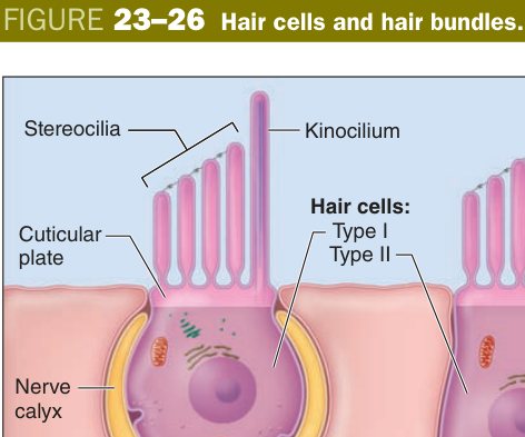
There is one row of inner hair cells and three rows of outer hair cells. The inner hair cells are the true sensory cells — they make most of the synapses with afferent fibres of the cochlear nerve and are responsible for the hearing signal sent to the brain. The outer hair cells are amplifiers: they are electromotile, changing length in response to voltage via a unique motor protein called prestin, and their dancing physically sharpens the tuning of the basilar membrane.
Each hair cell carries a bundle of stereocilia (modified microvilli, not true cilia) graded in height. Tip-links between adjacent stereocilia open mechanotransduction channels when the bundle is deflected. Deflection toward the tallest row depolarises the cell; deflection the other way hyperpolarises it. The tectorial membrane sits over the outer hair cell bundles and shears them as the basilar membrane moves.
The supporting cells are mainly the pillar cells (inner and outer pillars frame a triangular tunnel of Corti through the middle) and the phalangeal cells of Deiters (cup the bases of the outer hair cells and send a slender process up to form a reticular lamina at the apical surface).
| Cell family | Members & role |
|---|---|
| Supporting cells | Pillar cells (inner & outer — form the tunnel) + phalangeal cells (inner & outer — cup the hair cells) |
| Hair cells | Inner hair cells (1 row, true sensory) + outer hair cells (3 rows, electromotile amplifiers via prestin) — the receptor cells that convert sound vibration into electrical signals (mechanotransduction via stereocilia) |
Stretch the spiral cochlea out into a straight tube and the basilar membrane becomes a long, tapered keyboard. Each point along the keyboard resonates best to one frequency — high notes at the stiff narrow base near the oval window, low notes at the floppy wide apex. Pure tone arriving at the eardrum lights up just one spot. That spot's inner hair cells fire, the brain knows the pitch by knowing which fibre fired. Damage the basal end (noise, ageing, aminoglycosides) and high-frequency hearing goes first — presbycusis.
Corti = Hair cells (sensory) + Supporting cells. Supporting = Pillar (build the tunnel) + Phalangeal (hold the hair cells). The hair cells are the transducers; the tectorial membrane bends their stereocilia. 1 row of inner + 3 rows of outer hair cells.
Presbycusis: age-related high-frequency loss from basal cochlear damage. Aminoglycoside ototoxicity (gentamicin) and cisplatin selectively kill outer hair cells. Noise-induced hearing loss: mechanical injury to stereocilia bundles. Otosclerosis: abnormal bone fixes the stapes footplate at the oval window — conductive deafness. Meniere disease: endolymphatic hydrops → episodic vertigo + sensorineural loss + tinnitus.
The balance organs run on exactly the same principle as the cochlea — hair cells with stereocilia bathed in endolymph — but the mechanical stimulus is different. There are five vestibular receptor patches per ear: two maculae (one each in the utricle and saccule) and three cristae (one in the ampulla of each semicircular duct).
The maculae sense linear acceleration and the static pull of gravity. Hair cells project their stereocilia into the otolithic membrane, a gelatinous mat loaded with calcium-carbonate crystals (otoliths) that have more inertia than the surrounding fluid. When the head tilts or accelerates linearly, the otoliths lag, the gel slides over the hair cells, and stereocilia bend. The utricular macula is horizontal (senses sideways tilt and forward acceleration); the saccular macula is vertical (senses up-down tilt and vertical acceleration).
The cristae ampullares sense angular (rotational) acceleration. Each crista is a ridge of hair cells sitting in the swollen ampulla of a semicircular duct, with its stereocilia embedded in a tall gelatinous flap called the cupula that completely spans the lumen. When you rotate your head, endolymph in the duct lags by inertia, the cupula bends like a swinging door, and the stereocilia deflect.
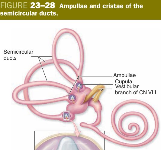
Maculae carry rocks (otoliths) — gravity-loaded sensors of linear motion. Cristae carry sails (cupulae) — current-driven sensors of rotation. Knock the rocks off their gel mat into the wrong semicircular canal and they swirl with head movement — that is benign paroxysmal positional vertigo (BPPV), the commonest cause of episodic spinning vertigo, fixed by the Epley repositioning manoeuvre.
- Maculae (in utricle & saccule): hair cells + otolithic membrane with otoliths — sense linear acceleration & gravity.
- Cristae ampullares (in semicircular ducts): hair cells + cupula — sense angular (rotational) acceleration.
Corneal endothelial failure → corneal oedema/clouding. Detachment of the retina from the pigment epithelium → vision loss. Damage to outer hair cells (noise, ototoxic drugs, ageing) → sensorineural deafness; dislodged otoliths → BPPV (benign positional vertigo). Meniere disease = endolymph excess → vertigo + SNHL + tinnitus.
TMU Exam Drill
📝 Open the full TMU Question Bank — 20 MCQ + 6 terms + 5 essays →
Authentic Tianjin Medical University past-paper questions (2021 Final & the multi-section Final with answer key) mapped to this unit, in the real exam format. Click Show answer to self-test.
□ Single best answer
- A. pigment epithelium
- B. bipolar cells
- C. ganglion cells
- D. Müller cells
- E. microglial cells
- A. pillar cells
- B. phalangeal cells
- C. hair cells
- D. bipolar cells
- E. cones
□ Fill in the blank
- (fill the cell + two supporting types)
- (fill four)
Eye & Ear complete
Cornea, retina's 4 cells & the organ of Corti mastered. Next: Male Reproductive.