Respiratory System
Two Functional Portions
Before you open a single slide, hold one idea in your head: the respiratory tract is two systems in one tube. The upper part is plumbing — it takes outside air, warms it to body temperature, humidifies it, filters out particles, and delivers it downstream. No gas crosses any wall up here. The lower part is the gas-exchange membrane — a microscopic interface where oxygen and carbon dioxide trade places between alveolar air and red cells. The whole architecture of the airway makes sense once you ask, at every level, “is this still plumbing, or is this exchange?”
The dividing line is precise and examinable. As long as a tube has a complete wall — no alveoli budding off — it is conducting. The moment alveoli appear on its wall, however few, it becomes respiratory. The last purely conducting segment is the terminal bronchiole; the first respiratory segment is the respiratory bronchiole. You will be asked which side of that line every named structure belongs on.
Think of an airport. The long corridors and security gates exist only to bring you safely to the boarding gate — they are conducting. The actual aircraft doors are where you cross from terminal to plane — that crossing is gas exchange. A respiratory tract without a long conducting zone would dump cold, dirty, dry air straight onto a fragile gas-exchange membrane. The two zones are not redundant; the conducting zone protects the respiratory zone.
| Portion | Parts | Job |
|---|---|---|
| Conducting | Nasal cavity, pharynx, larynx, trachea, bronchi, bronchioles, terminal bronchioles | Conduct, warm, moisten & clean air (no gas exchange) |
| Respiratory | Respiratory bronchioles, alveolar ducts, alveolar sacs, alveoli | Gas exchange |
“If it has alveoli, it's respiratory.” The terminal bronchiole is the LAST conducting segment; the respiratory bronchiole (first with alveoli budding off its wall) begins the respiratory portion.
Conducting Portion
Walk down the conducting tree and you are watching the same wall recipe being simplified at every step. Start at the nose, where the vestibule is lined by keratinised stratified squamous skin carrying coarse vibrissae — bouncers at the door for grit and insects. A little deeper, that skin gives way to respiratory epithelium: pseudostratified ciliated columnar with goblet cells, sitting on a thick basement membrane over a vascular lamina propria. This is the signature epithelium of essentially the entire conducting tree, and the olfactory patch in the roof of the nasal cavity is the one specialised exception — pseudostratified neurons, sustentacular cells, basal cells, with Bowman glands beneath them.
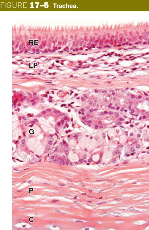
At the trachea you meet the classical textbook layers: respiratory epithelium on a strikingly thick basement membrane, a lamina propria of loose connective tissue, a submucosa packed with seromucous glands that pump out the airway fluid, and the famous C-shaped hyaline cartilage rings holding the lumen open. The open posterior face of each “C” is bridged by the trachealis smooth muscle — that gap exists so a swallowed bolus can bulge into the trachea without being blocked by rigid cartilage.
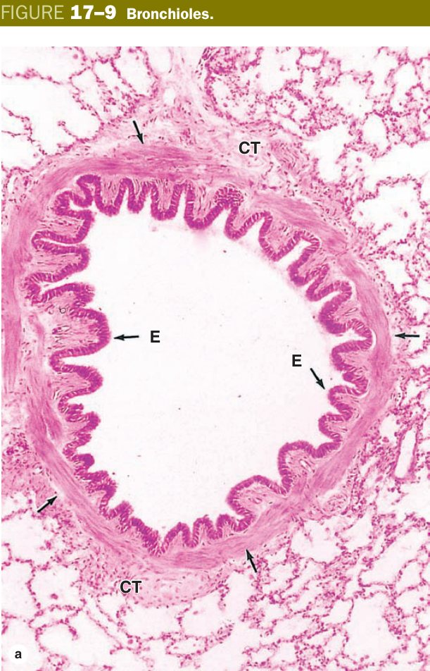
As you descend from trachea into bronchi, the rings break up into plates of cartilage, smooth muscle begins to form a continuous layer between epithelium and cartilage, and the seromucous glands gradually thin out. Then comes the most important transition in the whole tree: when the tube drops below roughly 1 mm in diameter, cartilage disappears completely and glands disappear with it. You are now in a bronchiole. The wall is essentially epithelium and a relatively thick band of smooth muscle — which is exactly why a bronchospasm in asthma squeezes here and nowhere else.
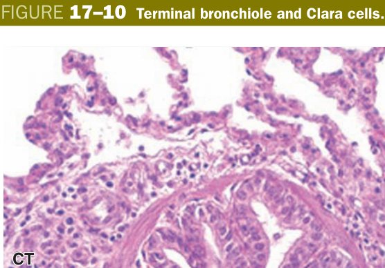
Inside the bronchioles the epithelium itself thins and changes character. Goblet cells vanish; ciliated cells shorten from columnar to cuboidal; and a new cell takes over — the club cell (Clara cell): non-ciliated, dome-shaped, with apical secretory granules. Club cells secrete a surfactant-like lipoprotein, the small protein CC16, and a battery of cytochrome P450 detoxifying enzymes that neutralise inhaled toxins. They also act as the local progenitor cell, regenerating both ciliated and club cells after injury. By the terminal bronchiole the lining is simple cuboidal with only occasional cilia — the final conducting checkpoint before gas exchange begins.
Picture the wall ingredients as a stack of toppings — epithelium, goblet cells, glands, cartilage, smooth muscle. As you travel distally you peel toppings off, not on. Goblet cells, glands and cartilage all leave together at the bronchus–bronchiole boundary. What remains is a thin tube whose only meaningful contractile element is smooth muscle. That is why “no cartilage, no glands” is the bronchiole’s exam fingerprint.
| Segment | Epithelium | Wall features |
|---|---|---|
| Trachea | Pseudostratified ciliated columnar + goblet | C-shaped hyaline cartilage rings; trachealis smooth muscle posteriorly |
| Bronchi | Pseudostratified ciliated columnar | Cartilage plates, smooth muscle, glands |
| Bronchioles | Simple columnar → cuboidal ciliated | No cartilage, no glands; relatively more smooth muscle |
| Terminal bronchiole | Simple cuboidal with Clara (club) cells + few ciliated | Last conducting segment; Clara cells secrete surfactant-like protein & detoxify |
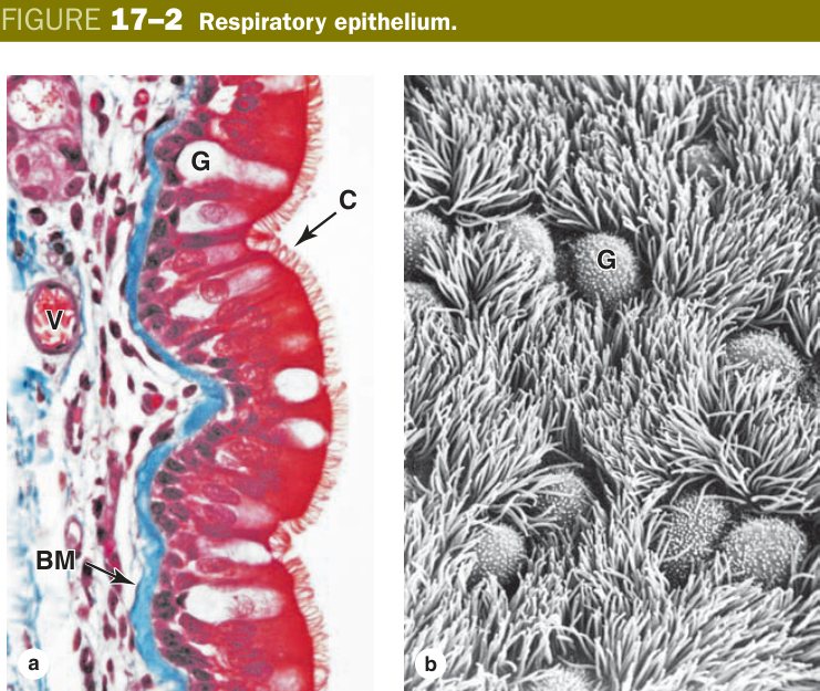
The mucociliary escalator is the single most important defence the conducting tree has, and it is built from just two cell types working against a two-phase fluid. Goblet cells, scattered through the pseudostratified epithelium like flask-shaped wineglasses, dump glycoprotein mucin onto the apical surface. There the mucin hydrates into a sticky gel layer that floats on top of a thinner, watery sol layer secreted by the submucosal seromucous glands. The cilia of the columnar cells are tuned exactly to this geometry — their shafts beat freely in the watery sol, but their tips just catch the underside of the gel during the power stroke, slapping the whole sticky blanket upward.
Each cilium is a microtubule cantilever with a 9+2 axoneme: nine peripheral doublets around a central pair, anchored to a basal body. The motor is dynein — little arms that walk one doublet against its neighbour. Because the doublets are tethered together by nexin links, that sliding is converted into a bending beat. Adjacent cells coordinate so the beat travels in waves, and the mucus blanket migrates from the bronchioles all the way up to the pharynx, where it is swallowed and harmlessly digested by gastric acid. You inhale a city’s worth of particulate every day; the escalator quietly delivers it to your stomach.
Imagine a conveyor belt in a factory. The belt itself is the watery sol layer — it moves smoothly because nothing sticky is touching the rollers. On top of the belt is a sheet of flypaper — the gel mucus — that catches every fleck of dust dropped onto it. The cilia are the rollers. Defects in any of these three — immotile rollers (Kartagener), dry belt (cystic fibrosis), or no flypaper at all (severe smoking) — jam the line and infection follows.
| Cell type | Structure | Function |
|---|---|---|
| Ciliated columnar cell | ~300 cilia per cell; cilia beat at ~12 Hz toward pharynx | Propels the mucous blanket upward |
| Goblet cell | Flask-shaped; abundant apical mucin granules | Secretes gel-phase mucus (sticky layer trapping particles & pathogens) |
| Serous cell | Seromucous gland cells (submucosal) | Watery sol-phase (low-viscosity periciliary fluid; ciliary tips beat in this layer) |
| Basal cell | Pyramidal, rest on BM; not ciliated | Stem cells for the epithelium |
The mucous blanket has two layers: (1) a thin sol phase (periciliary layer, low viscosity) in which ciliary shafts beat; (2) a thick gel phase (mucus layer, sticky) carried on top. Ciliary tips engage the gel phase during the power stroke, sweeping mucus (+ trapped particles) toward the pharynx where it is swallowed.
Primary ciliary dyskinesia (Kartagener's syndrome) — dynein arm defect → immotile cilia → recurrent sinopulmonary infections + situs inversus + male infertility. Cystic fibrosis — defective CFTR → thick, dehydrated mucus → impaired clearance → chronic infection. Smoking — ciliotoxic; goblet cell hyperplasia; chronic bronchitis (productive cough >3 months for >2 years).
Respiratory Portion
Cross the threshold into the respiratory portion and the wall starts to dissolve. The respiratory bronchiole still looks like a bronchiole at first — simple cuboidal lining of club cells and a few ciliated cells, smooth muscle in the wall — but every so often a pouch in that wall opens directly into an alveolus. Gas exchange has begun, intermittently, along a tube still trying to be a conducting airway. Further down, the wall gives up entirely: the alveolar duct is little more than a corridor flanked by continuous rows of alveoli, ringed by smooth-muscle “knobs” at the alveolar openings. Several alveolar ducts then open together into a shared chamber, the alveolar sac, which is simply a cluster of alveoli sharing a common atrium.
The terminal unit — the alveolus itself — is a 200µm polyhedral air pocket whose entire wall is the gas-exchange surface. There are roughly 300 million alveoli in an adult lung, giving a total exchange area of around 75 square metres — a tennis court folded into your chest. Every detail of alveolar histology — thin type I cells, fused basement membranes, dense capillary mesh, surfactant film — exists to keep that 75 m² thin enough for oxygen to diffuse across in less than a quarter of a second.
| Segment | Structure |
|---|---|
| Respiratory bronchiole | Simple cuboidal (Clara + ciliated); a few alveoli open off its wall (first gas exchange) |
| Alveolar duct | Simple squamous/cuboidal; wall is a row of alveolar openings with smooth-muscle “knobs” |
| Alveolar sac | Common space into which a cluster of alveoli open |
| Alveolus | Sac-like outpocket; site of gas exchange |
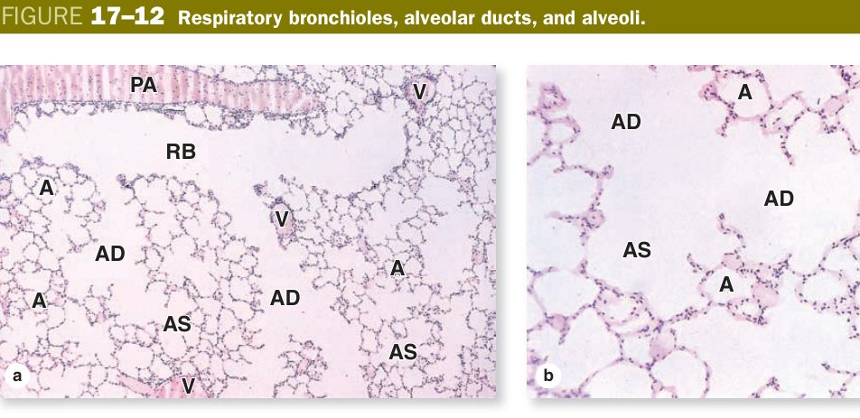
Lean a slide of healthy lung under the microscope and you will see two pneumocyte populations sharing the alveolar wall in a very lopsided division of labour. The type I pneumocyte is a simple squamous sheet so thin it almost vanishes — cytoplasm just a few hundred nanometres deep around a flattened nucleus. Only about 40% of the cells in the wall are type I, yet they cover roughly 95% of the surface area. That extreme thinness is the whole point: type I cells form the air-facing leaf of the blood-air barrier and let O₂ and CO₂ diffuse across in milliseconds. They are also post-mitotic — once damaged, they cannot replace themselves.
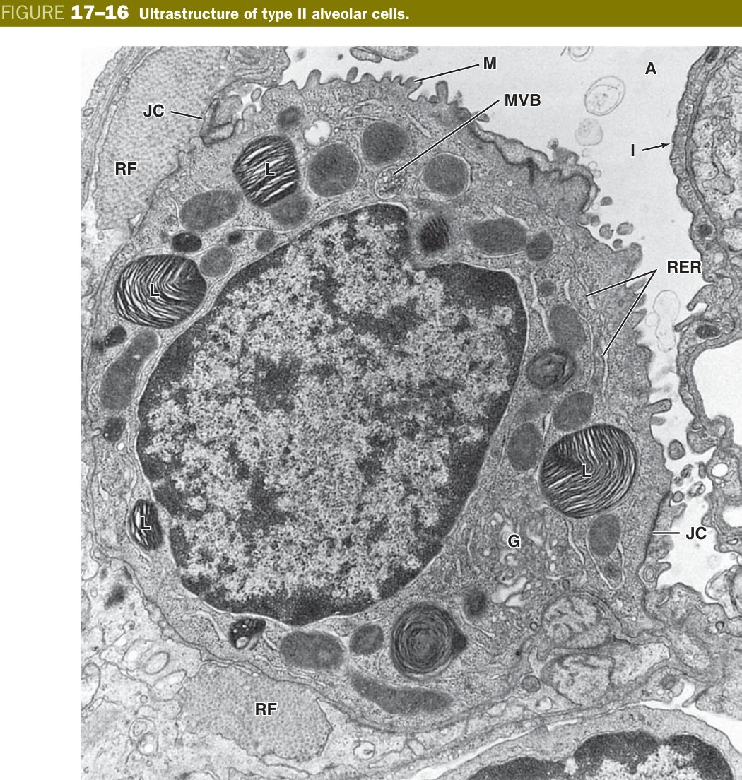
The type II pneumocyte is the opposite cell in every way. It is cuboidal, sits in the corners between adjacent type I sheets, and its cytoplasm is stuffed with characteristic lamellar bodies — whorled membrane-bound packets of phospholipid plus the surfactant proteins SP-A, SP-B, SP-C and SP-D. Lamellar bodies are exocytosed onto the alveolar surface where they unfold into a thin surfactant film dominated by dipalmitoylphosphatidylcholine (DPPC). Surfactant’s job is to lower the surface tension of the water lining the alveolus — without it, surface tension would collapse small alveoli into their larger neighbours every time you breathed out. Type II cells are also the alveolar stem cell: after a type I cell dies, a neighbouring type II divides and one daughter flattens out into a new type I.
In between the alveoli lies the interalveolar septum, which is far more than a wall — it is the meeting place of the two circulations of the lung. A dense capillary network runs through it, the capillary endothelium often pressing its basement membrane right against the type I cell’s basement membrane so that the two fuse into a single thin layer. The septum also carries elastic and reticular fibres (the source of the lung’s recoil), free alveolar macrophages drifting through, and the occasional pore of Kohn — a small hole in the septum that lets air bypass a blocked bronchiole by spilling sideways into the next alveolus.
Blow two soap bubbles of different sizes on the same straw and the small one empties into the big one — surface tension is higher in tighter curves (Laplace’s law). Your lungs face exactly this threat: 300 million alveoli at slightly different sizes, all connected. Surfactant fixes the maths. Because the same amount of surfactant gets crowded together in a small alveolus and stretched out in a big one, it lowers tension more in the small alveoli — equalising the pressures and stopping the cascade collapse. A premature baby missing surfactant cannot do this; alveoli pop one by one into hyaline-membrane RDS.
| Cell | Structure & function |
|---|---|
| Type I pneumocyte (Type I alveolar cell) | Simple squamous, extremely flat; covers ~95% of the alveolar surface; forms the blood-air barrier (gas exchange) |
| Type II pneumocyte (Type II alveolar cell) | Cuboidal, vacuolated cytoplasm with lamellar bodies; secretes surfactant (reduces surface tension); also the alveolar stem cell |
| Alveolar macrophage (dust cell) | Phagocytoses dust/debris in the alveolar lumen & septum |
Alveolar septum (interalveolar septum): the thin wall between two adjacent alveoli; it contains the alveolar (continuous) capillaries, elastic and reticular fibres, and alveolar macrophages — the elastic fibres give the lung its recoil.
Any particle small enough to bypass the mucociliary escalator and land on an alveolar surface meets the lung’s last line of defence: the alveolar macrophage, traditionally called the dust cell. These are mononuclear phagocytes derived from blood monocytes, recruited continuously from the bone marrow. They wander freely across the alveolar lumen and through the septal connective tissue, phagocytosing inhaled carbon, silica fragments, bacteria, viral particles and even old surfactant that the type II cells are recycling. Once loaded, most of them migrate up into the mucociliary escalator and ride out with the mucus; a few drain through the lymphatics.
The cytoplasm of an alveolar macrophage is a museum of what its host has inhaled. A city-dweller’s macrophages are sooty black from carbon — the condition called anthracosis, harmless on its own. A coal miner’s carry far more, and the same cell loaded with silica crystals triggers fibrogenic cytokines that scar the lung (silicosis). In left-sided heart failure the pulmonary capillaries leak red cells into the alveoli; macrophages eat them and digest the haemoglobin down to haemosiderin, which stains golden-brown in H&E. These haemosiderin-laden macrophages are the famous “heart failure cells”, found in the sputum and easily identified with a Prussian blue iron stain.
The septum has another, more architectural feature worth knowing: tiny holes called pores of Kohn, 5–15 µm across, that connect adjacent alveoli through their shared wall. They serve two functions. First, they permit collateral ventilation — if a small airway is plugged, the alveoli distal to it can still receive air sideways from a neighbour, preventing immediate collapse. Second, they let macrophages migrate between alveoli to find the dirty ones. The downside is that they also let pathogens and oedema fluid spread — classic lobar pneumonia spreads alveolus-to-alveolus through these very pores.
Think of the alveolar macrophage as a janitor pushing a transparent rubbish bag through 75 m² of corridor. Whatever they pick up stays visible through the bag — black carbon, white silica, golden iron — so the pathologist can reconstruct the patient’s exposure history simply by looking at their sputum.
| Structure | Details |
|---|---|
| Alveolar macrophage (dust cell) | Derived from blood monocytes; roam the alveolar lumen & septum; phagocytose inhaled dust, bacteria, and cellular debris; present antigens to lymphocytes in the septum |
| Heart failure cells | Alveolar macrophages that have phagocytosed haemosiderin (from extravasated RBCs in left heart failure); appear golden-brown in H&E; found in sputum |
| Pores of Kohn | Small openings (5–15 µm) in alveolar walls; allow collateral ventilation between adjacent alveoli — preventing collapse if a bronchiole is blocked |
Alveolar macrophage (dust cell): a mononuclear phagocyte derived from blood monocytes, residing in the alveolar lumen and interalveolar septum. It constitutes the first line of alveolar defence, phagocytosing particulate matter, pathogens, and surfactant debris. Its cytoplasm accumulates ingested carbon (black in city dwellers — anthracosis) or haemosiderin in left-sided heart failure (“heart failure cells”).
The Blood-Air Barrier
The blood-air barrier is the structure that justifies everything else in this unit. It is the actual diffusion membrane — the wall that an oxygen molecule must cross to reach a red cell, and that a CO₂ molecule must cross to escape. Anatomically it is astonishingly thin: 0.2 to 0.5 micrometres at its narrowest, less than one-fifth the diameter of a red blood cell. You can think of it as a sandwich with three layers. From the alveolar side: (1) a film of surfactant riding on a thin water layer, then the cytoplasm of a type I pneumocyte; (2) a single fused basement membrane — in most places the basal laminae of the pneumocyte and the endothelium press together so tightly that they merge into one electron-dense layer; (3) the capillary endothelial cytoplasm, with its own thin nucleus-free regions facing the alveolus.
The geometry is no accident. Fick’s law of diffusion says that flux is proportional to surface area divided by thickness — so evolution has maximised the area (75 m²) and minimised the thickness (a fraction of a micrometre). Anything that thickens the barrier — pulmonary oedema putting fluid into the septum, the hyaline membranes of acute respiratory distress syndrome, the collagen of idiopathic pulmonary fibrosis, the granulomas of sarcoidosis — produces clinically obvious hypoxaemia long before CO₂ retention shows up, because oxygen is the more diffusion-limited gas.
Picture two rooms separated by a sheet of cling-film stretched over a tennis-court-sized window frame. The film is so thin that perfume sprayed in one room is smelled in the other within seconds — not because air rushes through, but because individual molecules diffuse across an essentially-zero barrier. Now imagine taping a sheet of paper over the cling-film. The barrier is suddenly 100 times thicker; the perfume takes minutes to cross. Pulmonary oedema is that piece of paper.
Blood-air barrier: the thin structure (~0.2–0.5 µm) across which O₂ & CO₂ diffuse between alveolar air and capillary blood. Its layers are: (1) the surfactant film + Type I pneumocyte cytoplasm; (2) the fused basal laminae of the pneumocyte & endothelium; (3) the capillary endothelium.
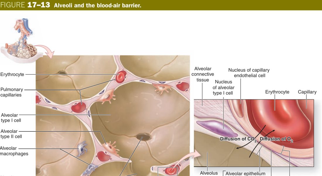
Three layers, lumen → blood: Type I pneumocyte (+ surfactant) → fused basal laminae → capillary endothelium. It must be thin — Type I cells, not Type II, line most of the alveolus precisely so the barrier is minimal.
Insufficient surfactant (premature Type II cells) → neonatal respiratory distress syndrome. Thickening of the blood-air barrier (oedema, fibrosis) impairs gas exchange. Loss of alveolar elastic fibres → emphysema. Dust cells laden with carbon → anthracosis.
Pleura & Lung Interstitium
Each lung is wrapped in a glistening serous sac — a closed double-walled balloon called the pleura. Both walls of this balloon are built from the same epithelial sheet, a simple squamous mesothelium derived embryologically from lateral plate mesoderm, sitting on a thin layer of vascular connective tissue. The two walls have different names depending on what they cover: the inner wall, glued to the lung surface and dipping into the fissures, is the visceral pleura; the outer wall, plastered against the inner chest wall, mediastinum and diaphragm, is the parietal pleura. The space between them is the pleural cavity, normally collapsed down to a capillary-thin film of serous fluid that the mesothelial cells secrete and reabsorb.
That fluid does two jobs at once. It lubricates — so the two pleural sheets glide over each other freely with every breath — and it couples, because the surface tension of a thin water layer is strong enough to hold two glass plates together. The lung is not stuck to the chest wall by glue; it is held there by the surface tension of pleural fluid, the same way two wet microscope slides resist being pulled apart. When air enters the pleural space (a pneumothorax) that coupling is broken, the lung’s elastic recoil collapses it inward, and the chest wall springs outward.
The two pleurae differ critically in one respect — their innervation. The parietal pleura is supplied by somatic intercostal and phrenic nerves and is exquisitely pain-sensitive; the visceral pleura is supplied only by autonomic fibres from the pulmonary plexus and has no pain perception at all. This is why pleurisy — inflammation of the parietal layer — produces sharp, breath-locked chest pain, while a lung tumour can grow silently against the visceral pleura until it finally reaches the parietal surface and announces itself.
Press two wet glass slides together and try to pull them apart straight — impossible, the water film resists you. Slide them sideways instead and they glide effortlessly. That is exactly how the pleurae work: the lung must slide against the chest wall every second of your life but never separate from it. The thin serous film delivers both at once.
| Layer | Epithelium | Notes |
|---|---|---|
| Visceral pleura | Simple squamous mesothelium + CT + elastic fibres + smooth muscle | Tightly adherent to the lung surface; has its own blood supply (bronchial arteries) |
| Parietal pleura | Simple squamous mesothelium + CT | Lines the thoracic cavity & diaphragm; somatic innervation (phrenic & intercostal nerves) → pain sensation |
| Pleural cavity | — | Potential space with small volume of serous fluid (lubricates the sliding surfaces) |
Pneumothorax — air enters the pleural cavity (trauma, ruptured bleb); lung collapses. Haemothorax — blood in the pleural cavity. Pleuritis (pleurisy) — inflammation of the pleura; friction rub on auscultation (parietal pleura has pain fibres; visceral does not). Mesothelioma — malignant tumour of pleural mesothelial cells, strongly associated with asbestos exposure.
TMU Exam Drill
📝 Open the full TMU Question Bank — 20 MCQ + 6 terms + 5 essays →
Authentic Tianjin Medical University past-paper questions (2021 Final & the multi-section Final with answer key) mapped to this unit, in the real exam format. Click Show answer to self-test.
□ Single best answer
- A. alveolus
- B. respiratory bronchiole
- C. alveolar duct
- D. alveolar sac
- E. terminal bronchiole
□ Fill in the blank
- (fill the four)
□ Explain the following terms
Respiratory system complete
Conducting vs respiratory portion, pneumocytes & the blood-air barrier mastered. Next: Eye & Ear.